



Anticancer agent 64
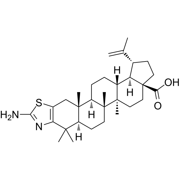
Anticancer agent 64 (compound 5m) shows cytotoxic activity in CCRF-CEM cells, with IC50 of 2.4 μM. Anticancer agent 64 shows good anticancer activity through apoptosis induction. Anticancer agent 64 induces caspase 3 and 7 activation and PARP cleavage. Anticancer agent 64 induces significant effect of mitochondria depolarization.
For research use only. We do not sell to patients.
- CAS No.: 2387902-92-1
- Formula: C31H46N2O2S
- Molecular Weight:510.77
-
Storage:
Please store the product under the recommended conditions in the Certificate of Analysis.

To place orders, for customer services and technical support, please contact: MedChemExpress USA
Tel: 609-228-6898 E-mail: [email protected] [email protected]


-
 Biological Activity
-
Chemical Information
- Protocols
- Purity & Documentation
- References
-
 Help & FAQs


All Caspase Isoforms
More

Biological Activity
Description
IC50 & Target
|
Caspase 3 |
Caspase-7 |
Bcl-2 |
Bax |
PARP |
Cellular Effect
|
Cell Line

|
Type | Value | Description | References |
|---|---|---|---|---|
| A549 | IC50 |
38.4 μM
Compound: 5m
|
Cytotoxicity against human A549 cells incubated for 72 hrs by MTS assay
Cytotoxicity against human A549 cells incubated for 72 hrs by MTS assay
|
[PMID: 31677446] |
| BJ | IC50 |
>50 μM
Compound: 5m
|
Cytotoxicity against human BJ cells incubated for 72 hrs by MTS assay
Cytotoxicity against human BJ cells incubated for 72 hrs by MTS assay
|
[PMID: 31677446] |
| CCRF-CEM | IC50 |
2.4 μM
Compound: 5m
|
Cytotoxicity against human CCRF-CEM cells incubated for 72 hrs by MTS assay
Cytotoxicity against human CCRF-CEM cells incubated for 72 hrs by MTS assay
|
[PMID: 31677446] |
| HCT-116 | IC50 |
36.5 μM
Compound: 5m
|
Cytotoxicity against human HCT116 cells incubated for 72 hrs by MTS assay
Cytotoxicity against human HCT116 cells incubated for 72 hrs by MTS assay
|
[PMID: 31677446] |
| HCT-116 | IC50 |
48.3 μM
Compound: 5m
|
Cytotoxicity against p53 knockout human HCT116 cells incubated for 72 hrs by MTS assay
Cytotoxicity against p53 knockout human HCT116 cells incubated for 72 hrs by MTS assay
|
[PMID: 31677446] |
| K562 | IC50 |
>50 μM
Compound: 5m
|
Cytotoxicity against human K562 cells incubated for 72 hrs by MTS assay
Cytotoxicity against human K562 cells incubated for 72 hrs by MTS assay
|
[PMID: 31677446] |
| MRC5 | IC50 |
>50 μM
Compound: 5m
|
Cytotoxicity against human MRC5 cells incubated for 72 hrs by MTS assay
Cytotoxicity against human MRC5 cells incubated for 72 hrs by MTS assay
|
[PMID: 31677446] |
| U2OS | IC50 |
48 μM
Compound: 5m
|
Cytotoxicity against human U2OS cells incubated for 72 hrs by MTS assay
Cytotoxicity against human U2OS cells incubated for 72 hrs by MTS assay
|
[PMID: 31677446] |
In Vitro
Anticancer agent 64 (compound 5m) increases BAX expression and decreases Bcl-2 protein expression[1].
.
Anticancer agent 64 shows significant increasement in cells accumulation in the G2/M phase of the cell cycle, while proportion of cells in S phase is reduced[1].
MedChemExpress (MCE) has not independently confirmed the accuracy of these methods. They are for reference only.


Chemical Information
-
CAS No. 2387902-92-1
-
Molecular Weight 510.77
-
Formula C31H46N2O2S
-
SMILES
OC([C@]12[C@@]([C@@H](CC2)C(C)=C)([H])[C@]3([H])[C@@](CC1)([C@]4([C@]([C@@]5([C@@](C(C)(C6=C(SC(N)=N6)C5)C)([H])CC4)C)([H])CC3)C)C)=O
-
Shipping
Room temperature in continental US; may vary elsewhere.
-
Storage
Please store the product under the recommended conditions in the Certificate of Analysis.


Purity & Documentation
References

Calculators
Concentration (start) × Volume (start) = Concentration (final) × Volume (final)


Anticancer agent 64 Related Classifications
HY-147514 Related Classifications
 Powered by Bioz