



DIF-3
Based on 8 publication(s) in Google Scholar
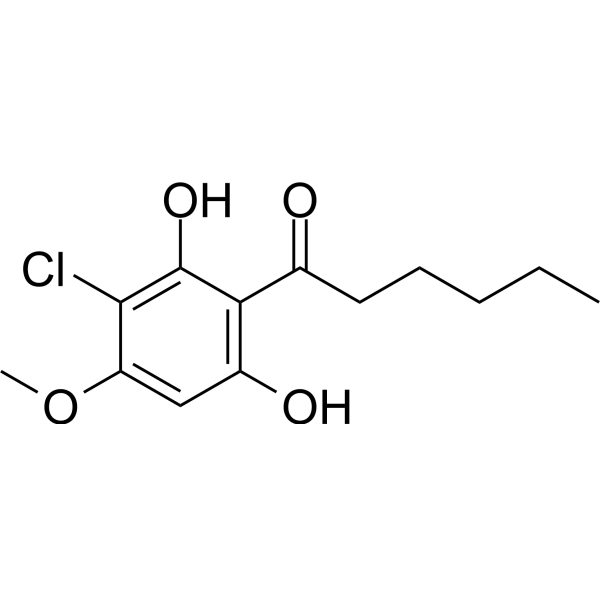
DIF-3 is an orally active anticancer agent. DIF-3 reduces the expression levels of cyclin D1 and c-Myc by facilitating their degradation via activation of GSK-3β. DIF-3 inhibits Wnt/β-catenin signaling pathway-related proteins in cells. DIF-3 induces reactive oxygen species (ROS) and autophagy. DIF suppresses the growth of Trypanosoma. cruzi in HT1080 cells. DIF-3 exerts antitumor effects both in vitro and in vivo.
For research use only. We do not sell to patients.
- Purity : 99.84%
- CAS No.: 113411-17-9
- Formula: C13H17ClO4
- Molecular Weight:272.72
-
Storage:
4°C, protect from light
* In solvent : -80°C, 6 months; -20°C, 1 month (protect from light)

To place orders, for customer services and technical support, please contact: MedChemExpress USA
Tel: 609-228-6898 E-mail: [email protected] [email protected]


-
 Biological Activity
-
Chemical Information
-
Solvent & Solubility
- Protocols
- Purity & Documentation
- References
-
 Help & FAQs


-

HY-10182
LaduviglusibGSK-3 Inhibitor -
HY-139324
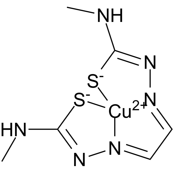
Cu(II)GTSMGSK3β/AβOs Inhibitor -
HY-N8423
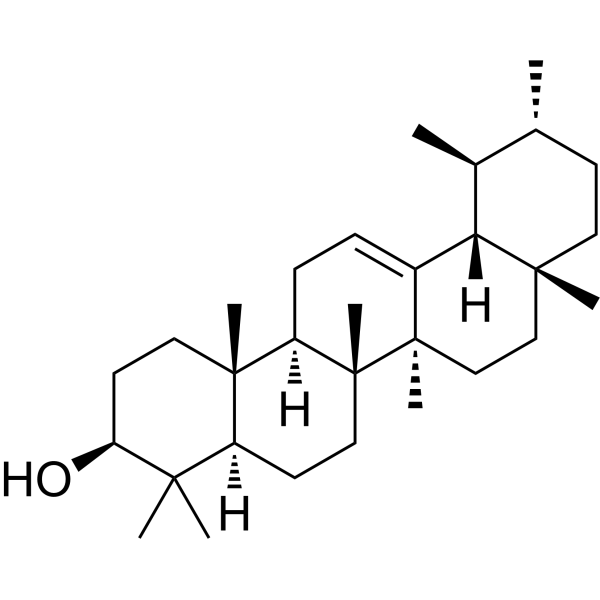
α-AmyrinPentacyclic Triterpenoid -
HY-12012
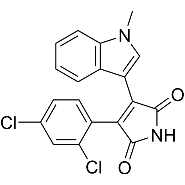
SB 216763Autophagy Inducer -
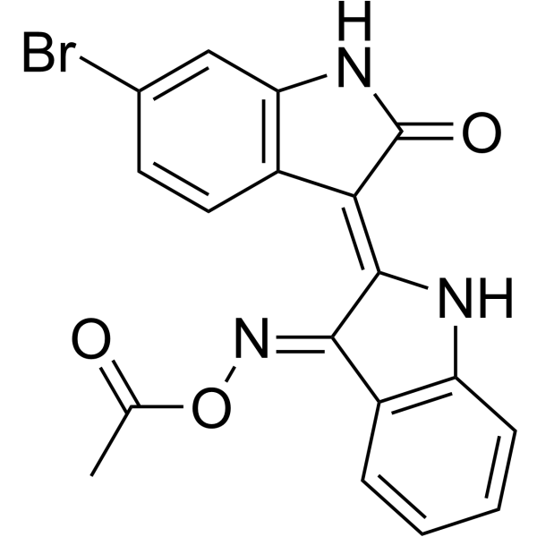
HY-15356
BIO-acetoxime -

HY-14648
DexamethasoneGR Agonist -
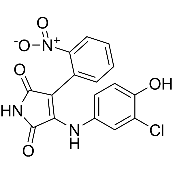
HY-15438
SB 415286Apoptosis Inducer -
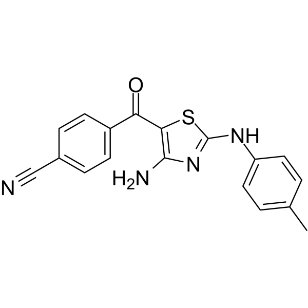
HY-100950
ABC1183GSK3/CDK9 Inhibitor -
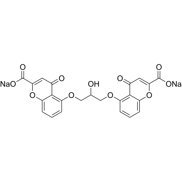
HY-B0320A
Cromolyn disodiumMast Cell Stabilizer -
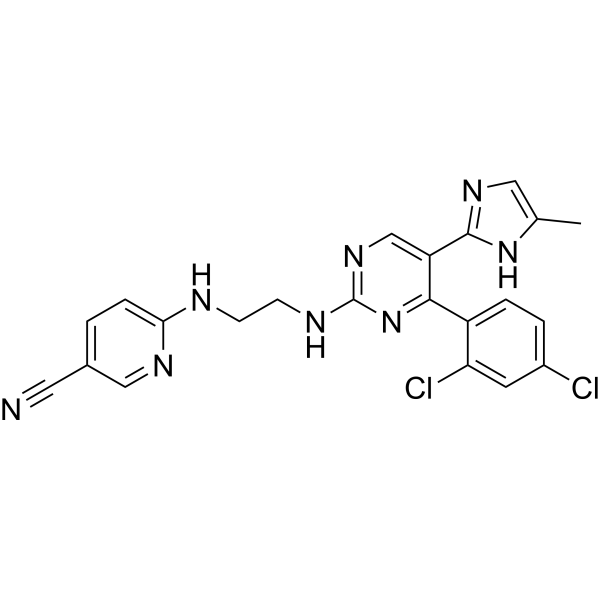
HY-10182G
Laduviglusib (GMP)GSK-3α/β Inhibitor (GMP) -
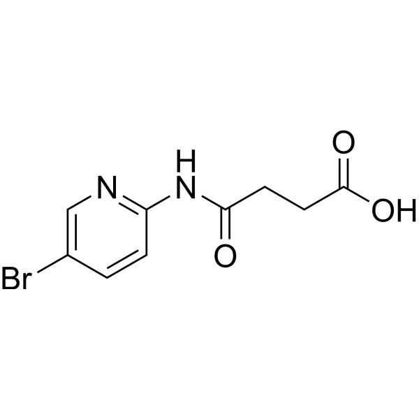
HY-12524
BikininGSK-3 Inhibitor -
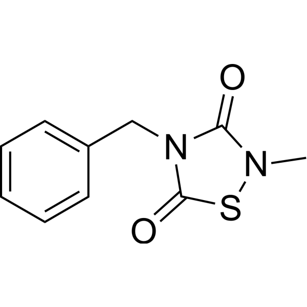
HY-11012
TDZD-8GSK-3 Inhibitor
Publications Citing Use of MedChemExpress (MCE) DIF-3
More- J Transl Med. 2026 Feb 9;24(1):361. [Abstract]
- Oncogene. 2023 Nov;42(47):3503-3513. [Abstract]
- Mol Med. 2024 Dec 20;30(1):260. [Abstract]
- Int J Oncol. 2026 Jan;68(1):8. [Abstract]
- mBio. 2025 Sep 25:e0037325. [Abstract]
- Diabetol Metab Syndr. 2025 Jun 5;17(1):193. [Abstract]
- Placenta. 2025 Oct 7:171:194-204. [Abstract]
- bioRxiv. 2025 Feb 8:2025.02.07.637189. [Abstract]


Biological Activity
Description
Cellular Effect
|
Cell Line

|
Type | Value | Description | References |
|---|---|---|---|---|
| A549 | IC50 |
2 μM
Compound: DIF-3
|
Growth inhibition of human A549 cells
Growth inhibition of human A549 cells
|
[PMID: 28814374] |
| S2 | IC50 |
4.6 μM
Compound: 2, DIF-3
|
Immunosuppressive activity in Drosophila S2 cells expressing att-luc reporter gene assessed as inhibition of peptidoglycan-induced attacin production pre-treated for 1.5 hrs before peptidoglycan stimulation for 8 hrs by luciferase reporter gene assay
Immunosuppressive activity in Drosophila S2 cells expressing att-luc reporter gene assessed as inhibition of peptidoglycan-induced attacin production pre-treated for 1.5 hrs before peptidoglycan stimulation for 8 hrs by luciferase reporter gene assay
|
[PMID: 26122773] |
In Vitro
DIF-3 (10-30 μM, 24-48 h) inhibits proliferation in a dose-dependent manner in human colon cancer cells[1].
DIF-3 (30 μM, 24 h) arrests the cell cycle at G0/G1 phase in human colon cancer cells[1].
DIF-3 (10-30 μM, 1-24 h) induces proteolysis of cyclin D1 and c-Myc in human colon cancer cells via activation of GSK-3β[1].
DIF-3 (10-30 μM, 1-24 h) inhibits Wnt/β-catenin signaling pathway-related proteins in human colon cancer cells[1].
DIF-3 (10 μM, 4 h) inhibits lysophosphatidic acide (LPA) stimulated migration of murine osteosarcoma LM8 cells[2].
DIF-3 (20 μM, 0.25-24 h) inhibits mTOR signaling and induces autophagy in K562 CML cells[3].
DIF-3 (20 μM) induces mitochondrial membrane potential (MMP) (0-10 min), a decrease of ATP content (6 h) and an induction of ROS production (0-30 min) in K562 cells[3].
DIF-3 (10 μM, 3 d) suppresses the growth of Trypanosoma. cruzi in HT1080 cells with an IC50 of 3.95 μM[4].
MedChemExpress (MCE) has not independently confirmed the accuracy of these methods. They are for reference only. Further protocols information, click here.
-
Cell Line:HCT-116, DLD-1
-
Concentration:10, 20, 30 μM
-
Incubation Time:24, 48 h
-
Result:Strongly inhibited proliferation in a dose-dependent manner in both cell lines.
HCT-116 cells were more sensitive to DIF-3 than DLD-1 cells.
-
Cell Line:HCT-116, DLD-1
-
Concentration:30 μM
-
Incubation Time:24 h
-
Result:Significantly increased the number of cells in G0/G1 phase and decreased those in S phase in both cell lines.
Also significantly increased the number of cells in G2 phase when HCT-116 cells were employed, this effect was not observed by using DLD-1 cells.
-
Cell Line:LM8
-
Concentration:10 μM
-
Incubation Time:4 h
-
Result:Suppressed LPA-induced cell migration, whereas did not significantly affected cell migration in the absence of LPA.
-
Cell Line:K562
-
Concentration:20 μM
-
Incubation Time:0.25, 0.5, 1, 3, 6, 24 h for mTOR study; 6, 16, 36, 48 h for Cathepsin B activity study
-
Result:Induced the rapid dephosphorylation of mTOR at Ser2481. Inhibited the mTOR pathway and leads to a rapid and robust conversion of LC3-I to LC3-II.
Increased autophagy was accompanied by the late activation of cathepsin B.
-
Cell Line:HCT-116
-
Concentration:10, 20, 30 μM
-
Incubation Time:1, 3, 6, 12, 24 h
-
Result:Induced a rapid and marked reduction in the amount of cyclin D1 protein and c-Myc protein in a time and dose-dependent manner.
Had no effect on the expression level of β-catenin (30 μM for 24 h).
Reduced the amount of TCF7L2 which increased gradually over time in control cells, in a time and dose-dependent manner.
Reduction of TCF7L2 was associated with the transcriptional inhibition of cyclin D1 mRNA in HCT-116 cells.
-
Cell Line:HCT-116
-
Concentration:30 μM
-
Incubation Time:1 h
-
Result:Effects could be attenuated by MG132 (HY-13259) and SB216763 (HY-12012), indicating the involvement of GSK-3β in degradation of cyclin D1 and c-Myc.
In Vivo
MedChemExpress (MCE) has not independently confirmed the accuracy of these methods. They are for reference only.
-
Animal Model:Mutyh-/- mice (KBrO3 dissolved in water at a concentration of 2 g/L was administered to 4-week-old mice for 12 weeks)[1]
-
Dosage:150 mg/kg
-
Administration:Oral gavage (p.o.); once a day for 5 days/week over 4 weeks
-
Result:Markedly reduced the number of intestinal tumors, especially the number of large tumors with a diameter of >2.0 mm.
No differences in the appearance, activity, body weight, or blood cell counts.
The numbers of TCF7L2-and cyclin D1-positive nuclei in tumors were significantly decreased.


Chemical Information
-
CAS No. 113411-17-9
-
Appearance Solid
-
Molecular Weight 272.72
-
Formula C13H17ClO4
-
Color White to off-white
-
SMILES
O=C(CCCCC)C1=C(C=C(C(Cl)=C1O)OC)O
-
Shipping
Room temperature in continental US; may vary elsewhere.
-
Storage
4°C, protect from light
* In solvent : -80°C, 6 months; -20°C, 1 month (protect from light)


Publications (8)
-
Journal Impact Factor
-
Most Recent
-
J Transl Med
MaR1 and NGF combine to inhibit autophagy through the GSK-3β/β-catenin pathway to promote sciatic nerve repair. [Abstract]2026 Feb 9;24(1):361. PMID: 41664203 -
Oncogene
D-mannose induces TFE3-dependent lysosomal degradation of EGFR and inhibits the progression of NSCLC. [Abstract]2023 Nov;42(47):3503-3513. PMID: 37845392 -
Mol Med
CGREF1 modulates osteosarcoma proliferation by regulating the cell cycle through the Wnt/β-catenin signaling pathway. [Abstract]2024 Dec 20;30(1):260. PMID: 39707194 -
Int J Oncol
Ephrin‑B2 promotes gastric cancer growth by inhibiting apoptosis and regulating the cell cycle via the Wnt/β‑catenin signaling pathway. [Abstract]2026 Jan;68(1):8. PMID: 41268595 -
mBio
The Streptococcus pyogenes Rgg2/Rgg3 quorum sensing system causes global suppression of macrophage inflammatory programs via an intranuclear mechanism. [Abstract]2025 Sep 25:e0037325. PMID: 40996031 -
Diabetol Metab Syndr
IL-37 inhibited inflammation to improve gestational diabetes mellitus through the GSK3/NF-κB pathway. [Abstract]2025 Jun 5;17(1):193. PMID: 40474280 -
Placenta
The downregulation of B2R inhibits spiral artery remodeling by reducing the autophagy of trophoblast cells. [Abstract]2025 Oct 7:171:194-204. PMID: 41076744 -
bioRxiv
Suppressed macrophage response to quorum-sensing-active Streptococcus pyogenes occurs at the level of the nucleus. [Abstract]2025 Feb 8:2025.02.07.637189. PMID: 39975246


Solvent & Solubility
In Vitro:
DMSO : 100 mg/mL (366.68 mM; Need ultrasonic; Hygroscopic DMSO has a significant impact on the solubility of product, please use newly opened DMSO)
Please refer to the solubility information to select the appropriate solvent. Once prepared, please aliquot and store the solution to prevent product inactivation from repeated freeze-thaw cycles.
Storage method and period of stock solution: -80°C, 6 months; -20°C, 1 month (protect from light). When stored at -80°C, please use it within 6 months. When stored at -20°C, please use it within 1 month.
Please refer to the solubility information to select the appropriate solvent. Once prepared, please aliquot and store the solution to prevent product inactivation from repeated freeze-thaw cycles.
Storage method and period of stock solution: -80°C, 6 months; -20°C, 1 month (protect from light). When stored at -80°C, please use it within 6 months. When stored at -20°C, please use it within 1 month.
Concentration (start) × Volume (start) = Concentration (final) × Volume (final)
In Vivo:
Select the appropriate dissolution method based on your experimental animal and administration route.
- For the following dissolution methods, please ensure to first prepare a clear stock solution using an In Vitro approach and then sequentially add co-solvents:
- To ensure reliable experimental results, the clarified stock solution can be appropriately stored based on storage conditions. As for the working solution for In Vivo experiments, it is recommended to prepare freshly and use it on the same day.
- The percentages shown for the solvents indicate their volumetric ratio in the final prepared solution. If precipitation or phase separation occurs during preparation, heat and/or sonication can be used to aid dissolution.
In Vivo Dissolution Calculator
Please enter the basic information of animal experiments:
-
-
-
-
Recommended: Prepare an additional quantity of animals to account for potential losses during experiments.
Please enter your animal formula composition:
-
%DMSO +
Recommended: Keep the proportion of DMSO in working solution below 2% if your animal is weak.
-
%+
-
+%Tween-80 + +
-
%Saline +
The co-solvents required include: DMSO, . All of co-solvents are available by MedChemExpress (MCE). , Tween 80. All of co-solvents are available by MedChemExpress (MCE).
Working solution concentration: 0.22 mg/mL
Method for preparing stock solution: mg drug dissolved in μL DMSO. Stock solution concentration: mg/mL. * In solvent : -80°C, 6 months; -20°C, 1 month (protect from light)
1. Take μL DMSO stock solution;
2. Add μL .
μL , mix evenly;
3. Then add μL Tween 80, mix evenly;
4. Then add μL
Please ensure that the stock solution in the first step is dissolved to a clear state, and add co-solvents in sequence. You can use ultrasonic heating (ultrasonic cleaner, recommended frequency 20-40 kHz), vortexing, etc. to assist dissolution.

Protocols
-
RNA extraction experimental
By lysing cells, releasing RNA, and removing impurities such as proteins and DNA, high-purity RNA products are finally obtained. The commonly used traditional method is the guanidine isothiocyanate/phenol/chloroform method (Trizol), which is suitable for a variety of animal materials including animal tissues, microorganisms, cultured cells, etc., and most plant materials.
-
Direct immunofluorescence on tissue biopsies
Direct immunofluorescence (DIF) detects tissue-bound immunoreactants in biopsy sections by applying fluorescein-labeled antibodies directly to frozen tissue; antigen-antibody binding is visualized as fluorescence under a fluorescence microscope, and the anatomic pattern of IgG, IgA, IgM, C3, or fibrin/fibrinogen deposition supports diagnosis of immune-mediated skin, mucosal, and renal disease. Classic interpretive examples include intercellular epidermal staining in pemphigus, linear basement-membrane-zone staining in pemphigoid-spectrum disorders, granular IgA in dermal papillae in dermatitis herpetiformis, and glomerular immune deposits in renal biopsies.
-
ROS/oxidative-stress fluorescent staining
ROS/oxidative-stress fluorescent staining uses cell-permeant fluorogenic probes that become fluorescent after oxidation inside cells or tissues; commonly used examples include DCFH-DA/DCFDA for broad cellular oxidant detection, DHE for superoxide-related signal detection, MitoSOX for mitochondrial superoxide-related signal detection, and CellROX probes for oxidative-stress-associated fluorescence readouts. The assay detects probe oxidation rather than a single ROS species unless the probe and analysis method have been chemically validated for that species. DCFH-DA enters cells, is deacetylated by intracellular esterases to DCFH, and produces fluorescent DCF after oxidation, so the readout is used as an operational measure of total cellular oxidative stress rather than a species-specific ROS measurement. DHE and MitoSOX can report superoxide-related oxidation, but red fluorescence alone can include non-specific ethidium-like oxidation products; HPLC or optimized spectral approaches are
-
Autophagy
Autophagy is a process in which eukaryotic cells use lysosomes to degrade their own cytoplasmic proteins and damaged organelles under the regulation of autophagy related gene (Atg). Microtubule-associated proteins light chain 3 (LC3) is recognized as autophagy marker, which transfers from cytoplasmic LC3 (LC3-I) to membrane type (LC3-II). LC3-II/I ratio could be detected by Western Blot and fluorescence microscopy.
-
Lysosome and acidic-vesicle live-cell staining
Lysosome and acidic-vesicle live-cell staining detects acidic intracellular compartments by using membrane-permeant acidotropic probes that accumulate in low-pH vesicles, including lysosomes, late endosomes, autolysosomes, and acidic phagosomes. LysoTracker staining is commonly used as an intensity-based readout of acidic lysosomal compartment abundance or enlargement, while acridine orange produces green fluorescence in less concentrated compartments and red fluorescence after concentration-dependent accumulation in acidic vesicular organelles. Loss or reduction of acridine-orange red signal can be used as a readout of lysosomal membrane permeabilization or reduced acidic-vesicle integrity. This protocol is designed for live cultured cells and can be adapted for fluorescence microscopy, high-content imaging, plate-reader readout, or flow cytometry when the selected literature supports the readout. Because these dyes report acidotropic accumulation rather than lysosome identity alone,
-
Macroautophagy Solutions
Macroautophagy is a conserved lysosome-dependent degradation pathway in which cytoplasmic material is sequestered into double-membrane autophagosomes and delivered to lysosomes for degradation and recycling. The pathway supports cellular homeostasis during nutrient limitation, organelle stress, protein-aggregate accumulation, infection, differentiation, and tissue remodeling by coupling cargo sequestration, autophagosome maturation, lysosomal fusion, and degradation of cargo-derived macromolecules. The core molecular sequence includes initiation by nutrient- and stress-regulated autophagy machinery, autophagosome nucleation, LC3/ATG8-family conjugation to autophagosomal membranes, cargo selection through receptors such as SQSTM1/p62, autophagosome-lysosome fusion, and lysosomal degradation. LC3 was identified as a mammalian homolog of yeast Atg8 that localizes to autophagosomal membranes after processing, and p62/SQSTM1 was shown to connect ubiquitinated cargo with autophagic degradati


Purity & Documentation
-
Data Sheet (282 KB)
-
SDS (252 KB)
- English - EN (252 KB)
- Français - FR (252 KB)
- Deutsch - DE (252 KB)
- Norwegian - NO (252 KB)
- Español - ES (252 KB)
- Swedish - SV (252 KB)
- Italian - IT (252 KB)
- Korean - KR (252 KB)
- Portuguese - PT (252 KB)
-
Handling Instructions (2659 KB)
References
[1]. Naoya Kubokura , et al. Differentiation-inducing factor-3 inhibits intestinal tumor growth in vitro and in vivo. J Pharmacol Sci. 2015 Apr;127(4):446-55. [Content Brief]
[2]. Kubohara Y, et al. Derivatives of Dictyostelium differentiation-inducing factors inhibit lysophosphatidic acid-stimulated migration of murine osteosarcoma LM8 cells. Biochem Biophys Res Commun. 2015 Aug 7;463(4):800-5. [Content Brief]
[3]. Dubois A, et al. Differentiation inducing factor 3 mediates its anti-leukemic effect through ROS-dependent DRP1-mediated mitochondrial fission and induction of caspase-independent cell death. Oncotarget. 2016 May 3;7(18):26120-36. [Content Brief]
[4]. Nakajima-Shimada J, et al. Derivatives of Dictyostelium discoideum differentiation-inducing factor-3 suppress the activities of Trypanosoma cruzi in vitro and in vivo. Biochem Pharmacol. 2013 Jun 1;85(11):1603-10. [Content Brief]


Complete Stock Solution Preparation Table
Please refer to the solubility information to select the appropriate solvent. Once prepared, please aliquot and store the solution to prevent product inactivation from repeated freeze-thaw cycles.
Storage method and period of stock solution: -80°C, 6 months; -20°C, 1 month (protect from light). When stored at -80°C, please use it within 6 months. When stored at -20°C, please use it within 1 month.
| Optional Solvent | Concentration Solvent Mass | 1 mg | 5 mg | 10 mg | 25 mg |
|---|---|---|---|---|---|
| DMSO | 1 mM | 3.6668 mL | 18.3338 mL | 36.6676 mL | 91.6691 mL |
| 5 mM | 0.7334 mL | 3.6668 mL | 7.3335 mL | 18.3338 mL | |
| 10 mM | 0.3667 mL | 1.8334 mL | 3.6668 mL | 9.1669 mL | |
| 15 mM | 0.2445 mL | 1.2223 mL | 2.4445 mL | 6.1113 mL | |
| 20 mM | 0.1833 mL | 0.9167 mL | 1.8334 mL | 4.5835 mL | |
| 25 mM | 0.1467 mL | 0.7334 mL | 1.4667 mL | 3.6668 mL | |
| 30 mM | 0.1222 mL | 0.6111 mL | 1.2223 mL | 3.0556 mL | |
| 40 mM | 0.0917 mL | 0.4583 mL | 0.9167 mL | 2.2917 mL | |
| 50 mM | 0.0733 mL | 0.3667 mL | 0.7334 mL | 1.8334 mL | |
| 60 mM | 0.0611 mL | 0.3056 mL | 0.6111 mL | 1.5278 mL | |
| 80 mM | 0.0458 mL | 0.2292 mL | 0.4583 mL | 1.1459 mL | |
| 100 mM | 0.0367 mL | 0.1833 mL | 0.3667 mL | 0.9167 mL |


DIF-3 Related Classifications
HY-145669 Related Classifications
 Powered by Bioz