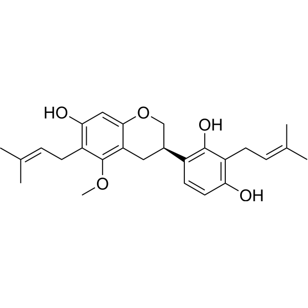
Licoricidin
Based on 1 Customer Validation
Licoricidin (LCD) is isolated from Glycyrrhiza uralensis Fisch, possesses anti-cancer activities. Licoricidin (LCD) inhibit SW480 cells (IC50=7.2 μM) by inducing cycle arrest, apoptosis and autophagy, and is a potential chemopreventive or chemotherapeutic agent against colorectal cancer. Licoricidin (LCD) inhibits Lung Metastasis by inhibition of tumor angiogenesis and lymphangiogenesis as well as changes in the local microenvironment of tumor tissues the anticarcinogenic effect. Licoricidin enhanced gemcitabine-induced cytotoxicity in Osteosarcoma (OS) cells by inactivation of the Akt and NF-κB pathways in vitro and in vivo. Licoricidin blocks UVA-induced photoaging via ROS scavenging, limits the activity of MMP-1, it can be considered as an active ingredient in new topically applied anti-ageing formulations.
Para uso exclusivo en investigación. No vendemos a pacientes.
- Pureza : 98.76%
- No. CAS: 30508-27-1
- Fòrmula: C26H32O5
- Peso molecular:424.53
-
Almacenamiento:
4°C, protect from light
* In solvent : -80°C, 6 months; -20°C, 1 month (protect from light)
Actividad biológica
Descripciòn
Cellular Effect
|
Cell Line
|
Type | Value | Description | References |
|---|---|---|---|---|
| A549 | IC50 |
6.9 μM
Compound: 71
|
Cytotoxicity against human A549 cells assessed as reduction in cell viability after 24 hrs by MTS assay
|
[PMID: 26841168] |
| HepG2 | IC50 |
0.3 μM
Compound: 71
|
Cytotoxicity against human HepG2 cells assessed as reduction in cell viability after 24 hrs by MTS assay
|
[PMID: 26841168] |
| MCF7 | IC50 |
5.2 μM
Compound: 71
|
Cytotoxicity against human MCF7 cells assessed as reduction in cell viability after 24 hrs by MTS assay
|
[PMID: 26841168] |
| SW480 | IC50 |
7.1 μM
Compound: 71
|
Cytotoxicity against human SW480 cells assessed as reduction in cell viability after 24 hrs by MTS assay
|
[PMID: 26841168] |
In Vitro
Licoricidin (LCD) (0-20 μM; 24 hours) dose-dependently inhibits the viability of colon cancer cell lines with various pathological and genetic characters, namely SW480, HCT116, SW620 and LoVo cells, with IC50 values of 7.2, 5.4, 4.5 and 5.1 μM, respectively[1]. Licoricidin (LCD) (0-20 μM; 0-12 hours) induces cell apoptosis was accompanied with the activation of caspase-3 by cleavage in a time- and dose-dependent manner[1]. Licoricidin (LCD) (0-20 μM; 0-12 hours) induces autophagy of SW480 cells, increases the cleavage of LC3-I to LC3-II and the degradation of p62 in a time and dose dependent manner[1]. Licoricidin (LCD) (0-5 μg/ml; 18 hours) inhibits cell migration, MMP-9 secretion, and VCAM expression in 4T1 cells[2].
MedChemExpress (MCE) has not independently confirmed the accuracy of these methods. They are for reference only. Further protocols information, click here.
-
Cell Line:SW480, HCT116, SW620 and LoVo cells
-
Concentration:0-20 μM
-
Incubation Time:24 hours
-
Result:Decreased colon cancer cell lines viability.
-
Cell Line:SW480 cells
-
Concentration:0 μM, 2.5 μM, 5 μM, 10 μM, 20 μM
-
Incubation Time:0 hours, 1 hour, 3 hours, 6 hours, 12 hours
-
Result:Induced cell apoptosis.
In Vivo
MedChemExpress (MCE) has not independently confirmed the accuracy of these methods. They are for reference only.
-
Animal Model:SW480 xenografted tumor growth in nude mice[1]
-
Dosage:5, 10, or 20 mg/kg
-
Administration:Intraperitoneal injection; once daily; 15 days
-
Result:Decreased tumor volumes.
-
Animal Model:BALB/c mouse orthotopic model[2]
-
Dosage:5, 10, or 20 mg/kg
-
Administration:Intraperitoneal injection; 5, 10, or 20 mg/kg; once daily; 32 days
-
Result:Inhibited Lung Metastasis of 4T1 Murine Mammary Carcinoma cells.
Chemical Information
-
No. CAS 30508-27-1
-
Appearance Solid
-
Peso molecular 424.53
-
Fòrmula C26H32O5
-
Color White to off-white
-
SMILES
COC1=C2C(OC[C@@H](C3=C(C(C/C=C(C)/C)=C(O)C=C3)O)C2)=CC(O)=C1C/C=C(C)/C
-
Structure Classification
-
Initial Source
-
Envío
Room temperature in continental US; may vary elsewhere.
-
Almacenamiento
4°C, protect from light
* In solvent : -80°C, 6 months; -20°C, 1 month (protect from light)
Solvente y solubilidad
In Vitro:
DMSO : 100 mg/mL (235.55 mM; Need ultrasonic; Hygroscopic DMSO has a significant impact on the solubility of product, please use newly opened DMSO)
Please refer to the solubility information to select the appropriate solvent. Once prepared, please aliquot and store the solution to prevent product inactivation from repeated freeze-thaw cycles.
Storage method and period of stock solution: -80°C, 6 months; -20°C, 1 month (protect from light). When stored at -80°C, please use it within 6 months. When stored at -20°C, please use it within 1 month.
Please refer to the solubility information to select the appropriate solvent. Once prepared, please aliquot and store the solution to prevent product inactivation from repeated freeze-thaw cycles.
Storage method and period of stock solution: -80°C, 6 months; -20°C, 1 month (protect from light). When stored at -80°C, please use it within 6 months. When stored at -20°C, please use it within 1 month.
Concentration (start) × Volume (start) = Concentration (final) × Volume (final)
Protocolo
-
Human pluripotent stem cell endothelial-cell differentiation
Human pluripotent stem cell endothelial differentiation is based on stepwise developmental patterning: early activation of WNT/GSK3β inhibition promotes mesodermal or vascular progenitor entry, followed by endothelial specification using VEGF-related signaling, BMP4, FGF2, Notch modulation, or cAMP depending on the published protocol. Endothelial differentiation is read out by acquisition of CD31, CD34, VE-cadherin/CD144, KDR/VEGFR2, vWF, Tie2, NOS3, acetylated LDL uptake, tube/network formation, barrier function, and in vivo vessel-forming capacity where tested.
-
Mitochondrial membrane-potential fluorescent assay
Mitochondrial membrane potential fluorescent assays estimate ΔΨm in living cells using lipophilic cationic dyes such as TMRM, TMRE, rhodamine 123, and JC-1, which accumulate in mitochondria according to membrane polarization; loss of signal after FCCP or CCCP treatment is interpreted as mitochondrial depolarization. TMRM/TMRE and rhodamine 123 are commonly used for semi-quantitative live-cell microscopy or flow cytometry, while JC-1 can report a shift from red aggregate fluorescence to green monomer fluorescence during depolarization; interpretation requires controls because dye concentration, quenching mode, cell type, dye efflux, and mitochondrial mass can affect fluorescence independently of ΔΨm.
-
Vascular/Branching Fractal Analysis
Vascular/branching fractal analysis quantifies the geometric complexity of vessel trees or vascular networks from segmented 2D images, commonly by converting vessels into binary and/or skeletonized maps and estimating fractal dimension using box-counting or related approaches. Fractal dimension is interpreted as an image-derived readout of vascular branching complexity, space filling, or density, and has been applied to retinal photographs, fluorescein angiography, OCT angiography, capillary perfusion maps, and in vitro Matrigel angiogenesis networks. The assay readout is generated from vessel-positive pixels after image preprocessing, vessel segmentation, binarization, and optional skeletonization; reported outputs include fractal dimension, vessel density, branchpoint density, endpoint density, vessel length density, tortuosity, and generation-based branching metrics when VESGEN-style analysis is used. The biological interpretation is limited to quantitative vascular patterning and s
-
Mammalian live/dead viability and cytotoxicity staining
Live/dead viability and cytotoxicity staining assays are based on the simultaneous detection of intracellular esterase activity in metabolically active (viable) cells and membrane integrity loss in non-viable cells. In commonly used dual-staining approaches, membrane-permeant fluorogenic substrates are converted by intracellular esterases into fluorescent products in live cells, while impermeant DNA-binding dyes selectively enter cells with compromised plasma membranes and label nucleic acids in dead or dying cells, enabling discrimination between viable and non-viable populations by fluorescence microscopy or flow cytometry.
-
Annexin V plus membrane-impermeant dye apoptosis staining
Annexin V-based apoptosis assays rely on the detection of phosphatidylserine (PS) externalization from the inner leaflet of the plasma membrane to the outer leaflet, an early biochemical hallmark of apoptosis. Fluorescently labeled Annexin V binds PS in a calcium-dependent manner, enabling identification of early apoptotic cells by flow cytometry or fluorescence microscopy. When combined with a membrane-impermeant DNA-binding dye (e. g. , propidium iodide), this approach allows discrimination between viable (Annexin V−/dye−), early apoptotic (Annexin V+/dye−), and late apoptotic or necrotic (Annexin V+/dye+) cell populations by assessing membrane integrity and PS exposure.
-
ROS/oxidative-stress fluorescent staining
ROS/oxidative-stress fluorescent staining uses cell-permeant fluorogenic probes that become fluorescent after oxidation inside cells or tissues; commonly used examples include DCFH-DA/DCFDA for broad cellular oxidant detection, DHE for superoxide-related signal detection, MitoSOX for mitochondrial superoxide-related signal detection, and CellROX probes for oxidative-stress-associated fluorescence readouts. The assay detects probe oxidation rather than a single ROS species unless the probe and analysis method have been chemically validated for that species. DCFH-DA enters cells, is deacetylated by intracellular esterases to DCFH, and produces fluorescent DCF after oxidation, so the readout is used as an operational measure of total cellular oxidative stress rather than a species-specific ROS measurement. DHE and MitoSOX can report superoxide-related oxidation, but red fluorescence alone can include non-specific ethidium-like oxidation products; HPLC or optimized spectral approaches are
-
Research Protocol for Inflammation-related Diseases
The NLRP3 inflammasome is a cytosolic innate immune signaling platform that integrates priming signals and danger-signal activation to promote caspase-1 activation, maturation of IL-1β and IL-18, and gasdermin D-mediated pyroptotic cell death. The core experimental logic is to determine whether inflammatory disease phenotypes are driven by increased NLRP3 expression, ASC-containing inflammasome assembly, caspase-1 cleavage, GSDMD cleavage, and extracellular release of IL-1β/IL-18 rather than by nonspecific cell injury alone. The pathway is strongly linked to inflammation-related disease phenotypes because monosodium urate crystals activate NALP3/NLRP3 inflammasome signaling in gout-like crystal inflammation, cholesterol crystals activate NLRP3 inflammasomes in atherogenesis models, and DSS-induced intestinal inflammation has been reported to involve NLRP3 inflammasome activity. However, experimental colitis studies also show context-dependent protective effects of NLRP3 inflammasome co
-
Cell Viability Determination by MTT Colorimetric Assay
The following protocol uses the MTT colorimetric assay as a classic literature-established method for assessing cell viability/metabolic activity in cultured mammalian cells. MTT[3-(4,5-dimethylthiazol-2-yl)-2,5-diphenyltetrazolium bromide] is reduced by metabolically active cells to a colored formazan product; the amount of formazan is quantified spectrophotometrically and provides an indirect measure of metabolically active viable cells. Importantly, MTT reduction reflects cellular oxidoreductase/metabolic activity rather than an absolute direct count of living cells, so changes in cellular metabolism can alter the signal independently of cell number.
-
Apoptosis
Apoptosis, also called programmed cell death, is generally characterized by distinct morphological characteristics.
-
TUNEL staining for apoptotic DNA fragmentation
TUNEL staining detects DNA strand breaks by using terminal deoxynucleotidyl transferase to add labeled nucleotides to exposed 3′-OH DNA termini, generating either microscopic staining in fixed cells or tissue sections, or fluorescence/cytometric signal in cell suspensions. TUNEL positivity reflects DNA fragmentation but should not be interpreted alone as definitive apoptosis, because TUNEL can also label necrotic, autolytic, mechanically damaged, or DNA-repair-associated DNA breaks.
-
Cell Cytotoxicity Assay
Cytotoxicity assays are usually based on the assessment of cell membrane damage, which can also be indirectly detected by measuring cell viability. Detection methods include MTT assay, CKK-8 assay, LDH assay and ATP assay, etc.
-
Autophagy
Autophagy is a process in which eukaryotic cells use lysosomes to degrade their own cytoplasmic proteins and damaged organelles under the regulation of autophagy related gene (Atg). Microtubule-associated proteins light chain 3 (LC3) is recognized as autophagy marker, which transfers from cytoplasmic LC3 (LC3-I) to membrane type (LC3-II). LC3-II/I ratio could be detected by Western Blot and fluorescence microscopy.
-
Tail-Vein Experimental Metastasis Xenograft
Tail-vein experimental metastasis xenograft models assess the ability of injected tumor cells to survive circulation, arrest in vascular beds, extravasate, and colonize distant organs, most commonly lung after lateral tail-vein injection; this model bypasses primary-tumor formation, local invasion, and intravasation, so the readout reflects late metastatic colonization rather than the full metastatic cascade. The main readouts are metastatic burden measured by bioluminescence imaging, gross metastatic nodules, histology, organ weight, survival, or ex vivo tumor-cell quantification; luciferase-labeled tumor cells permit longitudinal noninvasive monitoring, while histology confirms organ colonization and tissue localization.
-
Endothelial Tube Formation Assay
Endothelial tube formation assay evaluates the ability of endothelial cells to attach, migrate, align, and organize into capillary-like networks when cultured on gelled basement membrane extract or Matrigel; the readout is the morphology and quantity of tube-like networks, which reflects an in vitro endothelial morphogenesis step related to angiogenesis. Basement membrane extract/Matrigel provides laminin-rich extracellular matrix cues that support endothelial differentiation into capillary-like structures, but it can contain biologically active growth factors, so growth-factor-reduced matrix is preferred when testing defined angiogenic stimulators or inhibitors.
-
Mitochondrial membrane-potential and mitochondrial mass staining
Mitochondrial membrane potential staining measures the electrochemical polarization across the mitochondrial inner membrane in live cells using lipophilic cationic fluorescent probes; early rhodamine-based work showed that selective mitochondrial dye accumulation is lost when the mitochondrial transmembrane potential is dissipated. JC-1 reports mitochondrial polarization by shifting from green monomer fluorescence to red J-aggregate fluorescence as dye concentration increases within energized mitochondria; therefore, the red/green fluorescence ratio is used as a relative readout of mitochondrial membrane potential. TMRE or TMRM staining provides a single-channel relative readout because these cationic rhodamine esters accumulate in polarized mitochondria, and lower fluorescence indicates reduced mitochondrial polarization when acquisition and dye-loading conditions are controlled. Mitochondrial mass staining is commonly performed with MitoTracker Green FM or related MitoTracker dyes as
-
Apoptosis Solutions
Apoptosis is a regulated, generally non-lytic cell-death pathway that removes unwanted, damaged, infected, or abnormal cells through coordinated morphological changes, caspase activation, DNA fragmentation, and membrane remodeling. The intrinsic apoptosis pathway is controlled mainly by mitochondrial outer membrane permeabilization, BCL-2 family proteins, cytochrome c release, apoptosome formation, caspase-9 activation, and downstream executioner caspase-3/7 activation. The extrinsic apoptosis pathway is initiated by death receptors such as Fas, TNFR, and TRAIL receptors, which recruit adaptor proteins and activate caspase-8 before engaging executioner caspases or mitochondrial amplification through BID cleavage. Apoptosis is linked to many phenotypes, including cancer cell killing, tissue homeostasis, immune regulation, neurodegeneration, infection response, and treatment-induced cytotoxicity; unresolved questions include how apoptosis interacts with necroptosis, pyroptosis, ferroptos
-
Lysosome and acidic-vesicle live-cell staining
Lysosome and acidic-vesicle live-cell staining detects acidic intracellular compartments by using membrane-permeant acidotropic probes that accumulate in low-pH vesicles, including lysosomes, late endosomes, autolysosomes, and acidic phagosomes. LysoTracker staining is commonly used as an intensity-based readout of acidic lysosomal compartment abundance or enlargement, while acridine orange produces green fluorescence in less concentrated compartments and red fluorescence after concentration-dependent accumulation in acidic vesicular organelles. Loss or reduction of acridine-orange red signal can be used as a readout of lysosomal membrane permeabilization or reduced acidic-vesicle integrity. This protocol is designed for live cultured cells and can be adapted for fluorescence microscopy, high-content imaging, plate-reader readout, or flow cytometry when the selected literature supports the readout. Because these dyes report acidotropic accumulation rather than lysosome identity alone,
-
Macroautophagy Solutions
Macroautophagy is a conserved lysosome-dependent degradation pathway in which cytoplasmic material is sequestered into double-membrane autophagosomes and delivered to lysosomes for degradation and recycling. The pathway supports cellular homeostasis during nutrient limitation, organelle stress, protein-aggregate accumulation, infection, differentiation, and tissue remodeling by coupling cargo sequestration, autophagosome maturation, lysosomal fusion, and degradation of cargo-derived macromolecules. The core molecular sequence includes initiation by nutrient- and stress-regulated autophagy machinery, autophagosome nucleation, LC3/ATG8-family conjugation to autophagosomal membranes, cargo selection through receptors such as SQSTM1/p62, autophagosome-lysosome fusion, and lysosomal degradation. LC3 was identified as a mammalian homolog of yeast Atg8 that localizes to autophagosomal membranes after processing, and p62/SQSTM1 was shown to connect ubiquitinated cargo with autophagic degradati
-
Orthotopic Cell-Line Xenograft
Orthotopic cell-line xenograft models involve implantation of human cancer cell lines into the anatomically corresponding organ of immunodeficient mice to reproduce tumor growth within a native microenvironment, enabling more clinically relevant tumor behavior compared with subcutaneous models. These models are widely used because orthotopic placement better recapitulates tumor progression, including invasion and metastatic spread, which are often underrepresented in heterotopic implantation systems. Compared with conventional xenografts, orthotopic implantation is described as more technically complex but provides improved simulation of tumor-microenvironment interactions and metastatic behavior, making it particularly valuable for translational oncology research. Surgical orthotopic implantation approaches have been emphasized as enabling faithful reproduction of clinical cancer features, including metastasis and disease progression patterns that align with the tumor’s organ of origi
-
Endothelial Cell Migration/Angiogenic Sprouting Assay
Endothelial cell migration and angiogenic sprouting assays are in vitro (and partially ex vivo-adapted) functional models that quantify the ability of endothelial cells to undergo coordinated migration, extracellular matrix invasion, and multicellular organization into capillary-like sprouts in response to pro-angiogenic stimuli such as VEGF, bFGF, or conditioned microenvironments. These assays are used to model early angiogenic events including tip-cell formation, directional migration, and lumen-like sprout extension, which collectively reflect angiogenic activation and vascular morphogenesis processes observed in vivo.
Pureza y Documentación
-
Ficha de datos (277 KB)
-
SDS (252 KB)
- English - EN (252 KB)
- Français - FR (252 KB)
- Deutsch - DE (252 KB)
- Norwegian - NO (252 KB)
- Español - ES (252 KB)
- Swedish - SV (252 KB)
- Italian - IT (252 KB)
- Korean - KR (252 KB)
- Portuguese - PT (252 KB)
-
Instrucciones de manejo (2659 KB)
Referencias
[1]. Ji S, et al. Licoricidin inhibits the growth of SW480 human colorectal adenocarcinoma cells in vitro and in vivo by inducing cycle arrest, apoptosis and autophagy. Toxicol Appl Pharmacol. 2017 Jul 1;326:25-33. [Content Brief]
[2]. Park SY, et al. Licoricidin, an Active Compound in the Hexane/Ethanol Extract of Glycyrrhiza uralensis, Inhibits Lung Metastasis of 4T1 Murine Mammary Carcinoma Cells. Int J Mol Sci. 2016 Jun 14;17(6). [Content Brief]
[3]. Wang Y, et al. Licoricidin enhances gemcitabine-induced cytotoxicity in osteosarcoma cells by suppressing the Akt and NF-κB signal pathways. Chem Biol Interact. 2018 Jun 25;290:44-51. [Content Brief]
[4]. Kim KJ, et al. Licoricidin, an isoflavonoid isolated from Glycyrrhiza uralensis Fisher, prevents UVA-induced photoaging of human dermal fibroblasts. Int J Cosmet Sci. 2017 Apr;39(2):133-140. [Content Brief]
Complete Stock Solution Preparation Table
Please refer to the solubility information to select the appropriate solvent. Once prepared, please aliquot and store the solution to prevent product inactivation from repeated freeze-thaw cycles.
Storage method and period of stock solution: -80°C, 6 months; -20°C, 1 month (protect from light). When stored at -80°C, please use it within 6 months. When stored at -20°C, please use it within 1 month.
| Optional Solvent | Concentration Solvent Mass | 1 mg | 5 mg | 10 mg | 25 mg |
|---|---|---|---|---|---|
| DMSO | 1 mM | 2.3555 mL | 11.7777 mL | 23.5555 mL | 58.8887 mL |
| 5 mM | 0.4711 mL | 2.3555 mL | 4.7111 mL | 11.7777 mL | |
| 10 mM | 0.2356 mL | 1.1778 mL | 2.3555 mL | 5.8889 mL | |
| 15 mM | 0.1570 mL | 0.7852 mL | 1.5704 mL | 3.9259 mL | |
| 20 mM | 0.1178 mL | 0.5889 mL | 1.1778 mL | 2.9444 mL | |
| 25 mM | 0.0942 mL | 0.4711 mL | 0.9422 mL | 2.3555 mL | |
| 30 mM | 0.0785 mL | 0.3926 mL | 0.7852 mL | 1.9630 mL | |
| 40 mM | 0.0589 mL | 0.2944 mL | 0.5889 mL | 1.4722 mL | |
| 50 mM | 0.0471 mL | 0.2356 mL | 0.4711 mL | 1.1778 mL | |
| 60 mM | 0.0393 mL | 0.1963 mL | 0.3926 mL | 0.9815 mL | |
| 80 mM | 0.0294 mL | 0.1472 mL | 0.2944 mL | 0.7361 mL | |
| 100 mM | 0.0236 mL | 0.1178 mL | 0.2356 mL | 0.5889 mL |