



NecroIr2

Based on 1 Customer Validation
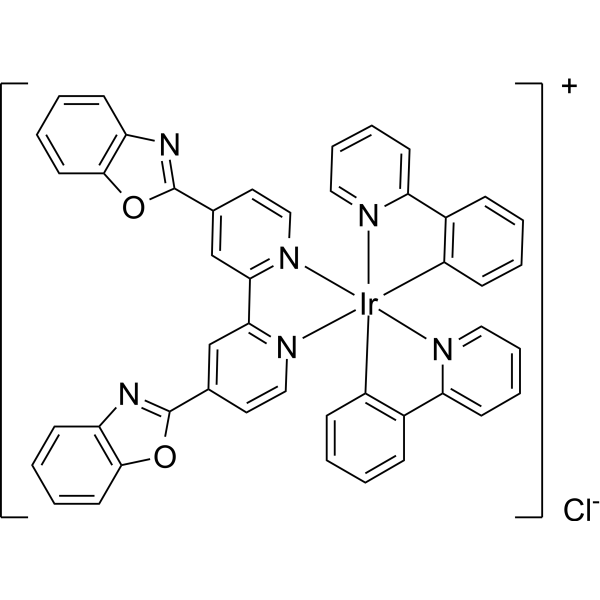
NecroIr2 is an iridium(III) complex, serves as necroptosis inducers in Cisplatin (HY-17394)-resistant lung cancer cells (A549R). NecroIr2 selectively accumulates in mitochondria, leading to oxidative stress and loss of mitochondrial membrane potential (MMP). NecroIr2 activates receptor-interacting serine-threonine kinase 3 (RIPK3) and mixed lineage kinase domain-like pseudokinase (MLKL), and regulates CDK4 expression.
For research use only. We do not sell to patients.
- Purity : 98.02%
- Formula: C46H30ClIrN6O2-
- Molecular Weight:926.44
-
Storage:
4°C, sealed storage, away from moisture and light
* In solvent : -80°C, 6 months; -20°C, 1 month (sealed storage, away from moisture and light)

To place orders, for customer services and technical support, please contact: MedChemExpress USA
Tel: 609-228-6898 E-mail: [email protected] [email protected]


-
 Biological Activity
-
Chemical Information
-
Solvent & Solubility
- Protocols
- Purity & Documentation
- References
-
 Help & FAQs




Biological Activity
Description
IC50 & Target
[1]|
CDK4 |
RIPK3 |
RIPK1 |
In Vitro
NecroIr2 (2 μM; 1-2 d) exerts subcellular distribution over 90% accumulated in mitochondria in non-labelled A549R and CFSE labelled L02 cells[1].
NecroIr2 (1.5 μM and 3 μM; 24 h) results ROS generation increasing and loss of mitochondrial membrane potential[1].
NecroIr2 (1.5 μM and 3 μM; 24 h) activates necroptosis proteins, increases the phosphorylation of RIPK1 and RIPK3[1].
NecroIr2 (0.75 μM and 1.5 μM; 24 h) induces necroptosis by arresting cell cycle at G0/G1[1].
NecroIr2 (0.375-1.5 μM; 24 h) inhibits A549R cells proliferation[1].
MedChemExpress (MCE) has not independently confirmed the accuracy of these methods. They are for reference only.
-
Cell Line:A549R cells
-
Concentration:1.5 μM and 3 μM
-
Incubation Time:24 hours
-
Result:Increased phospho-RIPK1 (p-PIPK1), total RIPK3, and phospho-RIPK3 (p-PIPK3) level.
-
Cell Line:A549R cells
-
Concentration:0 μM, 0.75 μM and 1.5 μM
-
Incubation Time:24 hours
-
Result:Arrested cell cycle at G0/G1 phase in a dose-dependent manner.
-
Cell Line:A549R cells
-
Concentration:0 μM, 0.375 μM, 0.75 μM and 1.5 μM
-
Incubation Time:24 hours
-
Result:Inhibits cell proliferation in a dose-dependent manner.


Chemical Information
-
Appearance Solid
-
Molecular Weight 926.44
-
Formula C46H30ClIrN6O2-
-
Color Brown to reddish brown
-
SMILES
C1(C2=NC(C=CC=C3)=C3O2)=CC(C4=[N]5C=CC(C6=NC(C=CC=C7)=C7O6)=C4)=[N]([Ir]89([N]%10=CC=CC=C%10C%11=C9C=CC=C%11)5[N]%12=CC=CC=C%12C%13=C8C=CC=C%13)C=C1.[Cl-]
-
Shipping
Room temperature in continental US; may vary elsewhere.
-
Storage
4°C, sealed storage, away from moisture and light
* In solvent : -80°C, 6 months; -20°C, 1 month (sealed storage, away from moisture and light)


Solvent & Solubility
In Vitro:
DMSO : 50 mg/mL (53.97 mM; Need ultrasonic; Hygroscopic DMSO has a significant impact on the solubility of product, please use newly opened DMSO)
Please refer to the solubility information to select the appropriate solvent. Once prepared, please aliquot and store the solution to prevent product inactivation from repeated freeze-thaw cycles.
Storage method and period of stock solution: -80°C, 6 months; -20°C, 1 month (sealed storage, away from moisture and light). When stored at -80°C, please use it within 6 months. When stored at -20°C, please use it within 1 month.
Please refer to the solubility information to select the appropriate solvent. Once prepared, please aliquot and store the solution to prevent product inactivation from repeated freeze-thaw cycles.
Storage method and period of stock solution: -80°C, 6 months; -20°C, 1 month (sealed storage, away from moisture and light). When stored at -80°C, please use it within 6 months. When stored at -20°C, please use it within 1 month.
Concentration (start) × Volume (start) = Concentration (final) × Volume (final)


Purity & Documentation
-
Data Sheet (283 KB)
-
SDS (252 KB)
- English - EN (252 KB)
- Français - FR (252 KB)
- Deutsch - DE (252 KB)
- Norwegian - NO (252 KB)
- Español - ES (252 KB)
- Swedish - SV (252 KB)
- Italian - IT (252 KB)
- Korean - KR (252 KB)
- Portuguese - PT (252 KB)
-
Handling Instructions (2659 KB)
References


Complete Stock Solution Preparation Table
Please refer to the solubility information to select the appropriate solvent. Once prepared, please aliquot and store the solution to prevent product inactivation from repeated freeze-thaw cycles.
Storage method and period of stock solution: -80°C, 6 months; -20°C, 1 month (sealed storage, away from moisture and light). When stored at -80°C, please use it within 6 months. When stored at -20°C, please use it within 1 month.
| Optional Solvent | Concentration Solvent Mass | 1 mg | 5 mg | 10 mg | 25 mg |
|---|---|---|---|---|---|
| DMSO | 1 mM | 1.0794 mL | 5.3970 mL | 10.7940 mL | 26.9850 mL |
| 5 mM | 0.2159 mL | 1.0794 mL | 2.1588 mL | 5.3970 mL | |
| 10 mM | 0.1079 mL | 0.5397 mL | 1.0794 mL | 2.6985 mL | |
| 15 mM | 0.0720 mL | 0.3598 mL | 0.7196 mL | 1.7990 mL | |
| 20 mM | 0.0540 mL | 0.2699 mL | 0.5397 mL | 1.3493 mL | |
| 25 mM | 0.0432 mL | 0.2159 mL | 0.4318 mL | 1.0794 mL | |
| 30 mM | 0.0360 mL | 0.1799 mL | 0.3598 mL | 0.8995 mL | |
| 40 mM | 0.0270 mL | 0.1349 mL | 0.2699 mL | 0.6746 mL | |
| 50 mM | 0.0216 mL | 0.1079 mL | 0.2159 mL | 0.5397 mL |


NecroIr2 Related Classifications
HY-148366 Related Classifications
 Powered by Bioz