



Pefloxacin-d5

Based on 1 Customer Validation
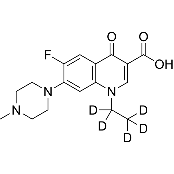
Pefloxacin-d5 (Pefloxacinium-d5) is the deuterium labeled Pefloxacin (HY-B0147). Pefloxacin (Pefloxacinium) is a broad spectrum antibiotic. Pefloxacin blocks DNA replication by inhibiting DNA gyrase. Pefloxacin inhibits DNA relaxation catalyzed by topoisomerase I with an IC50 of 45 μg/mL. Pefloxacin exhibits antibacterial activity against Escherichia coli, Pseudomonas aeruginosa, and Bacteroides fragilis with MIC90s of 0.12, 4, and 16 mg/L, respectively. Pefloxacin has anti-Plasmodium yoelii infection activity. Pefloxacin increase UVA-induced edema and immunesuppression. Pefloxacin can be used for infection studies.
For research use only. We do not sell to patients.
- Purity : 98.33%
- CAS No.: 1228182-51-1
- Formula: C17H15D5FN3O3
- Molecular Weight:338.39
-
Storage:Powder -20°C, 3 years , 4°C, 2 years ; In solvent -80°C, 6 months , -20°C, 1 month

To place orders, for customer services and technical support, please contact: MedChemExpress USA
Tel: 609-228-6898 E-mail: [email protected] [email protected]


-
 Biological Activity
-
Chemical Information
- Protocols
- Purity & Documentation
- References
-
 Help & FAQs


All Antibiotic Isoforms
MoreAll DNA/RNA Synthesis Isoforms
MoreAll Parasite Isoforms
MoreAll Topoisomerase Isoforms
More

Biological Activity
Description
In Vitro
Stable heavy isotopes of hydrogen, carbon, and other elements have been incorporated into drug molecules, largely as tracers for quantitation during the drug development process. Deuteration has gained attention because of its potential to affect the pharmacokinetic and metabolic profiles of drugs[1].
MedChemExpress (MCE) has not independently confirmed the accuracy of these methods. They are for reference only.
Application
1. This compound can be used as a tracer
2. This compound can be used as an internal standard for quantitative analysis by NMR, GC-MS, or LC-MS.


Chemical Information
-
CAS No. 1228182-51-1
-
Unlabeled CAS 70458-92-3
-
Appearance Solid
-
Molecular Weight 338.39
-
Formula C17H15D5FN3O3
-
Color White to off-white
-
SMILES
O=C(C1=CN(C([2H])([2H])C([2H])([2H])[2H])C2=C(C=C(F)C(N3CCN(C)CC3)=C2)C1=O)O
-
Synonyms
Pefloxacinium-d5
-
Shipping
Room temperature in continental US; may vary elsewhere.
-
Storage
Powder -20°C 3 years 4°C 2 years In solvent -80°C 6 months -20°C 1 month


Purity & Documentation
-
Data Sheet (284 KB)
-
SDS (252 KB)
- English - EN (252 KB)
- Français - FR (252 KB)
- Deutsch - DE (252 KB)
- Norwegian - NO (252 KB)
- Español - ES (252 KB)
- Swedish - SV (252 KB)
- Italian - IT (252 KB)
- Korean - KR (252 KB)
- Portuguese - PT (252 KB)
-
Handling Instructions (2659 KB)
References
[1]. Drlica K, et al. DNA gyrase, topoisomerase IV, and the 4-quinolones. Microbiol Mol Biol Rev. 1997 Sep;61(3):377-92. [Content Brief]
[2]. Hussy P, et al. Effect of 4-quinolones and novobiocin on calf thymus DNA polymerase alpha primase complex, topoisomerases I and II, and growth of mammalian lymphoblasts. Antimicrob Agents Chemother. 1986 Jun;29(6):1073-8. [Content Brief]
[3]. Clarke AM, et al. In-vitro activity of pefloxacin compared to enoxacin, norfloxacin, gentamicin and new beta-lactams. J Antimicrob Chemother. 1985 Jan;15(1):39-44. [Content Brief]
[4]. Jones BM, et al. Activity of pefloxacin and thirteen other antimicrobial agents in vitro against isolates from hospital and genitourinary infections. J Antimicrob Chemother. 1986 Jun;17(6):739-46. [Content Brief]
[5]. Tabary X, et al. Effect of DNA gyrase inhibitors pefloxacin, five other quinolones, novobiocin, and clorobiocin on Escherichia coli topoisomerase I. Antimicrob Agents Chemother. 1987 Dec;31(12):1925-8. [Content Brief]
[6]. Fantin B, et al. Correlation between in vitro and in vivo activity of antimicrobial agents against gram-negative bacilli in a murine infection model. Antimicrob Agents Chemother. 1991 Jul;35(7):1413-22. [Content Brief]
[7]. Sun YW, et al. Pefloxacin and ciprofloxacin increase UVA-induced edema and immune suppression. Photodermatol Photoimmunol Photomed. 2001 Aug;17(4):172-7. [Content Brief]
[8]. Truffot-Pernot C, et al. Activities of pefloxacin and ofloxacin against mycobacteria: in vitro and mouse experiments. Tubercle. 1991 Mar;72(1):57-64. [Content Brief]
[9]. Salmon D, et al. Activities of pefloxacin and ciprofloxacin against experimental malaria in mice. Antimicrob Agents Chemother. 1990 Dec;34(12):2327-30. [Content Brief]

Calculators
Concentration (start) × Volume (start) = Concentration (final) × Volume (final)


Pefloxacin-d5 Related Classifications
HY-B0147S Related Classifications
 Powered by Bioz