



Rhamnose monohydrate
Based on 1 publication(s) in Google Scholar
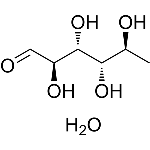
Rhamnose monohydrate (L-Rhamnose monohydrate) is an orally active deoxysugar. Rhamnose monohydrate can inhibit levels of pro-inflammatory interleukin and matrix metalloproteinases (MMPs) in skin aging models. Rhamnose can promote the phosphorylation levels of PKA substrates and HSL in SVF-derived adipocytes, stimulating PKA signaling. Rhamnose monohydrate can act against obesity in mice by stimulating fat dopamine receptors and inducing thermogenesis. Rhamnose monohydrate shows anti-aging effects. Rhamnose monohydrate can be used in the study of Ehrlich’s solid tumors and sarcomas.
For research use only. We do not sell to patients.
- Purity : 99.95%
- CAS No.: 10030-85-0
- Formula: C6H14O6
- Molecular Weight:182.17
-
Storage:Powder -20°C, 3 years , 4°C, 2 years ; In solvent -80°C, 2 years , -20°C, 1 year

To place orders, for customer services and technical support, please contact: MedChemExpress USA
Tel: 609-228-6898 E-mail: [email protected] [email protected]


-
 Biological Activity
-
Chemical Information
-
Solvent & Solubility
- Protocols
- Purity & Documentation
- References
-
 Help & FAQs


-
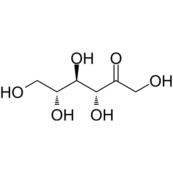
HY-N9914
D-PsicoseRare Sugar -

HY-N1420AR
Rhamnose monohydrate (Standard)Monosaccharide -
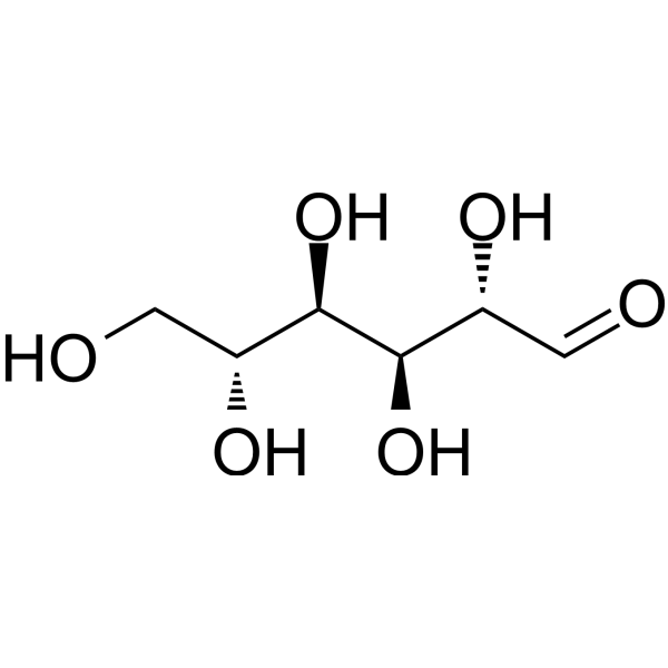
HY-N0379
D-MannoseEndogenous Metabolite -
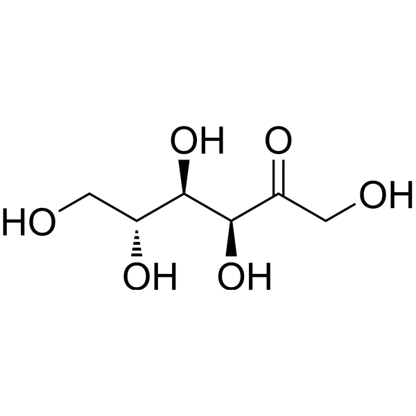
HY-N7092
D-FructoseEndogenous Metabolite -
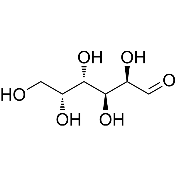
HY-N0210
D-GalactoseEndogenous Metabolite -
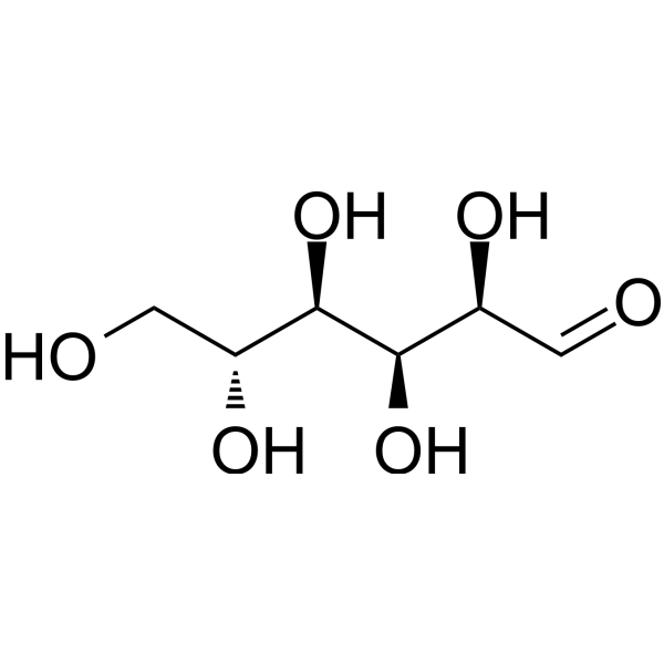
HY-B0389
D-GlucoseMonosaccharide -

HY-N0379R
D-Mannose (Standard)Endogenous Metabolite -

HY-N7092R
D-Fructose (Standard)Endogenous Metabolite -
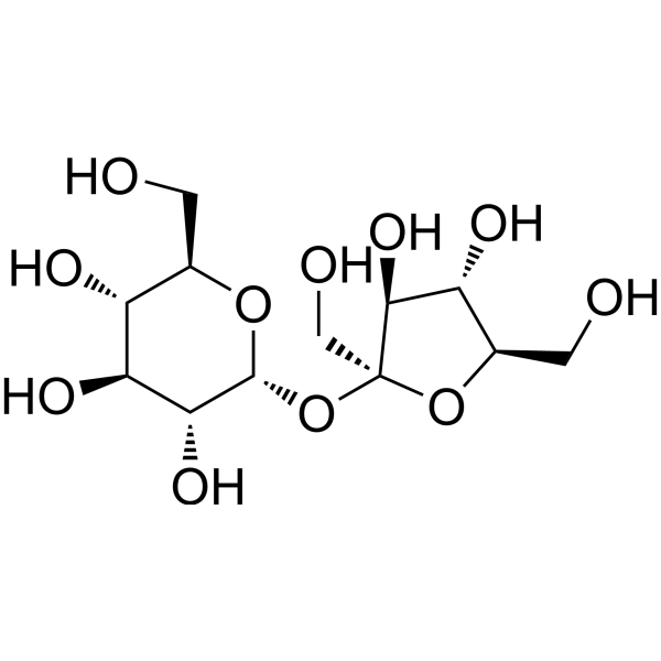
HY-B1779
SucroseDisaccharide -
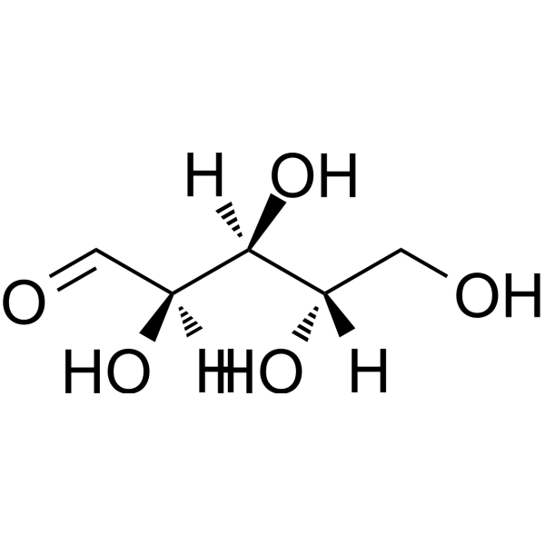
HY-W015611
L-(+)-Arabinose -
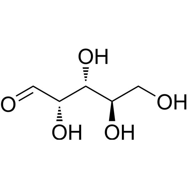
HY-N0059
D-ArabinoseAnti-parasite Agent -
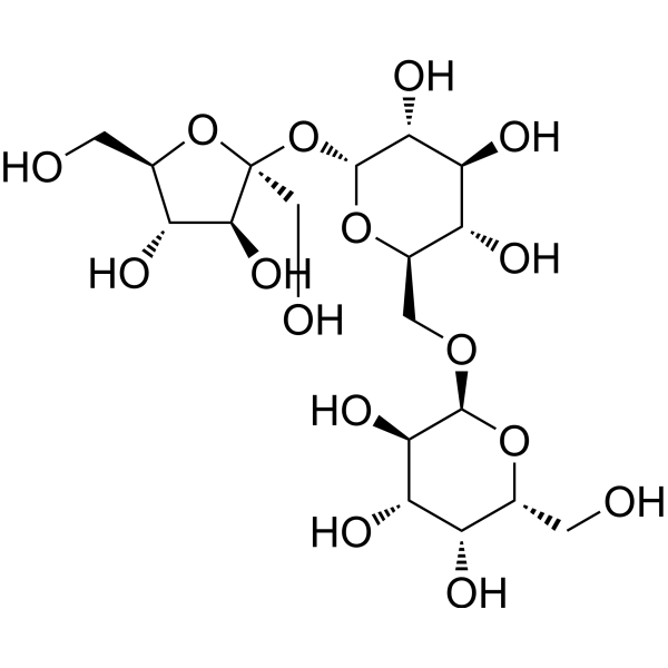
HY-N7088
RaffinoseNrf2 Activator
Publications Citing Use of MedChemExpress (MCE) Rhamnose monohydrate
MoreAll Endogenous Metabolite Isoforms
More

Biological Activity
Description
IC50 & Target
|
Microbial Metabolite |
In Vitro
Rhamnose (50 mM, 30 h) monohydrate can regulate the expression of Prha target genes in Listeria monocytogenes[1]. Rhamnose (1 mM) monohydrate can reduce the levels of pro-inflammatory interleukins and matrix metalloproteinases (MMPs) in a human dermal fibroblast (NHDF) skin aging model stimulated by advanced glycation end products (AGEs), thus possessing anti-aging potential[2]. Rhamnose (0-300 μM, 1 h) monohydrate promotes phosphorylation of PKA substrate and HSL in SVF-derived adipocytes, stimulates PKA signaling and induces thermogenesis[3].
MedChemExpress (MCE) has not independently confirmed the accuracy of these methods. They are for reference only. Further protocols information, click here.
-
Cell Line:L. monocytogenes 10403S
-
Concentration:50 mM
-
Incubation Time:30 h
-
Result:Promoted the growth of L. monocytogenes.
In Vivo
MedChemExpress (MCE) has not independently confirmed the accuracy of these methods. They are for reference only.
-
Animal Model:Diet induced obese mice obtained by administering high-fat diet (HFD) to 6-week-old male C57BL/6J mice for a total of 9-10 weeks[3]
-
Dosage:0.25, 0.5, 1 mg/mL (dissolve in water) or 300 mg/kg
-
Administration:Oral gavage (p.o.), 9 weeks; Intraperitoneal injection (i.p.), 7 days
-
Result:Reduced the total fat content of mice, improved glucose sensitivity, and promoted the expression of thermogenic genes.
-
Animal Model:Herston White mice injected intraperitoneally with S180 tumor cells, 30 g, aged 8-10 weeks[4]
-
Dosage:5, 10, 15 mg/kg
-
Administration:Intraperitoneal injection (i.p.); the first dose was given 0.5 h after administration of the S180 tumour cells and the remaining three doses were given at daily intervals
-
Result:Reduced the survival rate of mice in a dose-dependent manner in the presence of BEC.
-
Animal Model:NMRI female mouse model of subcutaneous inoculation of Ehrlich's solid tumor (SET)[5]
-
Dosage:1, 3, 5 g/kg
-
Administration:Intraperitoneal injection (i.p.); once daily; 14 days
-
Result:Inhibited tumor growth and prolonged the survival time of mice.
Clinical Trial
| NCT Number | Sponsor | Condition | Start Date |
Phase

|
|---|---|---|---|---|
| NCT01329991 | Plexxikon| | 2011-05 | PHASE1 |


Chemical Information
-
CAS No. 10030-85-0
-
Appearance Solid
-
Molecular Weight 182.17
-
Formula C6H14O6
-
Color White to off-white
-
SMILES
O=C[C@@H]([C@@H]([C@H]([C@H](C)O)O)O)O.O
-
Synonyms
L-Rhamnose monohydrate
-
Structure Classification
-
Initial Source
-
Shipping
Room temperature in continental US; may vary elsewhere.
-
Storage
Powder -20°C 3 years 4°C 2 years In solvent -80°C 2 years -20°C 1 year


Publications (1)
-
Journal Impact Factor
-
Most Recent
-
Ultrason Sonochem
Ultrasonic complexation with Lycium barbarum polysaccharide significantly enhances the aqueous solubility and bioavailability of curcumin. [Abstract]2025 Dec:123:107673. PMID: 41223460


Solvent & Solubility
In Vitro:
DMSO : 100 mg/mL (548.94 mM; Need ultrasonic; Hygroscopic DMSO has a significant impact on the solubility of product, please use newly opened DMSO)
Please refer to the solubility information to select the appropriate solvent. Once prepared, please aliquot and store the solution to prevent product inactivation from repeated freeze-thaw cycles.
Storage method and period of stock solution: -80°C, 2 years; -20°C, 1 year. When stored at -80°C, please use it within 2 years. When stored at -20°C, please use it within 1 year.
Please refer to the solubility information to select the appropriate solvent. Once prepared, please aliquot and store the solution to prevent product inactivation from repeated freeze-thaw cycles.
Storage method and period of stock solution: -80°C, 2 years; -20°C, 1 year. When stored at -80°C, please use it within 2 years. When stored at -20°C, please use it within 1 year.
Concentration (start) × Volume (start) = Concentration (final) × Volume (final)
In Vivo:
Select the appropriate dissolution method based on your experimental animal and administration route.
- For the following dissolution methods, please ensure to first prepare a clear stock solution using an In Vitro approach and then sequentially add co-solvents:
- To ensure reliable experimental results, the clarified stock solution can be appropriately stored based on storage conditions. As for the working solution for In Vivo experiments, it is recommended to prepare freshly and use it on the same day.
- The percentages shown for the solvents indicate their volumetric ratio in the final prepared solution. If precipitation or phase separation occurs during preparation, heat and/or sonication can be used to aid dissolution.
Add each solvent one by one: 10% DMSO 40% PEG300 5% Tween-80 45% Saline
Solubility: ≥ 2.5 mg/mL (13.72 mM); Clear solution
This protocol yields a clear solution of ≥ 2.5 mg/mL (saturation unknown).
Taking 1 mL working solution as an example, add 100 μL DMSO stock solution (25.0 mg/mL) to 400 μL PEG300, and mix evenly; then add 50 μL Tween-80 and mix evenly; then add 450 μL Saline to adjust the volume to 1 mL.
Preparation of Saline: Dissolve 0.9 g sodium chloride in ddH₂O and dilute to 100 mL to obtain a clear Saline solution.
Add each solvent one by one: 10% DMSO 90% (20% SBE-β-CD in Saline)
Solubility: ≥ 2.5 mg/mL (13.72 mM); Clear solution
This protocol yields a clear solution of ≥ 2.5 mg/mL (saturation unknown).
Taking 1 mL working solution as an example, add 100 μL DMSO stock solution (25.0 mg/mL) to 900 μL 20% SBE-β-CD in Saline, and mix evenly.
Preparation of 20% SBE-β-CD in Saline (4°C, storage for one week): 2 g SBE-β-CD powder is dissolved in 10 mL Saline, completely dissolve until clear.
In Vivo Dissolution Calculator
Please enter the basic information of animal experiments:
-
-
-
-
Recommended: Prepare an additional quantity of animals to account for potential losses during experiments.
Please enter your animal formula composition:
-
%DMSO +
Recommended: Keep the proportion of DMSO in working solution below 2% if your animal is weak.
-
%+
-
+%Tween-80 + +
-
%Saline +
The co-solvents required include: DMSO, . All of co-solvents are available by MedChemExpress (MCE). , Tween 80. All of co-solvents are available by MedChemExpress (MCE).
Working solution concentration: 0.22 mg/mL
Method for preparing stock solution: mg drug dissolved in μL DMSO. Stock solution concentration: mg/mL.
1. Take μL DMSO stock solution;
2. Add μL .
μL , mix evenly;
3. Then add μL Tween 80, mix evenly;
4. Then add μL
Please ensure that the stock solution in the first step is dissolved to a clear state, and add co-solvents in sequence. You can use ultrasonic heating (ultrasonic cleaner, recommended frequency 20-40 kHz), vortexing, etc. to assist dissolution.

Protocols
-
Kinase activity and phosphorylation assays
Kinase activity assays measure the ability of kinases to transfer phosphate groups from ATP to specific substrates, while phosphorylation assays detect the presence and levels of phosphorylated proteins. Common methods include radiolabeled ATP incorporation (e. g. ,), ADP release detection via bioluminescence (e. g. ,[3]), enzyme-linked immunosorbent assays (ELISA) for phospho-specific epitopes (e. g. ,[6]), and microtiter-based formats for high-throughput screening (e. g. ,[8]). The ADP-Glo assay quantifies kinase activity by measuring ADP produced during phosphorylation using a luciferase-based system. Radiometric assays involve autoradiography or scintillation counting after incorporation of 32P-labeled ATP into substrate proteins. ELISA-based approaches rely on phospho-specific antibodies to detect activated kinases in cell lysates or purified samples.
-
Western Blot
Western blotting (WB) is a commonly used experimental method in molecular biology, biochemistry, and immunogenetics for identifying and quantifying target proteins. It combines gel electrophoresis with immunoassay, enabling researchers to analyze protein expression, post-translational modifications, and molecular weight.
-
Large-size fat particle sorting
Large-size fat particle sorting is widely used to isolate cells up to 200 μm in diameter. Single-cell flow sorting will allow greater insight into adipocyte heterogeneity by identifying gene expression, protein composition, and metabolic signatures at the single-cell level.
-
Research Protocol for Inflammation-related Diseases
The NLRP3 inflammasome is a cytosolic innate immune signaling platform that integrates priming signals and danger-signal activation to promote caspase-1 activation, maturation of IL-1β and IL-18, and gasdermin D-mediated pyroptotic cell death. The core experimental logic is to determine whether inflammatory disease phenotypes are driven by increased NLRP3 expression, ASC-containing inflammasome assembly, caspase-1 cleavage, GSDMD cleavage, and extracellular release of IL-1β/IL-18 rather than by nonspecific cell injury alone. The pathway is strongly linked to inflammation-related disease phenotypes because monosodium urate crystals activate NALP3/NLRP3 inflammasome signaling in gout-like crystal inflammation, cholesterol crystals activate NLRP3 inflammasomes in atherogenesis models, and DSS-induced intestinal inflammation has been reported to involve NLRP3 inflammasome activity. However, experimental colitis studies also show context-dependent protective effects of NLRP3 inflammasome co
-
Protocol for Kinase activity and phosphorylation assays
Kinase activity assays measure transfer of phosphate from ATP to a protein or peptide substrate, generating phosphorylated substrate, ADP, or incorporated radiolabeled phosphate as the readout; phosphorylation assays measure site-specific phosphorylation in cells or tissues as a proxy for kinase-pathway activation, inhibition, or substrate regulation. Phosphorylation can be detected by phospho-specific Western blot, immunoprecipitation kinase assay, phospho-immunofluorescence, phospho-flow cytometry, luminescent ADP detection, radiolabeled ATP incorporation, or reporter-based pathway assays, and these readouts can be applied to cancer cells, primary neurons, mouse tumors, organoids, inflammatory macrophages, ferroptosis studies, and mitophagy studies when the kinase target is biologically relevant.
-
Research Protocol for Metabolic Diseases
AMP-activated protein kinase, AMPK, is a conserved cellular energy sensor that responds to reduced cellular energy status and coordinates metabolism by increasing ATP-generating catabolic pathways while suppressing ATP-consuming anabolic processes. In metabolic disease research, the AMPK pathway is experimentally relevant because it regulates hepatic lipid synthesis, fatty acid oxidation, glucose production, skeletal-muscle glucose disposal, mTORC1-linked biosynthesis, autophagy, mitochondrial homeostasis, and whole-body energy balance. The central pathway logic is that energy stress, metformin, exercise-like stimulation, or direct AMPK activators increase AMPKα Thr172 phosphorylation and downstream substrate phosphorylation, including ACC and RAPTOR. Phosphorylation of ACC suppresses lipogenesis and supports fatty acid oxidation, whereas phosphorylation of RAPTOR suppresses mTORC1 signaling and links cellular energy status to growth and protein synthesis control. The pathway is linked


Purity & Documentation
-
Data Sheet (283 KB)
-
SDS (396 KB)
- English - EN (396 KB)
- Français - FR (396 KB)
- Deutsch - DE (396 KB)
- Norwegian - NO (396 KB)
- Español - ES (396 KB)
- Swedish - SV (396 KB)
- Italian - IT (396 KB)
- Korean - KR (396 KB)
- Portuguese - PT (396 KB)
-
Handling Instructions (2659 KB)
References
[1]. Fieseler L, et al. Rhamnose-inducible gene expression in Listeria monocytogenes. PLoS One. 2012;7(8):e43444. [Content Brief]
[2]. Novotná R, et al. Hesperidin, Hesperetin, Rutinose, and Rhamnose Act as Skin Anti-Aging Agents. Molecules. 2023 Feb 11;28(4):1728. [Content Brief]
[3]. Lv S, et al. Rhamnose Displays an Anti-Obesity Effect Through Stimulation of Adipose Dopamine Receptors and Thermogenesis. Diabetes. 2023 Mar 1;72(3):326-335. [Content Brief]
[4]. Cham BE, et al. Selective cytotoxicity for cancer cells and inhibition of cytotoxicity by rhamnose in mice with sarcoma 180. Cancer Lett. 1990 Dec 17;55(3):221-5. [Content Brief]


Complete Stock Solution Preparation Table
Please refer to the solubility information to select the appropriate solvent. Once prepared, please aliquot and store the solution to prevent product inactivation from repeated freeze-thaw cycles.
Storage method and period of stock solution: -80°C, 2 years; -20°C, 1 year. When stored at -80°C, please use it within 2 years. When stored at -20°C, please use it within 1 year.
| Optional Solvent | Concentration Solvent Mass | 1 mg | 5 mg | 10 mg | 25 mg |
|---|---|---|---|---|---|
| DMSO | 1 mM | 5.4894 mL | 27.4469 mL | 54.8938 mL | 137.2345 mL |
| 5 mM | 1.0979 mL | 5.4894 mL | 10.9788 mL | 27.4469 mL | |
| 10 mM | 0.5489 mL | 2.7447 mL | 5.4894 mL | 13.7234 mL | |
| 15 mM | 0.3660 mL | 1.8298 mL | 3.6596 mL | 9.1490 mL | |
| 20 mM | 0.2745 mL | 1.3723 mL | 2.7447 mL | 6.8617 mL | |
| 25 mM | 0.2196 mL | 1.0979 mL | 2.1958 mL | 5.4894 mL | |
| 30 mM | 0.1830 mL | 0.9149 mL | 1.8298 mL | 4.5745 mL | |
| 40 mM | 0.1372 mL | 0.6862 mL | 1.3723 mL | 3.4309 mL | |
| 50 mM | 0.1098 mL | 0.5489 mL | 1.0979 mL | 2.7447 mL | |
| 60 mM | 0.0915 mL | 0.4574 mL | 0.9149 mL | 2.2872 mL | |
| 80 mM | 0.0686 mL | 0.3431 mL | 0.6862 mL | 1.7154 mL | |
| 100 mM | 0.0549 mL | 0.2745 mL | 0.5489 mL | 1.3723 mL |


Rhamnose monohydrate Related Classifications
HY-N1420A Related Classifications
 Powered by Bioz