



Sesamolin
Based on 2 publication(s) in Google Scholar
Sesamolin, isolated from Sesamum indicum, has antioxidative activity, Sesamolin inhibits lipid peroxidation and shows neuroprotection effect. Sesamolinl potently inhibits MAPK cascades by preventing phosphorylation of JNK, p38 MAPKs, and caspase-3 but not ERK-MAPK expression. Sesamolin is orally active.
For research use only. We do not sell to patients.
- Purity : 98.51%
- CAS No.: 526-07-8
- Formula: C20H18O7
- Molecular Weight:370.35
-
Storage:
4°C, protect from light
* In solvent : -80°C, 6 months; -20°C, 1 month (protect from light)

To place orders, for customer services and technical support, please contact: MedChemExpress USA
Tel: 609-228-6898 E-mail: [email protected] [email protected]


-
 Biological Activity
-
Chemical Information
-
Solvent & Solubility
- Protocols
- Purity & Documentation
- References
-
 Help & FAQs


-

HY-13259
MG-132Proteasome Inhibitor -
HY-N0924
(±)-StylopineAlkaloid -
HY-D1451
PKH 26Fluorescent -
HY-N0147
RutaecarpineCOX Inhibitor -
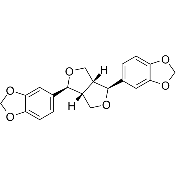
HY-N0121
SesaminDelta 5 Desaturase Inhibitor -

HY-N0809R
Sesamolin (Standard)Standard of Sesamolin -
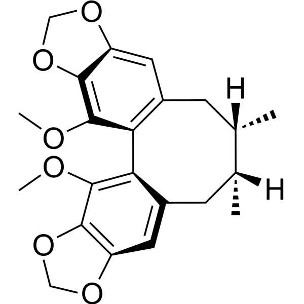
HY-N0690
Schisandrin CBioactive Lignan -
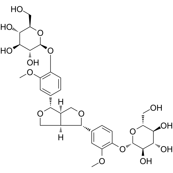
HY-N0657
Pinoresinol DiglucosideLignan -
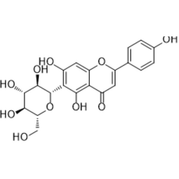
HY-N0773
IsovitexinNF-κB Inhibitor -
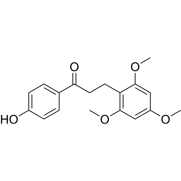
HY-N1504
Loureirin BPAI-1 Inhibitor -
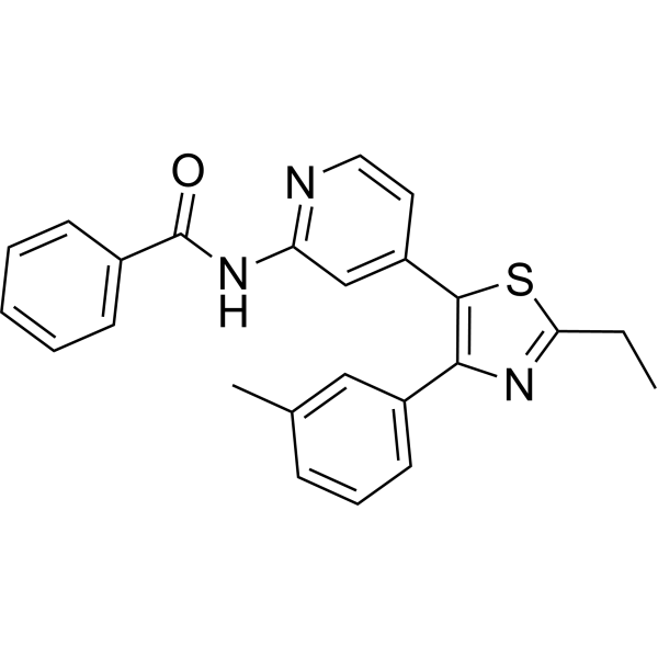
HY-10456
TAK-715p38 MAPK Inhibitor -
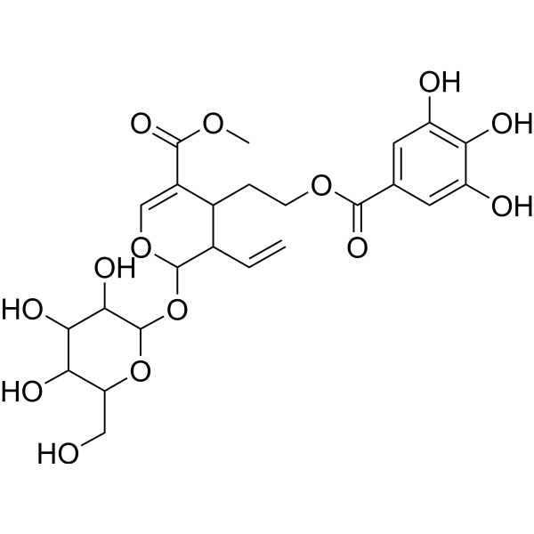
HY-N0631
CornusideMAPK/NF-κB Inhibitor
Publications Citing Use of MedChemExpress (MCE) Sesamolin
More Customer Validation & Images

-
RT-PCR

-
WB

-
Cell Migration/Invasion Assay

-
ELISA

-
Histological Imaging/Staining

All Caspase Isoforms
More

Biological Activity
Description
In Vitro
Sesamolin (0.5-50 μM, 1-2 h) reduces the release of LDH and ROS generation in H2O2 or hypoxia-stimulated PC12 cell, increases the cell viability, and exhibits antioxidant activity
Sesamolin (0-10 μM, 5 days) inhibits RANKL-induced formation and differentiation of osteoclast in bone marrow macrophages, inhibits the bone resorption of osteoclasts through inhibition of NF-κB/MAPK signaling pathway, and suppression of c-Fos and NFATc1 expressions[5].
MedChemExpress (MCE) has not independently confirmed the accuracy of these methods. They are for reference only. Further protocols information, click here.
-
Cell Line:PC12
-
Concentration:0.5-50 μM
-
Incubation Time:1-2 h
-
Result:Increased H2O2 or hypoxia-inhibited cell viability.
-
Cell Line:bone marrow macrophages
-
Concentration:10 μM
-
Incubation Time:5 days
-
Result:Inhibited levels of c-Fos and NFATc1.
In Vivo
Sesamolin (60 mg/kg, po, twice a day for 3 weeks) improves metabolic disorders, attenuates hepatic steatosis, alleviates endotoxemia and systemic inflammation, alters the composition of gut microbiota, changes serum metabolites and metabolomics pathways, thereby ameliorating high-fat and high-fructose diet -induced non-alcoholic fatty liver disease in mouse models[6].
MedChemExpress (MCE) has not independently confirmed the accuracy of these methods. They are for reference only.
-
Animal Model:Mouse OVX-induced osteoporosis models[5]
-
Dosage:5 mg/kg
-
Administration:ip, every two days for 42 days
-
Result:Reduced the bone loss and increased trabecular parameters.
-
Animal Model:High-fat and high-fructose diet -induced non-alcoholic fatty liver disease (NAFLD) in mouse models[6]
-
Dosage:60 mg/kg
-
Administration:po, twice a day for 3 weeks (from 9th week to 12th week)
-
Result:Inhibited weight gain, reduced the liver weight.
Reduced serum levels of glucose, insulin, HOMA-IR, total cholesterol, HDL-C, LDL-C, endotoxin, LBP, IL-6, TNF-α, AST and ALT.
Altered the composition of the gut flora, increased the relative abundance of beneficial bacteria and decreased the relative abundance of harmful bacteria.


Chemical Information
-
CAS No. 526-07-8
-
Appearance Solid
-
Molecular Weight 370.35
-
Formula C20H18O7
-
Color White to off-white
-
SMILES
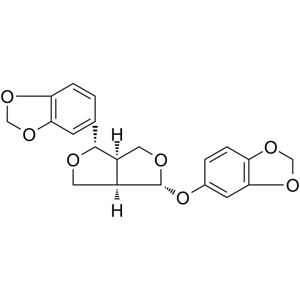
[H][C@@]12[C@@](CO[C@@H]2C3=CC(OCO4)=C4C=C3)([H])[C@H](OC1)OC5=CC(OCO6)=C6C=C5
-
Structure Classification
-
Initial Source
-
Shipping
Room temperature in continental US; may vary elsewhere.
-
Storage
4°C, protect from light
* In solvent : -80°C, 6 months; -20°C, 1 month (protect from light)


Publications (2)
-
Journal Impact Factor
-
Most Recent
-
Int Immunopharmacol
Hepatocyte-derived exosomes deliver H2AFJ to hepatic stellate cells and promote liver fibrosis via the MAPK/STMN1 axis activation. [Abstract]2023 Feb:115:109605. PMID: 36608439
Sesamolin purchased from MedChemExpress. Usage Cited in: Int Immunopharmacol. 2023 Feb:115:109605. [Abstract]
Expression of H2AFJ, JNK, c-jun, and STMN1 in HSCs determined by RT-qPCR treated with Sesamolin (5 μM).
Sesamolin purchased from MedChemExpress. Usage Cited in: Int Immunopharmacol. 2023 Feb:115:109605. [Abstract]
Expression of H2AFJ, STMN1, JNK, c-jun, and extent of phosphorylation of JNK and c-jun determined by Western blot analysis treated with Sesamolin (5 μM).
Sesamolin purchased from MedChemExpress. Usage Cited in: Int Immunopharmacol. 2023 Feb:115:109605. [Abstract]
HSC migration and invasion detected by determined by Transwell assay treated with Sesamolin (5 μM).
-
Heliyon
The topoisomerase inhibitor CPT-11 prevents the growth and metastasis of lung cancer cells in nude mice by inhibiting EGFR/MAPK signaling pathway. [Abstract]2023 Apr 27;9(5):e15805. PMID: 37251857
Sesamolin purchased from MedChemExpress. Usage Cited in: Heliyon. 2023 Apr 27;9(5):e15805. [Abstract]
The phosphorylation levels of MAPK pathway-related proteins in lung tissue in the metastatic tumor model upon EGFR overexpression or Sesamolin (5 mg/kg, i.p.) as determined by ELISA.
Sesamolin purchased from MedChemExpress. Usage Cited in: Heliyon. 2023 Apr 27;9(5):e15805. [Abstract]
The lung metastasis in nude mice upon EGFR overexpression or Sesamolin (5 mg/kg, i.p.) as observed using HE staining.

Sesamolin purchased from MedChemExpress. Usage Cited in: Heliyon. 2023 Apr 27;9(5):e15805. [Abstract]
Diagram of the body of subcutaneous xenograft tumor upon EGFR overexpression or Sesamolin (5 mg/kg, i.p.).


Solvent & Solubility
In Vitro:
DMSO : 100 mg/mL (270.01 mM; Need ultrasonic; Hygroscopic DMSO has a significant impact on the solubility of product, please use newly opened DMSO)
Please refer to the solubility information to select the appropriate solvent. Once prepared, please aliquot and store the solution to prevent product inactivation from repeated freeze-thaw cycles.
Storage method and period of stock solution: -80°C, 6 months; -20°C, 1 month (protect from light). When stored at -80°C, please use it within 6 months. When stored at -20°C, please use it within 1 month.
Please refer to the solubility information to select the appropriate solvent. Once prepared, please aliquot and store the solution to prevent product inactivation from repeated freeze-thaw cycles.
Storage method and period of stock solution: -80°C, 6 months; -20°C, 1 month (protect from light). When stored at -80°C, please use it within 6 months. When stored at -20°C, please use it within 1 month.
Concentration (start) × Volume (start) = Concentration (final) × Volume (final)
In Vivo:
Select the appropriate dissolution method based on your experimental animal and administration route.
- For the following dissolution methods, please ensure to first prepare a clear stock solution using an In Vitro approach and then sequentially add co-solvents:
- To ensure reliable experimental results, the clarified stock solution can be appropriately stored based on storage conditions. As for the working solution for In Vivo experiments, it is recommended to prepare freshly and use it on the same day.
- The percentages shown for the solvents indicate their volumetric ratio in the final prepared solution. If precipitation or phase separation occurs during preparation, heat and/or sonication can be used to aid dissolution.
Add each solvent one by one: 10% DMSO 40% PEG300 5% Tween-80 45% Saline
Solubility: 2.5 mg/mL (6.75 mM); Suspended solution; Need ultrasonic
This protocol yields a suspended solution of 2.5 mg/mL. Suspended solution can be used for oral and intraperitoneal injection.
Taking 1 mL working solution as an example, add 100 μL DMSO stock solution (25.0 mg/mL) to 400 μL PEG300, and mix evenly; then add 50 μL Tween-80 and mix evenly; then add 450 μL Saline to adjust the volume to 1 mL.
Preparation of Saline: Dissolve 0.9 g sodium chloride in ddH₂O and dilute to 100 mL to obtain a clear Saline solution.
Add each solvent one by one: 10% DMSO 90% (20% SBE-β-CD in Saline)
Solubility: ≥ 2.5 mg/mL (6.75 mM); Clear solution
This protocol yields a clear solution of ≥ 2.5 mg/mL (saturation unknown).
Taking 1 mL working solution as an example, add 100 μL DMSO stock solution (25.0 mg/mL) to 900 μL 20% SBE-β-CD in Saline, and mix evenly.
Preparation of 20% SBE-β-CD in Saline (4°C, storage for one week): 2 g SBE-β-CD powder is dissolved in 10 mL Saline, completely dissolve until clear.
In Vivo Dissolution Calculator
Please enter the basic information of animal experiments:
-
-
-
-
Recommended: Prepare an additional quantity of animals to account for potential losses during experiments.
Please enter your animal formula composition:
-
%DMSO +
Recommended: Keep the proportion of DMSO in working solution below 2% if your animal is weak.
-
%+
-
+%Tween-80 + +
-
%Saline +
The co-solvents required include: DMSO, . All of co-solvents are available by MedChemExpress (MCE). , Tween 80. All of co-solvents are available by MedChemExpress (MCE).
Working solution concentration: 0.22 mg/mL
Method for preparing stock solution: mg drug dissolved in μL DMSO. Stock solution concentration: mg/mL. * In solvent : -80°C, 6 months; -20°C, 1 month (protect from light)
1. Take μL DMSO stock solution;
2. Add μL .
μL , mix evenly;
3. Then add μL Tween 80, mix evenly;
4. Then add μL
Please ensure that the stock solution in the first step is dissolved to a clear state, and add co-solvents in sequence. You can use ultrasonic heating (ultrasonic cleaner, recommended frequency 20-40 kHz), vortexing, etc. to assist dissolution.

Protocols
-
Kinase activity and phosphorylation assays
Kinase activity assays measure the ability of kinases to transfer phosphate groups from ATP to specific substrates, while phosphorylation assays detect the presence and levels of phosphorylated proteins. Common methods include radiolabeled ATP incorporation (e. g. ,), ADP release detection via bioluminescence (e. g. ,[3]), enzyme-linked immunosorbent assays (ELISA) for phospho-specific epitopes (e. g. ,[6]), and microtiter-based formats for high-throughput screening (e. g. ,[8]). The ADP-Glo assay quantifies kinase activity by measuring ADP produced during phosphorylation using a luciferase-based system. Radiometric assays involve autoradiography or scintillation counting after incorporation of 32P-labeled ATP into substrate proteins. ELISA-based approaches rely on phospho-specific antibodies to detect activated kinases in cell lysates or purified samples.
-
Western Blot
Western blotting (WB) is a commonly used experimental method in molecular biology, biochemistry, and immunogenetics for identifying and quantifying target proteins. It combines gel electrophoresis with immunoassay, enabling researchers to analyze protein expression, post-translational modifications, and molecular weight.
-
RNA extraction experimental
By lysing cells, releasing RNA, and removing impurities such as proteins and DNA, high-purity RNA products are finally obtained. The commonly used traditional method is the guanidine isothiocyanate/phenol/chloroform method (Trizol), which is suitable for a variety of animal materials including animal tissues, microorganisms, cultured cells, etc., and most plant materials.
-
Apoptosis Solutions
Apoptosis is a regulated, generally non-lytic cell-death pathway that removes unwanted, damaged, infected, or abnormal cells through coordinated morphological changes, caspase activation, DNA fragmentation, and membrane remodeling. The intrinsic apoptosis pathway is controlled mainly by mitochondrial outer membrane permeabilization, BCL-2 family proteins, cytochrome c release, apoptosome formation, caspase-9 activation, and downstream executioner caspase-3/7 activation. The extrinsic apoptosis pathway is initiated by death receptors such as Fas, TNFR, and TRAIL receptors, which recruit adaptor proteins and activate caspase-8 before engaging executioner caspases or mitochondrial amplification through BID cleavage. Apoptosis is linked to many phenotypes, including cancer cell killing, tissue homeostasis, immune regulation, neurodegeneration, infection response, and treatment-induced cytotoxicity; unresolved questions include how apoptosis interacts with necroptosis, pyroptosis, ferroptos
-
Ferroptosis Solutions
Ferroptosis is an iron-dependent, non-apoptotic form of regulated cell death characterized by lethal lipid peroxidation and sensitivity to suppression by iron chelators or lipophilic radical-trapping antioxidants. The core pathway links cystine uptake through system Xc−, glutathione availability, GPX4-dependent detoxification of phospholipid hydroperoxides, iron-dependent oxidative reactions, and polyunsaturated-phospholipid metabolism into a cell-death program that is biochemically and morphologically distinct from apoptosis, necrosis, and autophagy. The ferroptosis pathway is experimentally linked to phenotype through chemical and genetic perturbation. Erastin induces ferroptosis by inhibiting cystine uptake through system Xc− and weakening antioxidant defenses, while GPX4 inhibition or depletion causes lipid peroxide accumulation and ferroptotic cancer-cell death. ACSL4 and oxidizable arachidonoyl- or adrenoyl-containing phosphatidylethanolamines shape ferroptosis sensitivity by con
-
Protocol for Kinase activity and phosphorylation assays
Kinase activity assays measure transfer of phosphate from ATP to a protein or peptide substrate, generating phosphorylated substrate, ADP, or incorporated radiolabeled phosphate as the readout; phosphorylation assays measure site-specific phosphorylation in cells or tissues as a proxy for kinase-pathway activation, inhibition, or substrate regulation. Phosphorylation can be detected by phospho-specific Western blot, immunoprecipitation kinase assay, phospho-immunofluorescence, phospho-flow cytometry, luminescent ADP detection, radiolabeled ATP incorporation, or reporter-based pathway assays, and these readouts can be applied to cancer cells, primary neurons, mouse tumors, organoids, inflammatory macrophages, ferroptosis studies, and mitophagy studies when the kinase target is biologically relevant.


Purity & Documentation
-
Data Sheet (279 KB)
-
SDS (393 KB)
- English - EN (393 KB)
- Français - FR (393 KB)
- Deutsch - DE (393 KB)
- Norwegian - NO (393 KB)
- Español - ES (393 KB)
- Swedish - SV (393 KB)
- Italian - IT (393 KB)
- Korean - KR (393 KB)
- Portuguese - PT (393 KB)
-
Handling Instructions (2659 KB)
References
[3]. Rolis Chien‐Wei Hou, et al. Protective effects of sesamin and sesamolin on hypoxic neuronal and PC12 cells. [Content Brief]
[4]. Lim JS, et al. Comparative analysis of sesame lignans (sesamin and sesamolin) in affecting hepatic fatty acid metabolism in rats. Br J Nutr. 2007 Jan;97(1):85-95. [Content Brief]
[5]. Yang X, et al., Sesamolin Protects Mice From Ovariectomized Bone Loss by Inhibiting Osteoclastogenesis and RANKL-Mediated NF-κB and MAPK Signaling Pathways. Front Pharmacol. 2021 Jun 14;12:664697. [Content Brief]
[6]. Yu J, et al., Sesamolin Alleviates Nonalcoholic Fatty Liver Disease through Modulating Gut Microbiota and Metabolites in High-Fat and High-Fructose Diet-Fed Mice. Int J Mol Sci. 2022 Nov 10;23(22):13853. [Content Brief]


Complete Stock Solution Preparation Table
Please refer to the solubility information to select the appropriate solvent. Once prepared, please aliquot and store the solution to prevent product inactivation from repeated freeze-thaw cycles.
Storage method and period of stock solution: -80°C, 6 months; -20°C, 1 month (protect from light). When stored at -80°C, please use it within 6 months. When stored at -20°C, please use it within 1 month.
| Optional Solvent | Concentration Solvent Mass | 1 mg | 5 mg | 10 mg | 25 mg |
|---|---|---|---|---|---|
| DMSO | 1 mM | 2.7001 mL | 13.5007 mL | 27.0015 mL | 67.5037 mL |
| 5 mM | 0.5400 mL | 2.7001 mL | 5.4003 mL | 13.5007 mL | |
| 10 mM | 0.2700 mL | 1.3501 mL | 2.7001 mL | 6.7504 mL | |
| 15 mM | 0.1800 mL | 0.9000 mL | 1.8001 mL | 4.5002 mL | |
| 20 mM | 0.1350 mL | 0.6750 mL | 1.3501 mL | 3.3752 mL | |
| 25 mM | 0.1080 mL | 0.5400 mL | 1.0801 mL | 2.7001 mL | |
| 30 mM | 0.0900 mL | 0.4500 mL | 0.9000 mL | 2.2501 mL | |
| 40 mM | 0.0675 mL | 0.3375 mL | 0.6750 mL | 1.6876 mL | |
| 50 mM | 0.0540 mL | 0.2700 mL | 0.5400 mL | 1.3501 mL | |
| 60 mM | 0.0450 mL | 0.2250 mL | 0.4500 mL | 1.1251 mL | |
| 80 mM | 0.0338 mL | 0.1688 mL | 0.3375 mL | 0.8438 mL | |
| 100 mM | 0.0270 mL | 0.1350 mL | 0.2700 mL | 0.6750 mL |


Sesamolin Related Classifications
HY-N0809 Related Classifications
 Powered by Bioz