



Skimmin
Based on 1 publication(s) in Google Scholar
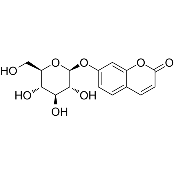
Skimmin (Umbelliferone glucoside) is a major pharmacologically active and orally active molecule present in Hydrangea paniculata, a medical herb used in traditional Chinese medicine as an anti-inflammatory agent. Skimmin has renal protective activity. Skimmin can improve creatinine clearance, and reduce plasma creatinine, and kidney injuries. Skimmin has good anti-amoebic activity against the HM1:IMMS strain of Entamoeba histolytica. Skimmin has anti-cancer and neuroprotective activities. Skimmin reduces cardiac fibrosis as well as decreasing TNF-α, IL-6, IL1β, and TGFβ1 in cardiac tissues. Skimmin can be studied in research for diabetes and diabetes-related diseases.
연구목적의 판매만을 진행합니다. 환자를 대상으로 한 판매는 하지 않습니다.
- Purity : 99.64%
- CAS No.: 93-39-0
- 화학식: C15H16O8
- 분자량:324.28
-
보관:
4°C, protect from light
* In solvent : -80°C, 6 months; -20°C, 1 month (protect from light)

To place orders, for customer services and technical support, please contact: MedChemExpress USA
Tel: 609-228-6898 E-mail: [email protected] [email protected]


-
 Biological Activity
-
Chemical Information
-
용액&용해도
- Protocol
- 순도&문서
- References
-
 Help & FAQs


Publications Citing Use of MedChemExpress (MCE) Skimmin
MoreAll Parasite Isoforms
More

Biological Activity
제품 설명
Cellular Effect
|
Cell Line

|
Type | Value | Description | References |
|---|---|---|---|---|
| SK-OV-3 | IC50 |
>100 μM
Compound: 3
|
Antiproliferative activity against human SK-OV-3 cells measured after 21 hrs by MTT assay
Antiproliferative activity against human SK-OV-3 cells measured after 21 hrs by MTT assay
|
[PMID: 38340509] |
In Vitro
In Vivo
Skimmin (15-30 mg/kg, i.g., 16 w) showed a certain effect on decreasing blood glucose in male SD rats[3].
Skimmin (7.5-30 mg/kg, p.o., 17 w) results in significantly less severe sclerosis in glomerulus segments especially at 15 mg/kg and 30 mg/kg dose in Wistar rats[4].
MedChemExpress (MCE) has not independently confirmed the accuracy of these methods. They are for reference only.
-
Animal Model:Female SD rats, 10 weeks old, (160-180 g)[1]
-
Dosage:15 or 30 mg/kg
-
Administration:Intragastric injection (i.g.) daily from days 37 to 66
-
Result:Reduced BUN whilst increased Ucr and urinary creatinine (Ccr) with 30 mg/kg dose.
Significantly reduced C-BSA-induced morphological changes including incidences of glomerular hypercellarity, glomerular basement membrane thickening or mesangial proliferation, and tubulointerstitial injuries.
Led to significantly less deposits of immune complexes in the mesangial areas and along the capillary walls.
Reduced the IgG positive staining area by 41% compared to model group.
Decreased the expression of IL-1β and IL-6 in glomeruli dose-dependently.
Reduced the CD20 mature B cell infiltration significantly.
-
Animal Model:Male Sprague Dawley rats (160-180 g, 6-8 w) with Streptozotocin (HY-13753) (60 mg/kg, i.p.)-induced type 1 diabetes mellitus[3]
-
Dosage:15 or 30 mg/kg
-
Administration:Intragastric injection (i.g.) daily for 16 w
-
Result:Lightly reduced the HR, SBP, DBP, and MAP.
Significantly improved cardiac function in diabetic rats dose dependently.
Significantly reduced the heart NO and malondialdehyde (MDA) concentration and increased GSH concentration in the heart.
Increased the enzyme activity of SOD and CAT in the heart tissues.
Showed anti-inflammatory activity by decreasing TNF-α, IL-6, IL1β, and TGFβ1 in the heart tissues from DCM rats.
Exerted a cardiac protective effect and inhibited the NLRP3 pathway in diabetic hearts by decreasing NLRP3, caspase-1, and IL-1β.
Reduced the P62 protein level in the heart tissues.
-
Animal Model:Adult male Wistar rats (180-200 g) with Streptozotocin (60 mg/kg)-induced diabetic nephropathy[4]
-
Dosage:7.5, 10, 30 mg/kg
-
Administration:Oral gavage (p.o.) for 17 weeks
-
Result:Reduced the incidence of tubule vacuolar degeneration and severity level.
Decreased TGF-β1 expression on mRNA level in the renal tissues dose dependently.


Chemical Information
-
CAS No. 93-39-0
-
Appearance Solid
-
분자량 324.28
-
화학식 C15H16O8
-
Color White to off-white
-
SMILES
O=C1C=CC2=CC=C(O[C@H]3[C@@H]([C@H]([C@@H]([C@@H](CO)O3)O)O)O)C=C2O1
-
Synonyms
Umbelliferone glucoside
-
Structure Classification
-
Initial Source
-
선적
Room temperature in continental US; may vary elsewhere.
-
보관
4°C, protect from light
* In solvent : -80°C, 6 months; -20°C, 1 month (protect from light)


Publications (1)
-
Journal Impact Factor
-
Most Recent


용액&용해도
In Vitro:
DMSO : 100 mg/mL (308.38 mM; Need ultrasonic; Hygroscopic DMSO has a significant impact on the solubility of product, please use newly opened DMSO)
Please refer to the solubility information to select the appropriate solvent. Once prepared, please aliquot and store the solution to prevent product inactivation from repeated freeze-thaw cycles.
Storage method and period of stock solution: -80°C, 6 months; -20°C, 1 month (protect from light). When stored at -80°C, please use it within 6 months. When stored at -20°C, please use it within 1 month.
Please refer to the solubility information to select the appropriate solvent. Once prepared, please aliquot and store the solution to prevent product inactivation from repeated freeze-thaw cycles.
Storage method and period of stock solution: -80°C, 6 months; -20°C, 1 month (protect from light). When stored at -80°C, please use it within 6 months. When stored at -20°C, please use it within 1 month.
Concentration (start) × Volume (start) = Concentration (final) × Volume (final)
In Vivo:
Select the appropriate dissolution method based on your experimental animal and administration route.
- For the following dissolution methods, please ensure to first prepare a clear stock solution using an In Vitro approach and then sequentially add co-solvents:
- To ensure reliable experimental results, the clarified stock solution can be appropriately stored based on storage conditions. As for the working solution for In Vivo experiments, it is recommended to prepare freshly and use it on the same day.
- The percentages shown for the solvents indicate their volumetric ratio in the final prepared solution. If precipitation or phase separation occurs during preparation, heat and/or sonication can be used to aid dissolution.
Add each solvent one by one: 10% DMSO 40% PEG300 5% Tween-80 45% Saline
Solubility: ≥ 2.5 mg/mL (7.71 mM); Clear solution
This protocol yields a clear solution of ≥ 2.5 mg/mL (saturation unknown).
Taking 1 mL working solution as an example, add 100 μL DMSO stock solution (25.0 mg/mL) to 400 μL PEG300, and mix evenly; then add 50 μL Tween-80 and mix evenly; then add 450 μL Saline to adjust the volume to 1 mL.
Preparation of Saline: Dissolve 0.9 g sodium chloride in ddH₂O and dilute to 100 mL to obtain a clear Saline solution.
Add each solvent one by one: 10% DMSO 90% (20% SBE-β-CD in Saline)
Solubility: ≥ 2.5 mg/mL (7.71 mM); Clear solution
This protocol yields a clear solution of ≥ 2.5 mg/mL (saturation unknown).
Taking 1 mL working solution as an example, add 100 μL DMSO stock solution (25.0 mg/mL) to 900 μL 20% SBE-β-CD in Saline, and mix evenly.
Preparation of 20% SBE-β-CD in Saline (4°C, storage for one week): 2 g SBE-β-CD powder is dissolved in 10 mL Saline, completely dissolve until clear.
In Vivo Dissolution Calculator
Please enter the basic information of animal experiments:
-
-
-
-
Recommended: Prepare an additional quantity of animals to account for potential losses during experiments.
Please enter your animal formula composition:
-
%DMSO +
Recommended: Keep the proportion of DMSO in working solution below 2% if your animal is weak.
-
%+
-
+%Tween-80 + +
-
%Saline +
The co-solvents required include: DMSO, . All of co-solvents are available by MedChemExpress (MCE). , Tween 80. All of co-solvents are available by MedChemExpress (MCE).
Working solution concentration: 0.22 mg/mL
Method for preparing stock solution: mg drug dissolved in μL DMSO. Stock solution concentration: mg/mL. * In solvent : -80°C, 6 months; -20°C, 1 month (protect from light)
1. Take μL DMSO stock solution;
2. Add μL .
μL , mix evenly;
3. Then add μL Tween 80, mix evenly;
4. Then add μL
Please ensure that the stock solution in the first step is dissolved to a clear state, and add co-solvents in sequence. You can use ultrasonic heating (ultrasonic cleaner, recommended frequency 20-40 kHz), vortexing, etc. to assist dissolution.

Protocol
-
Fibrosis/Collagen Morphometry
Fibrosis and collagen morphometry is based on the quantitative visualization of fibrillar collagen deposition in tissue sections using histochemical stains such as Sirius Red (Picrosirius Red) or Masson's trichrome, followed by image-based or polarization-enhanced analysis to estimate collagen proportional area as a surrogate of extracellular matrix accumulation during fibrotic remodeling. Sirius Red combined with polarized light microscopy enhances detection of collagen fibers due to birefringence properties, enabling more specific visualization of collagen type I and III fibrils compared to conventional bright-field histology, while whole-section or region-restricted digital morphometry reduces field-selection bias in fibrosis assessment. Alternative quantitative approaches include second harmonic generation (SHG) and two-photon excited fluorescence microscopy, which enable label-free detection of fibrillar collagen and have been validated against histological staining and biochemica
-
Collagen: Sirius Red Staining
Sirius Red or picrosirius red staining is a histochemical method for visualizing collagen-rich extracellular matrix in tissue sections, and collagen fibers are detected as red-stained structures under bright-field microscopy with enhanced birefringence under polarized light. Picrosirius red is useful for assessing total collagen organization, distribution, and fibrosis burden, but polarized color should not be interpreted as a definitive collagen type I versus type III readout because color is affected by fiber orientation, thickness, and packing.
-
Nephrotoxicity Study
This protocol assesses nephrotoxicity by combining functional kidney injury readouts, urinary/tissue injury biomarkers, and renal histopathology. Serum creatinine and BUN reflect impaired kidney function, while KIM-1, NGAL, clusterin, osteopontin, IL-18, cystatin C, nephrin, Oat5, urinary protein, glucose, and alkaline phosphatase have been used to detect tubular injury in cisplatin-, gentamicin-, and acetaminophen-induced nephrotoxicity models.
-
Connective Tissue: Masson's Trichrome/Collagen Trichrome Staining
Masson’s Trichrome (collagen/trichrome staining) is a histological technique that differentially stains tissue compartments using sequential acidic dyes to distinguish collagen from muscle and cytoplasmic components based on dye affinity and tissue permeability differences, enabling visualization of fibrosis and connective tissue architecture in histological sections. The classical formulation typically uses Weigert's iron hematoxylin for nuclear staining, Biebrich scarlet-acid fuchsin for cytoplasm and muscle, and aniline blue (or light green variants) for collagen, producing a characteristic blue/green collagen signal contrasted against red cytoplasm and dark nuclei. The staining principle relies on selective displacement of smaller dye molecules by larger anionic dyes in collagen-rich regions under controlled acidified conditions, which enhances collagen-specific dye retention. This property makes the method widely used for fibrosis assessment in organs such as heart, liver, lung, a
-
Protocol for Pharmacokinetic Study
Pharmacokinetic studies quantify how an organism handles a drug over time through absorption, distribution, metabolism, and excretion, and the core experimental readout is the concentration-time profile of parent drug and, when relevant, metabolites in biological matrices such as plasma, whole blood, urine, bile, or tissue. Pharmacokinetic analysis links dose, route, exposure, clearance, half-life, distribution, bioavailability, and systemic exposure to drug efficacy and toxicity hypotheses rather than measuring a signaling pathway directly. The literature links pharmacokinetics to drug-development phenotypes by showing that drug metabolism and pharmacokinetics influence compound progression, exposure-response interpretation, safety margins, dosing strategy, and failure risk during discovery and development. DMPK science contributes to compound optimization by integrating physicochemical properties, in vitro metabolism, transporter behavior, in vivo exposure, and pharmacodynamic contex
-
Research Protocol for Inflammation-related Diseases
The NLRP3 inflammasome is a cytosolic innate immune signaling platform that integrates priming signals and danger-signal activation to promote caspase-1 activation, maturation of IL-1β and IL-18, and gasdermin D-mediated pyroptotic cell death. The core experimental logic is to determine whether inflammatory disease phenotypes are driven by increased NLRP3 expression, ASC-containing inflammasome assembly, caspase-1 cleavage, GSDMD cleavage, and extracellular release of IL-1β/IL-18 rather than by nonspecific cell injury alone. The pathway is strongly linked to inflammation-related disease phenotypes because monosodium urate crystals activate NALP3/NLRP3 inflammasome signaling in gout-like crystal inflammation, cholesterol crystals activate NLRP3 inflammasomes in atherogenesis models, and DSS-induced intestinal inflammation has been reported to involve NLRP3 inflammasome activity. However, experimental colitis studies also show context-dependent protective effects of NLRP3 inflammasome co
-
Research Protocol for Metabolic Diseases
AMP-activated protein kinase, AMPK, is a conserved cellular energy sensor that responds to reduced cellular energy status and coordinates metabolism by increasing ATP-generating catabolic pathways while suppressing ATP-consuming anabolic processes. In metabolic disease research, the AMPK pathway is experimentally relevant because it regulates hepatic lipid synthesis, fatty acid oxidation, glucose production, skeletal-muscle glucose disposal, mTORC1-linked biosynthesis, autophagy, mitochondrial homeostasis, and whole-body energy balance. The central pathway logic is that energy stress, metformin, exercise-like stimulation, or direct AMPK activators increase AMPKα Thr172 phosphorylation and downstream substrate phosphorylation, including ACC and RAPTOR. Phosphorylation of ACC suppresses lipogenesis and supports fatty acid oxidation, whereas phosphorylation of RAPTOR suppresses mTORC1 signaling and links cellular energy status to growth and protein synthesis control. The pathway is linked


순도&문서
-
Data Sheet (280 KB)
-
SDS (393 KB)
- English - EN (393 KB)
- Français - FR (393 KB)
- Deutsch - DE (393 KB)
- Norwegian - NO (393 KB)
- Español - ES (393 KB)
- Swedish - SV (393 KB)
- Italian - IT (393 KB)
- Korean - KR (393 KB)
- Portuguese - PT (393 KB)
-
Handling Instructions (2659 KB)
References
[1]. Zhang S, et al. Skimmin, a Coumarin from Hydrangea paniculata, Slows down the Progression of Membranous Glomerulonephritis by Anti-Inflammatory Effects and Inhibiting Immune Complex Deposition. Evid Based Complement Alternat Med. 2013;2013:819296. [Content Brief]
[2]. Lou, Y., et al., (2020). Determination and pharmacokinetic study of skimmin by UHPLC-MS/MS in rat plasma. Journal of pharmaceutical and biomedical analysis, 179, 112969. [Content Brief]
[3]. Liang, R. K., et al., (2021). Skimmin protects diabetic cardiomyopathy in streptozotocin-induced diabetic rats. The Kaohsiung journal of medical sciences, 37(2), 136–144. [Content Brief]
[4]. Zhang, S., et al., (2012). Skimmin, a coumarin, suppresses the streptozotocin-induced diabetic nephropathy in wistar rats. European journal of pharmacology, 692(1-3), 78–83. [Content Brief]


Complete Stock Solution Preparation Table
Please refer to the solubility information to select the appropriate solvent. Once prepared, please aliquot and store the solution to prevent product inactivation from repeated freeze-thaw cycles.
Storage method and period of stock solution: -80°C, 6 months; -20°C, 1 month (protect from light). When stored at -80°C, please use it within 6 months. When stored at -20°C, please use it within 1 month.
| Optional Solvent | Concentration Solvent Mass | 1 mg | 5 mg | 10 mg | 25 mg |
|---|---|---|---|---|---|
| DMSO | 1 mM | 3.0838 mL | 15.4188 mL | 30.8375 mL | 77.0939 mL |
| 5 mM | 0.6168 mL | 3.0838 mL | 6.1675 mL | 15.4188 mL | |
| 10 mM | 0.3084 mL | 1.5419 mL | 3.0838 mL | 7.7094 mL | |
| 15 mM | 0.2056 mL | 1.0279 mL | 2.0558 mL | 5.1396 mL | |
| 20 mM | 0.1542 mL | 0.7709 mL | 1.5419 mL | 3.8547 mL | |
| 25 mM | 0.1234 mL | 0.6168 mL | 1.2335 mL | 3.0838 mL | |
| 30 mM | 0.1028 mL | 0.5140 mL | 1.0279 mL | 2.5698 mL | |
| 40 mM | 0.0771 mL | 0.3855 mL | 0.7709 mL | 1.9273 mL | |
| 50 mM | 0.0617 mL | 0.3084 mL | 0.6168 mL | 1.5419 mL | |
| 60 mM | 0.0514 mL | 0.2570 mL | 0.5140 mL | 1.2849 mL | |
| 80 mM | 0.0385 mL | 0.1927 mL | 0.3855 mL | 0.9637 mL | |
| 100 mM | 0.0308 mL | 0.1542 mL | 0.3084 mL | 0.7709 mL |


Skimmin Related Classifications
HY-N2263 Related Classifications
 Powered by Bioz