



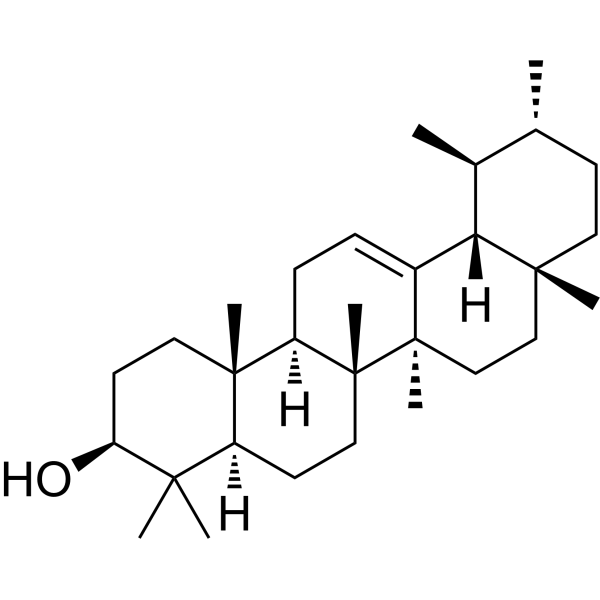
α-Amyrin
Based on 2 publication(s) in Google Scholar
α-Amyrin is a pentacyclic triterpenoid compound with oral activity. α-Amyrin activates the ERK and GSK-3β signaling pathways. α-Amyrin can inhibit cancer cells proliferation and induce apoptosis. α-Amyrin shows anti-bacterial and anti-inflammation activity. α-Amyrin can reduce blood glucose level. α-Amyrin can be used for the researches of cancer, infection, inflammation, metabolic disease and neurological disease, such as breast cancer, Streptococcus oralis infection, skin inflammation and diabetes.
For research use only. We do not sell to patients.
- Purity : 99.99%
- CAS No.: 638-95-9
- Formula: C30H50O
- Molecular Weight:426.72
-
Storage:
4°C, protect from light
* In solvent : -80°C, 6 months; -20°C, 1 month (protect from light)

To place orders, for customer services and technical support, please contact: MedChemExpress USA
Tel: 609-228-6898 E-mail: [email protected] [email protected]


-
 Biological Activity
-
Chemical Information
-
Solvent & Solubility
- Protocols
- Purity & Documentation
- References
-
 Help & FAQs


-
HY-N2922
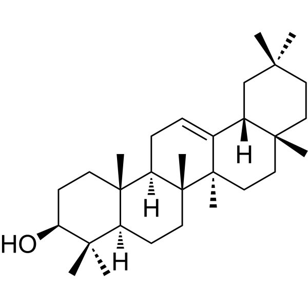
β-AmyrinAnti-inflammatory compound -
HY-N0162
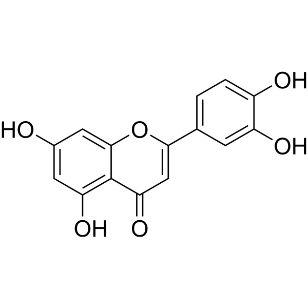
LuteolinNrf2 Inhibitor -
HY-N0790
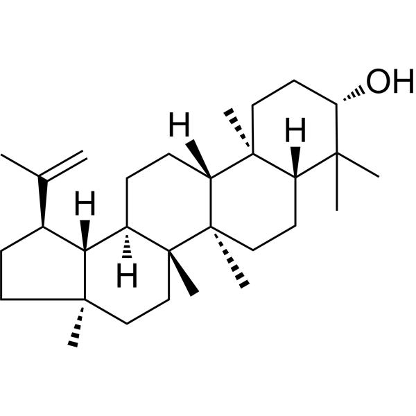
LupeolAndrogen Receptor Inhibitor -
HY-14590
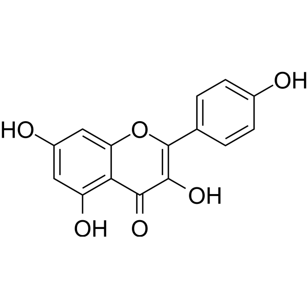
KaempferolEstrogen Receptor α Inhibitor -
HY-B0573B
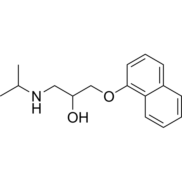
Propranololβ-Adrenoceptor Blocker -
HY-18085
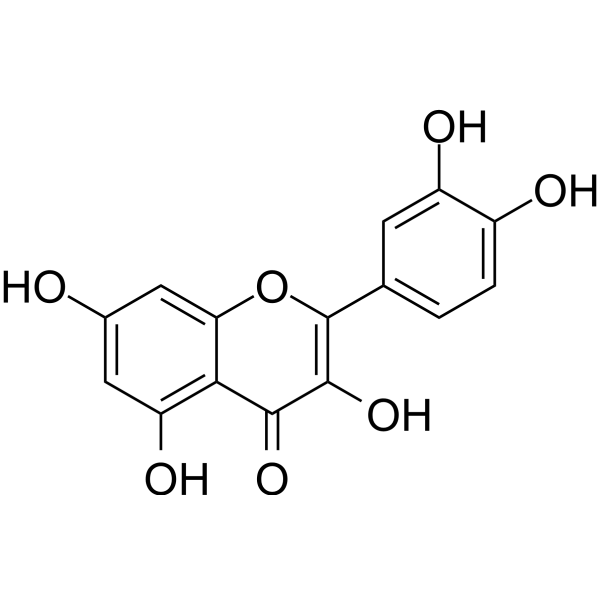
QuercetinFlavonoid Antioxidant -
HY-13417
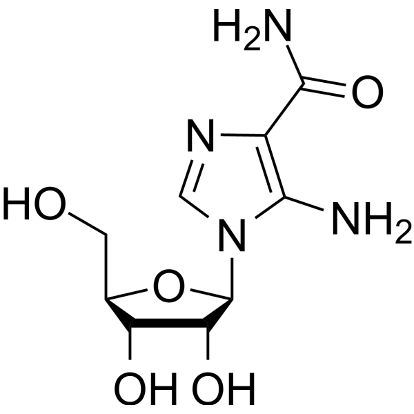
AICARAMPK Activator -
HY-18258
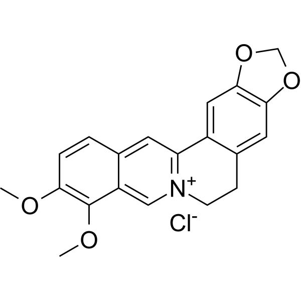
Berberine chlorideAntibiotic -
HY-14872
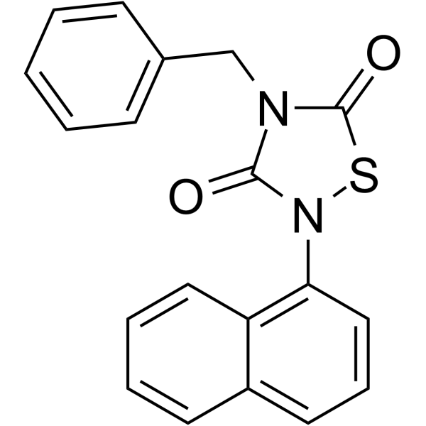
TideglusibGSK-3 Inhibitor -
HY-145669
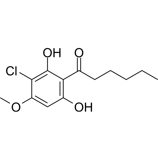
DIF-3Cyclin D1 Inhibitor -

HY-N8423R
α-Amyrin (Standard)Pentacyclic Triterpenoid -
HY-W013812
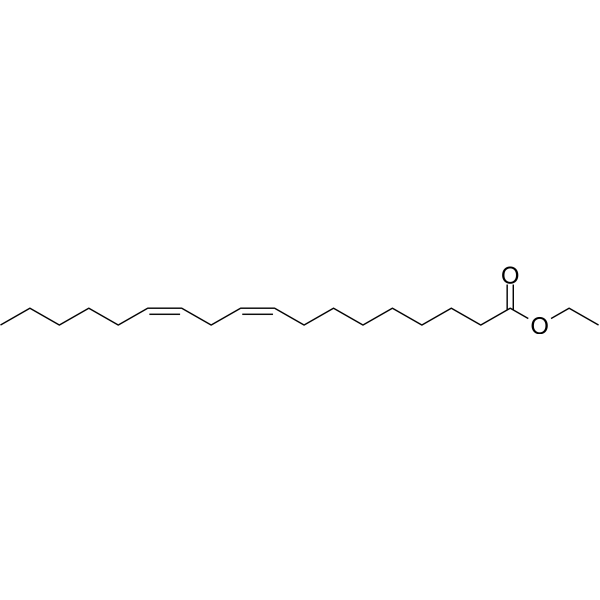
Ethyl linoleateAntiinflammatory Agent
Publications Citing Use of MedChemExpress (MCE) α-Amyrin
MoreAll Caspase Isoforms
More

Biological Activity
Description
IC50 & Target
[1]|
GSK-3β |
Caspase 3 |
COX-2 |
Cellular Effect
|
Cell Line

|
Type | Value | Description | References |
|---|---|---|---|---|
| A2780 | IC50 |
20.6 μg/mL
Compound: alpha-amyrin
|
Cytotoxicity against human A2780 cells
Cytotoxicity against human A2780 cells
|
[PMID: 15165160] |
| A549 | GI50 |
10.54 μM
Compound: 58
|
Cytotoxicity against human A549 cells after 48 hrs by WST8 assay
Cytotoxicity against human A549 cells after 48 hrs by WST8 assay
|
[PMID: 20371180] |
| B16 | ED50 |
>50 μM
Compound: 19
|
Induction of melanogenesis in mouse B16 2F2 cells assessed as intracellular melanin content after 3 days
Induction of melanogenesis in mouse B16 2F2 cells assessed as intracellular melanin content after 3 days
|
[PMID: 12027734] |
| B16 | IC50 |
50 μM
Compound: 19
|
Growth inhibition of mouse B16 2F2 cells after 3 days
Growth inhibition of mouse B16 2F2 cells after 3 days
|
[PMID: 12027734] |
| CHO | EC50 |
>10 μM
Compound: Alpha-amyrin
|
Agonist activity at TGR5 expressed in CHO cells by CRE-driven luciferase reporter gene assay
Agonist activity at TGR5 expressed in CHO cells by CRE-driven luciferase reporter gene assay
|
[PMID: 19911773] |
| COS-1 | EC50 |
0 μM
Compound: Alpha-amyrin
|
Agonist activity at human FXR expressed in COS1 cells by luciferase reporter gene assay
Agonist activity at human FXR expressed in COS1 cells by luciferase reporter gene assay
|
[PMID: 19911773] |
| HepG2 | IC50 |
>100 μM
Compound: 8
|
Cytotoxicity against human HepG2 cells assessed as growth inhibition after 72 hrs by neutral red assay
Cytotoxicity against human HepG2 cells assessed as growth inhibition after 72 hrs by neutral red assay
|
[PMID: 28318944] |
| HepG2 | IC50 |
220 μM
Compound: 8
|
Cytotoxicity against human Hep G2 cells after 48 hrs by WST-8 assay
Cytotoxicity against human Hep G2 cells after 48 hrs by WST-8 assay
|
[PMID: 15730243] |
| J774 | IC50 |
128.5 μM
Compound: 1
|
Cytotoxicity against mouse J774 cells by alamar blue assay
Cytotoxicity against mouse J774 cells by alamar blue assay
|
[PMID: 17637068] |
| KB | IC50 |
>100 μM
Compound: 8
|
Cytotoxicity against human KB cells assessed as growth inhibition after 72 hrs by neutral red assay
Cytotoxicity against human KB cells assessed as growth inhibition after 72 hrs by neutral red assay
|
[PMID: 28318944] |
| Lu1 | IC50 |
>100 μM
Compound: 8
|
Cytotoxicity against human Lu1 cells assessed as growth inhibition after 72 hrs by neutral red assay
Cytotoxicity against human Lu1 cells assessed as growth inhibition after 72 hrs by neutral red assay
|
[PMID: 28318944] |
| MCF7 | IC50 |
>100 μM
Compound: 8
|
Cytotoxicity against human MCF7 cells assessed as growth inhibition after 72 hrs by neutral red assay
Cytotoxicity against human MCF7 cells assessed as growth inhibition after 72 hrs by neutral red assay
|
[PMID: 28318944] |
| PANC-1 | GI50 |
12.8 μM
Compound: 58
|
Cytotoxicity against human PANC1 cells after 48 hrs by WST8 assay
Cytotoxicity against human PANC1 cells after 48 hrs by WST8 assay
|
[PMID: 20371180] |
| PC-3 | GI50 |
7.66 μM
Compound: 58
|
Cytotoxicity against human PC3 cells after 48 hrs by WST8 assay
Cytotoxicity against human PC3 cells after 48 hrs by WST8 assay
|
[PMID: 20371180] |
| SH-SY5Y | EC50 |
>50 μM
Compound: 37
|
Neuroprotective activity in human SH-SY5Y cells assessed as reduction in 1-methyl-4-phenylpyridinium ion-induced cell death after 48 hrs by MTT assay
Neuroprotective activity in human SH-SY5Y cells assessed as reduction in 1-methyl-4-phenylpyridinium ion-induced cell death after 48 hrs by MTT assay
|
[PMID: 27420919] |
| SH-SY5Y | EC50 |
13.3 μM
Compound: 37
|
Neuroprotective activity in human SH-SY5Y cells assessed as reduction in oxygen glucose deprivation-induced cell death incubated for 16 hrs under hypoxic condition followed by incubation for 24 hrs under normoxic condition by MTT assay
Neuroprotective activity in human SH-SY5Y cells assessed as reduction in oxygen glucose deprivation-induced cell death incubated for 16 hrs under hypoxic condition followed by incubation for 24 hrs under normoxic condition by MTT assay
|
[PMID: 27420919] |
| WI-38 | IC50 |
12.7 μM
Compound: 8
|
Cytotoxicity against human WI 38 cells after 48 hrs by WST-8 assay
Cytotoxicity against human WI 38 cells after 48 hrs by WST-8 assay
|
[PMID: 15730243] |
| WI-38 VA13 | IC50 |
>235 μM
Compound: 8
|
Cytotoxicity against human VA13 cells after 48 hrs by WST-8 assay
Cytotoxicity against human VA13 cells after 48 hrs by WST-8 assay
|
[PMID: 15730243] |
In Vitro
α-Amyrin (10 μM, 24 h) induces AMPK and PPARδ/ γ-mediated GLUT4 translocation in C2C12 myoblasts[1].
α-Amyrin (acetate) (2.5-10 μg/mL, 0-72 h) inhibits proliferation and colony formation in MDA-MB-231 cells[6].
α-Amyrin (acetate) (5-10 μg/mL, 24 h) induces apoptosis and increases caspase-3 activity in MDA-MB-231 cells[6].
α-Amyrin (acetate) (10 μg/mL, 24 h) promotes the cleavage of Poly polymerase, increased E-cadherin, and inhibits vimentin and phosphorylated extracellular signal-regulated activation in MDA-MB-231 cells[6].
α-Amyrin (10-1000 μg/mL, 24 h) reduces percentage of viability of Streptococcus salivarius, Streptococcus sanguinis and Streptococcus oralis[7].
MedChemExpress (MCE) has not independently confirmed the accuracy of these methods. They are for reference only. Further protocols information, click here.
-
Cell Line:C2C12 cells
-
Concentration:10 μM
-
Incubation Time:24 h
-
Result:Increased the mRNA and protein expression of PPARs, GLUT4 and FATP.
-
Cell Line:MDA-MB-231 cells
-
Concentration:5 and 10 μg/mL
-
Incubation Time:24 h
-
Result:Increased apoptotic cells.
Increased caspase-3 activity.
In Vivo
α-Amyrin (2-4 mg/kg, p.o.) improves cognitive dysfunction caused by low cholinergic neurotransmission in mice by activating ERK and GSK-3β signaling pathways[3].
α-Amyrin (acetate) (50 mg/kg, p.o., daily for 1-10 days) reduces the blood glucose level in diabetic rats[4].
α-Amyrin (0.1-1.0 mg/ear, topical application) inhibits inflammation in 12-O-tetradecanoylphorbol-acetate (TPA)-induced skin inflammation of mice[5].
MedChemExpress (MCE) has not independently confirmed the accuracy of these methods. They are for reference only.
-
Animal Model:High-fructose diet (HFD)-induced metabolic syndrome in rats[2]
-
Dosage:50, 100 and 200 mg/kg
-
Administration:Orally administration
-
Result:Decreased systolic blood pressure, plasma glucose, total cholesterol, and plasma triglycerides.
Attenuated hepatic oxidative stress as well as micro- and macrovesicular fatty changes in hepatocytes caused by HFD.
-
Animal Model:Scopolamine-Induced Memory Impairment Mice[3]
-
Dosage:2 and 4 mg/kg
-
Administration:Orally administration
-
Result:Increased the expression levels of phosphorylated extracellular signal-regulated kinase 1/2 (pERK) and phosphorylated glycogen synthase kinase-3β (pGSK-3β).
-
Animal Model:12-O-tetradecanoylphorbol-acetate (TPA)-induced skin inflammation of mice[5]
-
Dosage:0.1, 0.3 and 1.0 mg/ear, topical application
-
Administration:Topically application
-
Result:Inhibited skin inflammation.
Reduced COX-2 levels and activation of NF-κB and MAPKs.


Chemical Information
-
CAS No. 638-95-9
-
Appearance Solid
-
Molecular Weight 426.72
-
Formula C30H50O
-
Color White to off-white
-
SMILES
C[C@]12[C@]3(C([C@@]4([H])[C@@](CC3)(CC[C@H]([C@@H]4C)C)C)=CC[C@]1([H])[C@@]5([C@@](C(C)([C@H](CC5)O)C)([H])CC2)C)C
-
Structure Classification
-
Initial Source
-
Shipping
Room temperature in continental US; may vary elsewhere.
-
Storage
4°C, protect from light
* In solvent : -80°C, 6 months; -20°C, 1 month (protect from light)


Publications (2)
-
Journal Impact Factor
-
Most Recent
-
Phytomedicine
Study of α-Amyrin targeting Sirt1/PGC-1α/PPARα axis to regulate fatty acid metabolism against liver fibrosis. [Abstract]2026 Jun 13:159:158432. PMID: 42322765 -
Trop Med Infect Dis
Lupeol Acetate and α-Amyrin Terpenes Activity against Trypanosoma cruzi: Insights into Toxicity and Potential Mechanisms of Action. [Abstract]2023 May 3;8(5):263. PMID: 37235311


Solvent & Solubility
In Vitro:
DMSO : 5 mg/mL (11.72 mM; Need ultrasonic; Hygroscopic DMSO has a significant impact on the solubility of product, please use newly opened DMSO)
Methanol : 1 mg/mL (2.34 mM; ultrasonic and warming and heat to 60°C)
Please refer to the solubility information to select the appropriate solvent. Once prepared, please aliquot and store the solution to prevent product inactivation from repeated freeze-thaw cycles.
Storage method and period of stock solution: -80°C, 6 months; -20°C, 1 month (protect from light). When stored at -80°C, please use it within 6 months. When stored at -20°C, please use it within 1 month.
Please refer to the solubility information to select the appropriate solvent. Once prepared, please aliquot and store the solution to prevent product inactivation from repeated freeze-thaw cycles.
Storage method and period of stock solution: -80°C, 6 months; -20°C, 1 month (protect from light). When stored at -80°C, please use it within 6 months. When stored at -20°C, please use it within 1 month.
Concentration (start) × Volume (start) = Concentration (final) × Volume (final)

Protocols
-
Research Protocol for Infectious Diseases
Infectious-disease experiments test how pathogens interact with host barriers, innate immune receptors, inflammatory signaling, pathogen replication, and tissue injury; pattern-recognition receptors such as TLRs, RIG-I-like receptors, NOD-like receptors, and inflammasomes detect microbial molecules and activate NF-κB, interferon, and cytokine responses. The central hypothesis is that infection severity reflects the balance between pathogen burden and host response: protective inflammation restricts pathogen growth, whereas excessive or mislocalized inflammation contributes to tissue damage and disease phenotype. Unresolved questions include which host pathways are protective versus pathogenic, why some infection models fail to translate to human disease, and which combined readouts best predict clinically relevant infection outcomes.
-
Annexin V plus membrane-impermeant dye apoptosis staining
Annexin V-based apoptosis assays rely on the detection of phosphatidylserine (PS) externalization from the inner leaflet of the plasma membrane to the outer leaflet, an early biochemical hallmark of apoptosis. Fluorescently labeled Annexin V binds PS in a calcium-dependent manner, enabling identification of early apoptotic cells by flow cytometry or fluorescence microscopy. When combined with a membrane-impermeant DNA-binding dye (e. g. , propidium iodide), this approach allows discrimination between viable (Annexin V−/dye−), early apoptotic (Annexin V+/dye−), and late apoptotic or necrotic (Annexin V+/dye+) cell populations by assessing membrane integrity and PS exposure.
-
Research Protocol for Inflammation-related Diseases
The NLRP3 inflammasome is a cytosolic innate immune signaling platform that integrates priming signals and danger-signal activation to promote caspase-1 activation, maturation of IL-1β and IL-18, and gasdermin D-mediated pyroptotic cell death. The core experimental logic is to determine whether inflammatory disease phenotypes are driven by increased NLRP3 expression, ASC-containing inflammasome assembly, caspase-1 cleavage, GSDMD cleavage, and extracellular release of IL-1β/IL-18 rather than by nonspecific cell injury alone. The pathway is strongly linked to inflammation-related disease phenotypes because monosodium urate crystals activate NALP3/NLRP3 inflammasome signaling in gout-like crystal inflammation, cholesterol crystals activate NLRP3 inflammasomes in atherogenesis models, and DSS-induced intestinal inflammation has been reported to involve NLRP3 inflammasome activity. However, experimental colitis studies also show context-dependent protective effects of NLRP3 inflammasome co
-
Research Protocol for Neurological Diseases
PINK1/Parkin-mediated mitophagy pathway is a mitochondrial quality-control signaling axis in which mitochondrial depolarization stabilizes PINK1 on damaged mitochondria, activates Parkin recruitment and E3 ubiquitin ligase activity, promotes ubiquitination of outer mitochondrial membrane proteins, recruits selective autophagy adaptors, and drives lysosomal degradation of damaged mitochondria. In neurological disease research, this pathway is experimentally important because neurons, especially dopaminergic neurons, are highly dependent on mitochondrial integrity, and defective mitochondrial turnover can lead to mitochondrial dysfunction, oxidative stress, impaired neuronal survival, α-synuclein accumulation, and neuroinflammatory damage-associated signals. The genetic disease link is strongest in Parkinson’s disease because mutations in PRKN/parkin cause autosomal recessive juvenile parkinsonism, mutations in PINK1 cause hereditary early-onset Parkinson’s disease, and Drosophila studie
-
Research Protocol for Metabolic Diseases
AMP-activated protein kinase, AMPK, is a conserved cellular energy sensor that responds to reduced cellular energy status and coordinates metabolism by increasing ATP-generating catabolic pathways while suppressing ATP-consuming anabolic processes. In metabolic disease research, the AMPK pathway is experimentally relevant because it regulates hepatic lipid synthesis, fatty acid oxidation, glucose production, skeletal-muscle glucose disposal, mTORC1-linked biosynthesis, autophagy, mitochondrial homeostasis, and whole-body energy balance. The central pathway logic is that energy stress, metformin, exercise-like stimulation, or direct AMPK activators increase AMPKα Thr172 phosphorylation and downstream substrate phosphorylation, including ACC and RAPTOR. Phosphorylation of ACC suppresses lipogenesis and supports fatty acid oxidation, whereas phosphorylation of RAPTOR suppresses mTORC1 signaling and links cellular energy status to growth and protein synthesis control. The pathway is linked
-
Breast Cancer Modeling
Breast cancer is a heterogeneous cancer, and it has been distinguished into four subtypes: luminal A, luminal B, HER2-positive and basal-like. Molecular mutations, epigenetic alterations, hormone exposure and immune microenvironment are related to the progression of breast cancer.
-
Cell Viability Determination by MTT Colorimetric Assay
The following protocol uses the MTT colorimetric assay as a classic literature-established method for assessing cell viability/metabolic activity in cultured mammalian cells. MTT[3-(4,5-dimethylthiazol-2-yl)-2,5-diphenyltetrazolium bromide] is reduced by metabolically active cells to a colored formazan product; the amount of formazan is quantified spectrophotometrically and provides an indirect measure of metabolically active viable cells. Importantly, MTT reduction reflects cellular oxidoreductase/metabolic activity rather than an absolute direct count of living cells, so changes in cellular metabolism can alter the signal independently of cell number.
-
Apoptosis
Apoptosis, also called programmed cell death, is generally characterized by distinct morphological characteristics.
-
TUNEL staining for apoptotic DNA fragmentation
TUNEL staining detects DNA strand breaks by using terminal deoxynucleotidyl transferase to add labeled nucleotides to exposed 3′-OH DNA termini, generating either microscopic staining in fixed cells or tissue sections, or fluorescence/cytometric signal in cell suspensions. TUNEL positivity reflects DNA fragmentation but should not be interpreted alone as definitive apoptosis, because TUNEL can also label necrotic, autolytic, mechanically damaged, or DNA-repair-associated DNA breaks.
-
Imiquimod-Induced Psoriasiform Dermatitis
Imiquimod (IMQ)-induced psoriasiform dermatitis is a widely used murine model in which topical application of IMQ, a Toll-like receptor 7 (TLR7) agonist, triggers innate immune activation in the skin and induces a psoriasis-like inflammatory cascade characterized by epidermal hyperplasia, immune cell infiltration, and cytokine production dominated by the IL-23/IL-17 axis. This inflammatory response is mediated through activation of dendritic cells and downstream induction of IL-23, IL-17A, IL-22, and related pro-inflammatory mediators, recapitulating key features of human plaque psoriasis and enabling mechanistic and therapeutic studies. The model is commonly induced using Aldara (5% IMQ cream) applied topically to murine skin, resulting in rapid onset of erythema, scaling, and thickening that can be quantified as disease severity indices and validated histologically.
-
TPA/Croton Oil Ear Edema and Dermatitis
The TPA (12-O-tetradecanoylphorbol-13-acetate) and croton oil-induced mouse ear edema model is a well-established acute cutaneous inflammation system used to evaluate topical anti-inflammatory activity by measuring edema formation, neutrophil infiltration, vascular permeability, and cytokine-mediated skin responses in vivo. The inflammatory response is triggered by topical application of phorbol esters (TPA) or croton oil constituents, leading to rapid activation of protein kinase C signaling, leukocyte recruitment, and increased vascular permeability, which can be quantified by ear thickness, weight, dye extravasation, and biochemical markers such as myeloperoxidase (MPO) activity and pro-inflammatory mediators in ear tissue homogenates. This model is widely used for screening anti-inflammatory agents, where reductions in edema and inflammatory biomarkers reflect suppression of acute dermal inflammation and immune cell infiltration. Histological evaluation typically confirms epidermal
-
Apoptosis Solutions
Apoptosis is a regulated, generally non-lytic cell-death pathway that removes unwanted, damaged, infected, or abnormal cells through coordinated morphological changes, caspase activation, DNA fragmentation, and membrane remodeling. The intrinsic apoptosis pathway is controlled mainly by mitochondrial outer membrane permeabilization, BCL-2 family proteins, cytochrome c release, apoptosome formation, caspase-9 activation, and downstream executioner caspase-3/7 activation. The extrinsic apoptosis pathway is initiated by death receptors such as Fas, TNFR, and TRAIL receptors, which recruit adaptor proteins and activate caspase-8 before engaging executioner caspases or mitochondrial amplification through BID cleavage. Apoptosis is linked to many phenotypes, including cancer cell killing, tissue homeostasis, immune regulation, neurodegeneration, infection response, and treatment-induced cytotoxicity; unresolved questions include how apoptosis interacts with necroptosis, pyroptosis, ferroptos
-
Contact Hypersensitivity Dermatitis
Contact hypersensitivity (CHS) dermatitis is a T cell-mediated delayed-type (Type IV) immune reaction in which low-molecular-weight haptens applied to the skin bind host proteins to form complete antigens, triggering sensitization followed by a secondary inflammatory response upon re-exposure (elicitation phase), which is commonly quantified by ear swelling as a readout of skin inflammation in murine models. This model is widely used to study allergic contact dermatitis because it is antigen-specific, reproducible, and reflects key immunological events including dendritic cell activation, T cell priming in draining lymph nodes, and effector T cell-driven tissue inflammation. DNFB- and oxazolone-induced CHS models are standard systems for evaluating both acute and chronic T cell-dependent skin inflammation and for testing immunomodulatory interventions.


Purity & Documentation
-
Data Sheet (285 KB)
-
SDS (392 KB)
- English - EN (392 KB)
- Français - FR (392 KB)
- Deutsch - DE (392 KB)
- Norwegian - NO (392 KB)
- Español - ES (392 KB)
- Swedish - SV (392 KB)
- Italian - IT (392 KB)
- Korean - KR (392 KB)
- Portuguese - PT (392 KB)
-
Handling Instructions (2659 KB)
References
[1]. Giacoman-Martínez A, et al. α-Amyrin induces GLUT4 translocation mediated by AMPK and PPARδ/γ in C2C12 myoblasts. Can J Physiol Pharmacol. 2021 Sep;99(9):935-942. [Content Brief]
[2]. Prabhakar P, et al. α-Amyrin attenuates high fructose diet-induced metabolic syndrome in rats. Appl Physiol Nutr Metab. 2017 Jan;42(1):23-32. [Content Brief]
[3]. Park SJ, et al. Amyrin attenuates scopolamine-induced cognitive impairment in mice. Biol Pharm Bull. 2014;37(7):1207-13. [Content Brief]
[4]. Singh AB, et al. Antihyperglycaemic activity of alpha-amyrin acetate in rats and db/db mice. Nat Prod Res. 2009;23(9):876-82. [Content Brief]
[5]. Medeiros R, et al. Mechanisms underlying the inhibitory actions of the pentacyclic triterpene alpha-amyrin in the mouse skin inflammation induced by phorbol ester 12-O-tetradecanoylphorbol-13-acetate. Eur J Pharmacol. 2007 Mar 22;559(2-3):227-35. [Content Brief]
[7]. Díaz-Ruiz G, et al. Growth inhibition of Streptococcus from the oral cavity by α-amyrin esters. Molecules. 2012 Oct 25;17(11):12603-11. [Content Brief]


Complete Stock Solution Preparation Table
Please refer to the solubility information to select the appropriate solvent. Once prepared, please aliquot and store the solution to prevent product inactivation from repeated freeze-thaw cycles.
Storage method and period of stock solution: -80°C, 6 months; -20°C, 1 month (protect from light). When stored at -80°C, please use it within 6 months. When stored at -20°C, please use it within 1 month.
| Optional Solvent | Concentration Solvent Mass | 1 mg | 5 mg | 10 mg | 25 mg |
|---|---|---|---|---|---|
| Methanol / DMSO | 1 mM | 2.3435 mL | 11.7173 mL | 23.4346 mL | 58.5864 mL |
| DMSO | 5 mM | 0.4687 mL | 2.3435 mL | 4.6869 mL | 11.7173 mL |
| 10 mM | 0.2343 mL | 1.1717 mL | 2.3435 mL | 5.8586 mL |


α-Amyrin Related Classifications
HY-N8423 Related Classifications
 Powered by Bioz