



Acenocoumarol

Based on 1 Customer Validation
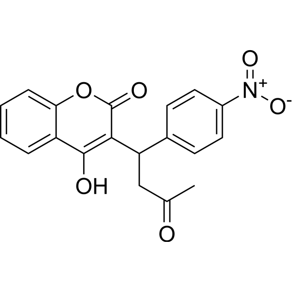
Acenocoumarol is an anticoagulant that functions as a Vitamin K antagonist. Acenocoumarol inhibits MAPK/ERK/JNK signaling pathway, reduces the nuclear translocation of NF-κB p65, activates Akt/GSK3β signaling pathway. Acenocoumarol induces apoptosis in cell A549, arrests cell cycle at S phase.
For research use only. We do not sell to patients.
- Purity : 99.17%
- CAS No.: 152-72-7
- Formula: C19H15NO6
- Molecular Weight:353.33
-
Storage:Powder -20°C, 3 years ; In solvent -80°C, 6 months , -20°C, 1 month

To place orders, for customer services and technical support, please contact: MedChemExpress USA
Tel: 609-228-6898 E-mail: [email protected] [email protected]


-
 Biological Activity
-
Chemical Information
-
Solvent & Solubility
- Protocols
- Purity & Documentation
- References
-
 Help & FAQs


-
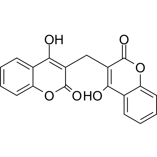
HY-N0645
DicoumarolPDHK Inhibitor -

HY-B1014R
Acenocoumarol (Standard)Anticoagulant -
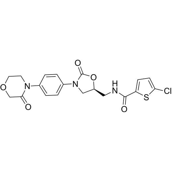
HY-50903
RivaroxabanFactor Xa Inhibitor -
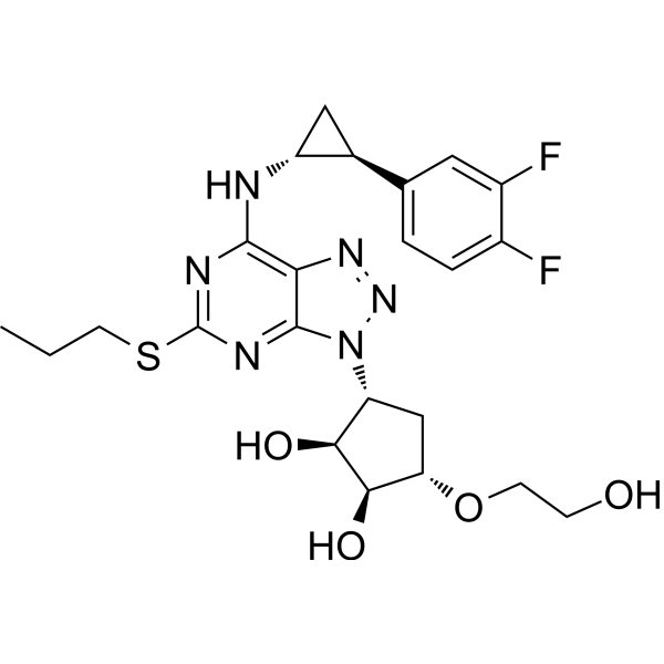
HY-10064
TicagrelorP2Y12 Antagonist -
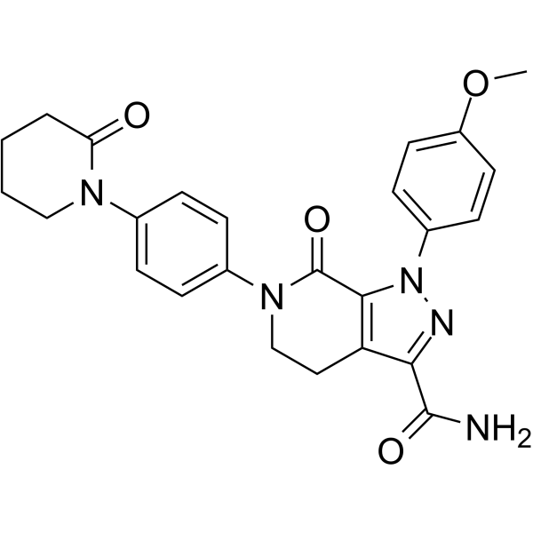
HY-50667
ApixabanSelective Factor Xa Inhibitor -

HY-50903R
Rivaroxaban (Standard)Factor Xa Inhibitor -

HY-50667R
Apixaban (Standard)Selective Factor Xa Inhibitor -
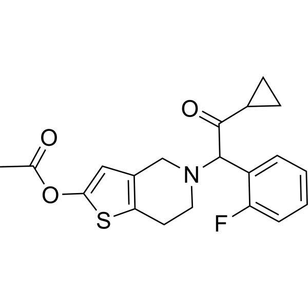
HY-15284
PrasugrelP2Y12 Receptor Antagonist -
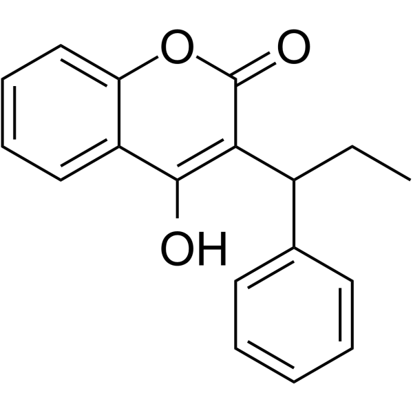
HY-A0145
PhenprocoumonVitamin K Antagonist -

HY-A0145R
Phenprocoumon (Standard)Vitamin K Antagonist -

HY-10064R
Ticagrelor (Standard)P2Y12 Antagonist -
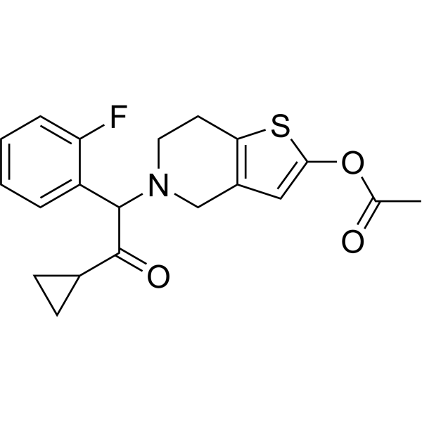
HY-15284R
Prasugrel (Standard)P2Y12 Receptor Antagonist


Biological Activity
Description
In Vitro
Acenocoumarol (30-500 μM, 24 h) inhibits the expression of iNOS and COX-2, inhibits the expression of NO, prostaglandin E2, TNF-α, IL-6 and IL-1β in LPS (HY-D1056)-stimulated RAW 264.7 cell[1].
Acenocoumarol (10-60 μM, 72 h) reduces the expression of tyrosinase, tyrosinase-related protein (TRP)-1, TRP-2 in αMSH-stimulated B16F10 melanoma cell, blocks melanin synthesis through PKA/JNK inhibition and Akt/GSK3β activation[2].
Acenocoumarol (0-100 μM, 24 h) exhibits cytotoxicity in cancer cell A549 and IMR-90 with IC50 of 28.75 μM and 74.11 μM[4].
MedChemExpress (MCE) has not independently confirmed the accuracy of these methods. They are for reference only.
-
Cell Line:B16F10
-
Concentration:10-60 μM
-
Incubation Time:22 h
-
Result:Inhibited the expression of pPKA, pJNK, increased the expression of TRP-1 p-p38, pAKT and pGSK3β.
-
Cell Line:A549 and IMR-90
-
Concentration:0-100 μM
-
Incubation Time:24 h
-
Result:Inhibited cell viability.
In Vivo
Acenocoumarol (0.2 mg/kg, po, once daily for 21 days) inhibits tumor growth, reduces cellular infiltration and inflammation in tumor tissues in A549-Xenograft mouse models[4].
MedChemExpress (MCE) has not independently confirmed the accuracy of these methods. They are for reference only.
-
Animal Model:ischemia/reperfusion-induced acute pancreatitis in Wistar rats models[3]
-
Dosage:50-150 µg/kg
-
Administration:po, once daily for 14 days
-
Result:Reduced pancreatic edema, necrosis, and hemorrhages, decreased pancreatic inflammatory infiltration and vacuolization of pancreatic acinar cells.
-
Animal Model:A549-Xenograft mouse models[4]
-
Dosage:0.2 mg/kg
-
Administration:po, once daily for 21 days
-
Result:Inhibited tumor growth with a TGI of 45.3%, reduced cellular infiltration and inflammation in tumor tissues.
- Cytoplasmic-Nuclear Fractionated Protein Extraction
- EdU Incorporation Assay (Click Chemistry-Based DNA Synthesis Measurement)
- Detection of Apoptosis
- TUNEL staining for apoptotic DNA fragmentation
- Flow cytometric DNA-content cell-cycle staining
- Annexin V plus membrane-impermeant dye apoptosis staining
- BrdU Incorporation Assay
- Apoptosis Solutions
- Protocol for Cell Cycle
- Research Protocol for Inflammation-related Diseases
- Directly Induced Neuron Culture
Clinical Trial
| NCT Number | Sponsor | Condition | Start Date |
Phase

|
|---|---|---|---|---|
| NCT01329991 | 2011-05 | PHASE1 |


Chemical Information
-
CAS No. 152-72-7
-
Appearance Solid
-
Molecular Weight 353.33
-
Formula C19H15NO6
-
Color White to off-white
-
SMILES
O=C1C(C(C2=CC=C([N+]([O-])=O)C=C2)CC(C)=O)=C(O)C3=CC=CC=C3O1
-
Shipping
Room temperature in continental US; may vary elsewhere.
-
Storage
Powder -20°C 3 years In solvent -80°C 6 months -20°C 1 month


Solvent & Solubility
In Vitro:
DMSO : 100 mg/mL (283.02 mM; Need ultrasonic; Hygroscopic DMSO has a significant impact on the solubility of product, please use newly opened DMSO)
Please refer to the solubility information to select the appropriate solvent. Once prepared, please aliquot and store the solution to prevent product inactivation from repeated freeze-thaw cycles.
Storage method and period of stock solution: -80°C, 6 months; -20°C, 1 month. When stored at -80°C, please use it within 6 months. When stored at -20°C, please use it within 1 month.
Please refer to the solubility information to select the appropriate solvent. Once prepared, please aliquot and store the solution to prevent product inactivation from repeated freeze-thaw cycles.
Storage method and period of stock solution: -80°C, 6 months; -20°C, 1 month. When stored at -80°C, please use it within 6 months. When stored at -20°C, please use it within 1 month.
Concentration (start) × Volume (start) = Concentration (final) × Volume (final)
In Vivo:
Select the appropriate dissolution method based on your experimental animal and administration route.
- For the following dissolution methods, please ensure to first prepare a clear stock solution using an In Vitro approach and then sequentially add co-solvents:
- To ensure reliable experimental results, the clarified stock solution can be appropriately stored based on storage conditions. As for the working solution for In Vivo experiments, it is recommended to prepare freshly and use it on the same day.
- The percentages shown for the solvents indicate their volumetric ratio in the final prepared solution. If precipitation or phase separation occurs during preparation, heat and/or sonication can be used to aid dissolution.
Add each solvent one by one: 10% DMSO 90% (20% SBE-β-CD in Saline)
Solubility: ≥ 2.5 mg/mL (7.08 mM); Clear solution
This protocol yields a clear solution of ≥ 2.5 mg/mL (saturation unknown).
Taking 1 mL working solution as an example, add 100 μL DMSO stock solution (25.0 mg/mL) to 900 μL 20% SBE-β-CD in Saline, and mix evenly.
Preparation of 20% SBE-β-CD in Saline (4°C, storage for one week): 2 g SBE-β-CD powder is dissolved in 10 mL Saline, completely dissolve until clear.
Add each solvent one by one: 10% DMSO 40% PEG300 5% Tween-80 45% Saline
Solubility: ≥ 2.08 mg/mL (5.89 mM); Clear solution
This protocol yields a clear solution of ≥ 2.08 mg/mL (saturation unknown).
Taking 1 mL working solution as an example, add 100 μL DMSO stock solution (20.8 mg/mL) to 400 μL PEG300, and mix evenly; then add 50 μL Tween-80 and mix evenly; then add 450 μL Saline to adjust the volume to 1 mL.
Preparation of Saline: Dissolve 0.9 g sodium chloride in ddH₂O and dilute to 100 mL to obtain a clear Saline solution.
In Vivo Dissolution Calculator
Please enter the basic information of animal experiments:
-
-
-
-
Recommended: Prepare an additional quantity of animals to account for potential losses during experiments.
Please enter your animal formula composition:
-
%DMSO +
Recommended: Keep the proportion of DMSO in working solution below 2% if your animal is weak.
-
%+
-
+%Tween-80 + +
-
%Saline +
The co-solvents required include: DMSO, . All of co-solvents are available by MedChemExpress (MCE). , Tween 80. All of co-solvents are available by MedChemExpress (MCE).
Working solution concentration: 0.22 mg/mL
Method for preparing stock solution: mg drug dissolved in μL DMSO. Stock solution concentration: mg/mL.
1. Take μL DMSO stock solution;
2. Add μL .
μL , mix evenly;
3. Then add μL Tween 80, mix evenly;
4. Then add μL
Please ensure that the stock solution in the first step is dissolved to a clear state, and add co-solvents in sequence. You can use ultrasonic heating (ultrasonic cleaner, recommended frequency 20-40 kHz), vortexing, etc. to assist dissolution.


Purity & Documentation
-
Data Sheet (280 KB)
-
SDS (393 KB)
- English - EN (393 KB)
- Français - FR (393 KB)
- Deutsch - DE (393 KB)
- Norwegian - NO (393 KB)
- Español - ES (393 KB)
- Swedish - SV (393 KB)
- Italian - IT (393 KB)
- Korean - KR (393 KB)
- Portuguese - PT (393 KB)
-
Handling Instructions (2659 KB)
References
[1]. Han HJ, et al., Acenocoumarol Exerts Anti-Inflammatory Activity via the Suppression of NF-κB and MAPK Pathways in RAW 264.7 Cells. Molecules. 2023 Feb 22;28(5):2075. [Content Brief]
[2]. Han H, et al., Acenocoumarol, an Anticoagulant Drug, Prevents Melanogenesis in B16F10 Melanoma Cells. Pharmaceuticals (Basel). 2023 Apr 17;16(4):604. [Content Brief]
[3]. Warzecha Z, et al., Therapeutic Effect of Low Doses of Acenocoumarol in the Course of Ischemia/Reperfusion-Induced Acute Pancreatitis in Rats. Int. J. Mol. Sci.2017, 18, 882. Int J Mol Sci. 2019 Jun 24;20(12):3086. [Content Brief]


Complete Stock Solution Preparation Table
Please refer to the solubility information to select the appropriate solvent. Once prepared, please aliquot and store the solution to prevent product inactivation from repeated freeze-thaw cycles.
Storage method and period of stock solution: -80°C, 6 months; -20°C, 1 month. When stored at -80°C, please use it within 6 months. When stored at -20°C, please use it within 1 month.
| Optional Solvent | Concentration Solvent Mass | 1 mg | 5 mg | 10 mg | 25 mg |
|---|---|---|---|---|---|
| DMSO | 1 mM | 2.8302 mL | 14.1511 mL | 28.3022 mL | 70.7554 mL |
| 5 mM | 0.5660 mL | 2.8302 mL | 5.6604 mL | 14.1511 mL | |
| 10 mM | 0.2830 mL | 1.4151 mL | 2.8302 mL | 7.0755 mL | |
| 15 mM | 0.1887 mL | 0.9434 mL | 1.8868 mL | 4.7170 mL | |
| 20 mM | 0.1415 mL | 0.7076 mL | 1.4151 mL | 3.5378 mL | |
| 25 mM | 0.1132 mL | 0.5660 mL | 1.1321 mL | 2.8302 mL | |
| 30 mM | 0.0943 mL | 0.4717 mL | 0.9434 mL | 2.3585 mL | |
| 40 mM | 0.0708 mL | 0.3538 mL | 0.7076 mL | 1.7689 mL | |
| 50 mM | 0.0566 mL | 0.2830 mL | 0.5660 mL | 1.4151 mL | |
| 60 mM | 0.0472 mL | 0.2359 mL | 0.4717 mL | 1.1793 mL | |
| 80 mM | 0.0354 mL | 0.1769 mL | 0.3538 mL | 0.8844 mL | |
| 100 mM | 0.0283 mL | 0.1415 mL | 0.2830 mL | 0.7076 mL |


Acenocoumarol Related Classifications
HY-B1014 Related Classifications
 Powered by Bioz