



Lixisenatide
Based on 4 publication(s) in Google Scholar
Lixisenatide is a glucagon-like peptide-1 (GLP-1) receptor agonist. Lixisenatide inhibits the inflammatory response through down regulation of pro-inflammatory cytokines, and suppresses of the Akt-MEK1/2 signaling pathway. Lixisenatide can inhibit oxidative stress, mitochondrial dysfunction and apoptosis. Lixisenatide can be used for the researches of inflammation, metabolic disease, neurological disease and cardiovascular disease, such as rheumatoid arthritis, diabetes, Alzheimer's disease and atherosclerosis.
For research use only. We do not sell to patients.
- Purity : 99.84%
- CAS No.: 320367-13-3
- Formula: C215H347N61O65S
- Molecular Weight:4858.49
-
Storage:
Sealed storage, away from moisture.
Powder -80°C, 2 years , -20°C, 1 year* In solvent : -80°C, 6 months; -20°C, 1 month (sealed storage, away from moisture)


To place orders, for customer services and technical support, please contact: MedChemExpress USA
Tel: 609-228-6898 E-mail: [email protected] [email protected]


-
 Biological Activity
-
Chemical Information
-
Solvent & Solubility
- Protocols
- Purity & Documentation
- References
-
 Help & FAQs


-

HY-114118
SemaglutideGLP-1R Agonist -

HY-P0014
LiraglutideGLP-1R Agonist -

HY-13443
Exendin-4GLP-1 Receptor Agonist -

HY-P0120
DulaglutideGLP-1 Receptor Agonist -
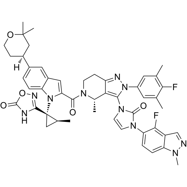
HY-112185
OrforglipronGLP-1R Agonist -

HY-P0264
Exendin(9-39) amideGLP-1R Antagonist -

HY-P5311
AlbiglutideGLP-1 Receptor Agonist -

HY-114118CP
Semaglutide (crude)Crude Semaglutide -
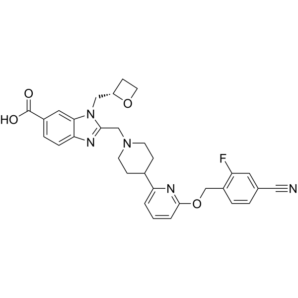
HY-125824
DanuglipronGLP-1R Agonist -

HY-P3506
RetatrutideGCGR/GIPR/GLP-1R Agonist -
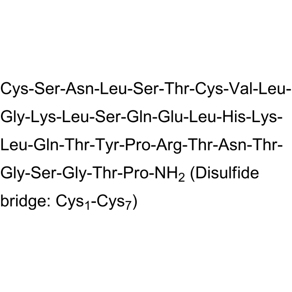
HY-P0090
Calcitonin (salmon)CGRP Receptor Agonist
Publications Citing Use of MedChemExpress (MCE) Lixisenatide
More Customer Validation & Images

-
In Vivo Efficacy Study

-
WB

-
IHC

All MEK Isoforms
More

Biological Activity
Description
IC50 & Target
[1]|
GLP-1 |
MEK1 |
MEK2 |
MMP-1 |
MMP-3 |
MMP13 |
In Vitro
Lixisenatide (100 μM, 24 h) inhibits the Aβ25-35 (HY-P0128)-induced cytotoxicity on cultured hippocampal cells. [1].
Lixisenatide (100 μM, 24 h) relieves the Aβ25-35-induced suppression of the Akt-MEK1/2 signaling pathway on cultured hippocampal cells [1].
Lixisenatide (10-20 μM, 48 h) ameliorates IL-1β-induced oxidative stress, mitochondrial dysfunction, and apoptosis in fibroblast-like synoviocytes (FLSs) [3].
Lixisenatide (10-20 μM, 48 h) reduces IL-1β-induced expression of MMPs and inhibits activation of proinflammatory pathways by IL-1β in FLSs[3].
Lixisenatide (10-20 μM, 6 h) reduced oxygen-glucose deprivation/reperfusion (OGD/R)-induced generation of ROS in HUVECs[5].
MedChemExpress (MCE) has not independently confirmed the accuracy of these methods. They are for reference only. Further protocols information, click here.
-
Cell Line:Fibroblast-like synoviocytes
-
Concentration:10 and 20 μM
-
Incubation Time:48 h
-
Result:Significantly reduced expression of MMP-1, MMP-3, and MMP-13 at both the mRNA and protein levels in a dose-dependent manner.
In Vivo
Lixisenatide (1 nmol/kg, Intraperitoneal injection, once a day for 14 days) shows renoprotective effects on experimental early diabetic nephropathy in diabetic rats [4].
Lixisenatide (1-10 mg/kg, i.p., daily for 10 weeks) improves cognitive ability in APP/PS1 mice[6].
MedChemExpress (MCE) has not independently confirmed the accuracy of these methods. They are for reference only.
-
Animal Model:Apoe−/− Irs2+/− mice models[2]
-
Dosage:10 μg/kg
-
Administration:Subcutaneously injection, daily for a month
-
Result:Exhibited smaller atheromas in the aortic arch region.
Reduced the lesion size in cross-sections of hearts.
-
Animal Model:Diabetic rats[4]
-
Dosage:1 nmol/kg
-
Administration:Intraperitoneally injection, once a day for a month
-
Result:Showed a significant amelioration on the elevated renal parameters.
Showed significant mitigation in renal MDA and total NOx− (by 50.3 and 79.9%, respectively) and 43.9% elevation in renal total antioxidant capacity.
Averted the observed increments in iNOS and COX-2 expressions in the renal tissues of the diabetic group.
Decreased the level of TGF-β protein expression.
-
Animal Model:APP/PS1 mice[6]
-
Dosage:1 and 10 mg/kg
-
Administration:Intraperitoneally injection, daily for 10 weeks
-
Result:Improved cognitive ability.
Prevented the reduction of synapse numbers.
Reduced amyloid plaque load and chronic inflammation response (microglial activation).
Clinical Trial
| NCT Number | Sponsor | Condition | Start Date |
Phase

|
|---|---|---|---|---|
| NCT01329991 | Plexxikon| | 2011-05 | PHASE1 |


Chemical Information
-
CAS No. 320367-13-3
-
Appearance Solid
-
Molecular Weight 4858.49
-
Formula C215H347N61O65S
-
Color White to pink
-
Sequence
His-Gly-Glu-Gly-Thr-Phe-Thr-Ser-Asp-Leu-Ser-Lys-Gln-Met-Glu-Glu-Glu-Ala-Val-Arg-Leu-Phe-Ile-Glu-Trp-Leu-Lys-Asn-Gly-Gly-Pro-Ser-Ser-Gly-Ala-Pro-Pro-Ser-Lys-Lys-Lys-Lys-Lys-Lys-NH2
-
Sequence Shortening
HGEGTFTSDLSKQMEEEAVRLFIEWLKNGGPSSGAPPSKKKKKK-NH2
-
Shipping
Room temperature in continental US; may vary elsewhere.
-
Storage
Sealed storage, away from moisture
Powder -80°C 2 years -20°C 1 year * In solvent : -80°C, 6 months; -20°C, 1 month (sealed storage, away from moisture)


Publications (4)
-
Journal Impact Factor
-
Most Recent
-
Biomacromolecules
Poly(l-threonine- co-l-threonine Succinate) Thermogels for Sustained Release of Lixisenatide. [Abstract]2024 Aug 12;25(8):4946-4955. PMID: 38949062 -
-
J Mol Neurosci
Lixisenatide Reduced Damage in Hippocampus CA1 Neurons in a Rat Model of Cerebral Ischemia-Reperfusion Possibly Via the ERK/P38 Signaling Pathway. [Abstract]2020 Jul;70(7):1026-1037. PMID: 32040827
Lixisenatide purchased from MedChemExpress. Usage Cited in: J Mol Neurosci. 2020 Jul;70(7):1026-1037. [Abstract]
Two different doses of lixisenatide (Lixi: 0.7 and 7 nmol/kg) were given to rats for 2 weeks before induction of cerebral I/R. High-dose of Lixi was more effective than the lower dose.
Lixisenatide purchased from MedChemExpress. Usage Cited in: J Mol Neurosci. 2020 Jul;70(7):1026-1037. [Abstract]
Effects of lixisenatide (Lixi: 7 nmol/kg) on MAPK. Western blot and statistical analysis of pERK.
Lixisenatide purchased from MedChemExpress. Usage Cited in: J Mol Neurosci. 2020 Jul;70(7):1026-1037. [Abstract]
Effects of lixisenatide (Lixi: 7 nmol/kg) on proapoptotic and antiapoptotic markers. Immunohistochemistry analysis of bax, bcl-2, and caspase-3 in hippocampus CA1 pyramidal neurons.
-


Solvent & Solubility
In Vitro:
H2O : 100 mg/mL (20.58 mM; Need ultrasonic)
Please refer to the solubility information to select the appropriate solvent. Once prepared, please aliquot and store the solution to prevent product inactivation from repeated freeze-thaw cycles.
Storage method and period of stock solution: -80°C, 6 months; -20°C, 1 month (sealed storage, away from moisture). When stored at -80°C, please use it within 6 months. When stored at -20°C, please use it within 1 month.
* Note: If you choose water as the stock solution, please dilute it to the working solution, then filter and sterilize it with a 0.22 μm filter before use.
Please refer to the solubility information to select the appropriate solvent. Once prepared, please aliquot and store the solution to prevent product inactivation from repeated freeze-thaw cycles.
Storage method and period of stock solution: -80°C, 6 months; -20°C, 1 month (sealed storage, away from moisture). When stored at -80°C, please use it within 6 months. When stored at -20°C, please use it within 1 month.
* Note: If you choose water as the stock solution, please dilute it to the working solution, then filter and sterilize it with a 0.22 μm filter before use.
Concentration (start) × Volume (start) = Concentration (final) × Volume (final)

Protocols
-
Annexin V plus membrane-impermeant dye apoptosis staining
Annexin V-based apoptosis assays rely on the detection of phosphatidylserine (PS) externalization from the inner leaflet of the plasma membrane to the outer leaflet, an early biochemical hallmark of apoptosis. Fluorescently labeled Annexin V binds PS in a calcium-dependent manner, enabling identification of early apoptotic cells by flow cytometry or fluorescence microscopy. When combined with a membrane-impermeant DNA-binding dye (e. g. , propidium iodide), this approach allows discrimination between viable (Annexin V−/dye−), early apoptotic (Annexin V+/dye−), and late apoptotic or necrotic (Annexin V+/dye+) cell populations by assessing membrane integrity and PS exposure.
-
ROS/oxidative-stress fluorescent staining
ROS/oxidative-stress fluorescent staining uses cell-permeant fluorogenic probes that become fluorescent after oxidation inside cells or tissues; commonly used examples include DCFH-DA/DCFDA for broad cellular oxidant detection, DHE for superoxide-related signal detection, MitoSOX for mitochondrial superoxide-related signal detection, and CellROX probes for oxidative-stress-associated fluorescence readouts. The assay detects probe oxidation rather than a single ROS species unless the probe and analysis method have been chemically validated for that species. DCFH-DA enters cells, is deacetylated by intracellular esterases to DCFH, and produces fluorescent DCF after oxidation, so the readout is used as an operational measure of total cellular oxidative stress rather than a species-specific ROS measurement. DHE and MitoSOX can report superoxide-related oxidation, but red fluorescence alone can include non-specific ethidium-like oxidation products; HPLC or optimized spectral approaches are
-
Research Protocol for Cardiovascular Diseases
Cardiovascular disease can be modeled as maladaptive cardiac remodeling, where ischemic injury or pressure overload activates inflammatory signaling, fibroblast activation, extracellular-matrix deposition, cardiomyocyte hypertrophy, vascular remodeling, and progressive ventricular dysfunction. The TGF-β/SMAD axis is a central profibrotic pathway after myocardial injury and pressure overload, while innate immune and cytokine pathways regulate leukocyte recruitment, scar formation, and adverse remodeling. Key unresolved questions include which inflammatory signals are reparative versus harmful, when fibrosis is protective versus maladaptive, and whether pathway inhibition improves function without weakening necessary infarct healing or compensatory remodeling.
-
Research Protocol for Inflammation-related Diseases
The NLRP3 inflammasome is a cytosolic innate immune signaling platform that integrates priming signals and danger-signal activation to promote caspase-1 activation, maturation of IL-1β and IL-18, and gasdermin D-mediated pyroptotic cell death. The core experimental logic is to determine whether inflammatory disease phenotypes are driven by increased NLRP3 expression, ASC-containing inflammasome assembly, caspase-1 cleavage, GSDMD cleavage, and extracellular release of IL-1β/IL-18 rather than by nonspecific cell injury alone. The pathway is strongly linked to inflammation-related disease phenotypes because monosodium urate crystals activate NALP3/NLRP3 inflammasome signaling in gout-like crystal inflammation, cholesterol crystals activate NLRP3 inflammasomes in atherogenesis models, and DSS-induced intestinal inflammation has been reported to involve NLRP3 inflammasome activity. However, experimental colitis studies also show context-dependent protective effects of NLRP3 inflammasome co
-
Research Protocol for Neurological Diseases
PINK1/Parkin-mediated mitophagy pathway is a mitochondrial quality-control signaling axis in which mitochondrial depolarization stabilizes PINK1 on damaged mitochondria, activates Parkin recruitment and E3 ubiquitin ligase activity, promotes ubiquitination of outer mitochondrial membrane proteins, recruits selective autophagy adaptors, and drives lysosomal degradation of damaged mitochondria. In neurological disease research, this pathway is experimentally important because neurons, especially dopaminergic neurons, are highly dependent on mitochondrial integrity, and defective mitochondrial turnover can lead to mitochondrial dysfunction, oxidative stress, impaired neuronal survival, α-synuclein accumulation, and neuroinflammatory damage-associated signals. The genetic disease link is strongest in Parkinson’s disease because mutations in PRKN/parkin cause autosomal recessive juvenile parkinsonism, mutations in PINK1 cause hereditary early-onset Parkinson’s disease, and Drosophila studie
-
Research Protocol for Metabolic Diseases
AMP-activated protein kinase, AMPK, is a conserved cellular energy sensor that responds to reduced cellular energy status and coordinates metabolism by increasing ATP-generating catabolic pathways while suppressing ATP-consuming anabolic processes. In metabolic disease research, the AMPK pathway is experimentally relevant because it regulates hepatic lipid synthesis, fatty acid oxidation, glucose production, skeletal-muscle glucose disposal, mTORC1-linked biosynthesis, autophagy, mitochondrial homeostasis, and whole-body energy balance. The central pathway logic is that energy stress, metformin, exercise-like stimulation, or direct AMPK activators increase AMPKα Thr172 phosphorylation and downstream substrate phosphorylation, including ACC and RAPTOR. Phosphorylation of ACC suppresses lipogenesis and supports fatty acid oxidation, whereas phosphorylation of RAPTOR suppresses mTORC1 signaling and links cellular energy status to growth and protein synthesis control. The pathway is linked
-
Alzheimer’s Disease Modeling
Alzheimer’s Disease (AD) is a neurodegenerative disorder characterized by a progressive decline in cognitive functions and loss of specific types of neurons and synapses. Alzheimer's symptoms can be simulated in mice by injecting drugs (such as Aβ) or genetically modified.
-
Collagen-Induced Arthritis
Collagen-induced arthritis (CIA) is an autoimmune murine model of rheumatoid arthritis in which immunization with type II collagen (CII) emulsified in an adjuvant induces a T cell- and autoantibody-driven inflammatory arthritis characterized by synovial hyperplasia, immune cell infiltration, and joint destruction. The model typically relies on genetically susceptible mouse strains (e. g. , DBA/1) and reproduces key features of human rheumatoid arthritis, including anti-collagen immune responses and progressive joint inflammation. Disease onset generally occurs within ~3-4 weeks after immunization, depending on antigen/adjuvant combinations and protocol variation. The immunopathology is driven by adaptive immune activation against CII, leading to systemic and local joint inflammation mediated by pro-inflammatory cytokines and effector immune cells, making CIA a standard preclinical platform for evaluating immunomodulatory and anti-arthritic interventions.
-
Apoptosis
Apoptosis, also called programmed cell death, is generally characterized by distinct morphological characteristics.
-
TUNEL staining for apoptotic DNA fragmentation
TUNEL staining detects DNA strand breaks by using terminal deoxynucleotidyl transferase to add labeled nucleotides to exposed 3′-OH DNA termini, generating either microscopic staining in fixed cells or tissue sections, or fluorescence/cytometric signal in cell suspensions. TUNEL positivity reflects DNA fragmentation but should not be interpreted alone as definitive apoptosis, because TUNEL can also label necrotic, autolytic, mechanically damaged, or DNA-repair-associated DNA breaks.
-
LPS-Induced Endotoxemia/Systemic Inflammation
Lipopolysaccharide (LPS)-induced endotoxemia is a widely used in vivo model of acute systemic inflammation in which LPS, a Gram-negative bacterial endotoxin, activates innate immune signaling primarily through TLR4, leading to rapid and transient induction of pro-inflammatory cytokines such as TNF-α, IL-6, and IL-1β in circulation and tissues. This cytokine surge is commonly used as a measurable readout of systemic inflammatory activation and immune dysregulation, and is typically assessed within hours after intraperitoneal LPS administration in mouse models of endotoxemia. The model captures key features of systemic inflammatory response syndrome, including cytokine release, immune cell activation, and downstream tissue responses, and has been used to evaluate anti-inflammatory interventions such as cytokine modulation, lipid mediators, and immune cell-targeting therapies.
-
Mitochondrial membrane-potential and mitochondrial mass staining
Mitochondrial membrane potential staining measures the electrochemical polarization across the mitochondrial inner membrane in live cells using lipophilic cationic fluorescent probes; early rhodamine-based work showed that selective mitochondrial dye accumulation is lost when the mitochondrial transmembrane potential is dissipated. JC-1 reports mitochondrial polarization by shifting from green monomer fluorescence to red J-aggregate fluorescence as dye concentration increases within energized mitochondria; therefore, the red/green fluorescence ratio is used as a relative readout of mitochondrial membrane potential. TMRE or TMRM staining provides a single-channel relative readout because these cationic rhodamine esters accumulate in polarized mitochondria, and lower fluorescence indicates reduced mitochondrial polarization when acquisition and dye-loading conditions are controlled. Mitochondrial mass staining is commonly performed with MitoTracker Green FM or related MitoTracker dyes as
-
Apoptosis Solutions
Apoptosis is a regulated, generally non-lytic cell-death pathway that removes unwanted, damaged, infected, or abnormal cells through coordinated morphological changes, caspase activation, DNA fragmentation, and membrane remodeling. The intrinsic apoptosis pathway is controlled mainly by mitochondrial outer membrane permeabilization, BCL-2 family proteins, cytochrome c release, apoptosome formation, caspase-9 activation, and downstream executioner caspase-3/7 activation. The extrinsic apoptosis pathway is initiated by death receptors such as Fas, TNFR, and TRAIL receptors, which recruit adaptor proteins and activate caspase-8 before engaging executioner caspases or mitochondrial amplification through BID cleavage. Apoptosis is linked to many phenotypes, including cancer cell killing, tissue homeostasis, immune regulation, neurodegeneration, infection response, and treatment-induced cytotoxicity; unresolved questions include how apoptosis interacts with necroptosis, pyroptosis, ferroptos


Purity & Documentation
-
Data Sheet (315 KB)
-
SDS (419 KB)
- English - EN (419 KB)
- Français - FR (419 KB)
- Deutsch - DE (419 KB)
- Norwegian - NO (419 KB)
- Español - ES (419 KB)
- Swedish - SV (419 KB)
- Italian - IT (419 KB)
- Korean - KR (419 KB)
- Portuguese - PT (419 KB)
-
Handling Instructions (2659 KB)
References
[1]. Cai HY, et al. Lixisenatide attenuates the detrimental effects of amyloid β protein on spatial working memory and hippocampal neurons in rats. Behav Brain Res. 2017 Feb 1;318:28-35. [Content Brief]
[2]. Vinué Á, et al. The GLP-1 analogue lixisenatide decreases atherosclerosis in insulin-resistant mice by modulating macrophage phenotype. Diabetologia. 2017 Sep;60(9):1801-1812. [Content Brief]
[3]. Du X, et al. The protective effects of lixisenatide against inflammatory response in human rheumatoid arthritis fibroblast-like synoviocytes. Int Immunopharmacol. 2019 Oct;75:105732. [Content Brief]
[4]. Abdel-Latif RG, et al. Low-dose lixisenatide protects against early-onset nephropathy induced in diabetic rats. Life Sci. 2020 Dec 15;263:118592. [Content Brief]
[5]. Xiao M, et al. The protective effects of GLP-1 receptor agonist lixisenatide on oxygen-glucose deprivation/reperfusion (OGD/R)-induced deregulation of endothelial tube formation. RSC Adv. 2020 Mar 10;10(17):10245-10253. [Content Brief]
[6]. McClean PL, et al. Lixisenatide, a drug developed to treat type 2 diabetes, shows neuroprotective effects in a mouse model of Alzheimer's disease. Neuropharmacology. 2014 Nov;86:241-58. [Content Brief]


Complete Stock Solution Preparation Table
Please refer to the solubility information to select the appropriate solvent. Once prepared, please aliquot and store the solution to prevent product inactivation from repeated freeze-thaw cycles.
Storage method and period of stock solution: -80°C, 6 months; -20°C, 1 month (sealed storage, away from moisture). When stored at -80°C, please use it within 6 months. When stored at -20°C, please use it within 1 month.
| Optional Solvent | Concentration Solvent Mass | 1 mg | 5 mg | 10 mg | 25 mg |
|---|---|---|---|---|---|
| H2O | 1 mM | 0.2058 mL | 1.0291 mL | 2.0583 mL | 5.1456 mL |
| 5 mM | 0.0412 mL | 0.2058 mL | 0.4117 mL | 1.0291 mL | |
| 10 mM | 0.0206 mL | 0.1029 mL | 0.2058 mL | 0.5146 mL | |
| 15 mM | 0.0137 mL | 0.0686 mL | 0.1372 mL | 0.3430 mL | |
| 20 mM | 0.0103 mL | 0.0515 mL | 0.1029 mL | 0.2573 mL |
* Note: If you choose water as the stock solution, please dilute it to the working solution, then filter and sterilize it with a 0.22 μm filter before use.


Lixisenatide Related Classifications
HY-P0119 Related Classifications
 Powered by Bioz