



Apoptosis inducer 34

Based on 1 Customer Validation
Apoptosis inducer 34 (Compound 4) is a small molecule compound that induces apoptosis by directly activating the intrinsic apoptotic pathway. Apoptosis inducer 34 promotes Apaf-1 oligomerization to form mature apoptosomes, thereby activating caspase-9 and caspase-3. It significantly activates the apoptotic pathway in Jurkat cells by enhancing the cytochrome c-dependent apoptotic signaling pathway, inducing PARP cleavage and chromosomal DNA fragmentation. Furthermore, Apoptosis inducer 34 exhibits low toxicity to normal cells, demonstrating potential for selective targeting of cancer cells. Apoptosis inducer 34 is a promising candidate for studying cancer related to apoptotic pathways.
Nos produits utilisent uniquement pour la recherche. Nous ne vendons pas aux patients.
- Pureté : 99.9%
- CAS No.: 582314-48-5
- Formule: C17H15Cl2NO2
- Masse moléculaire:336.21
-
Stockage:Powder -20°C, 3 years ; In solvent -80°C, 6 months , -20°C, 1 month


To place orders, for customer services and technical support, please contact: MedChemExpress USA
Tel: 609-228-6898 E-mail: [email protected] [email protected]


-
 Activité biologique
-
Chemical Information
- Protocole
- Pureté et documentation
- Références
-
 Help & FAQs


-
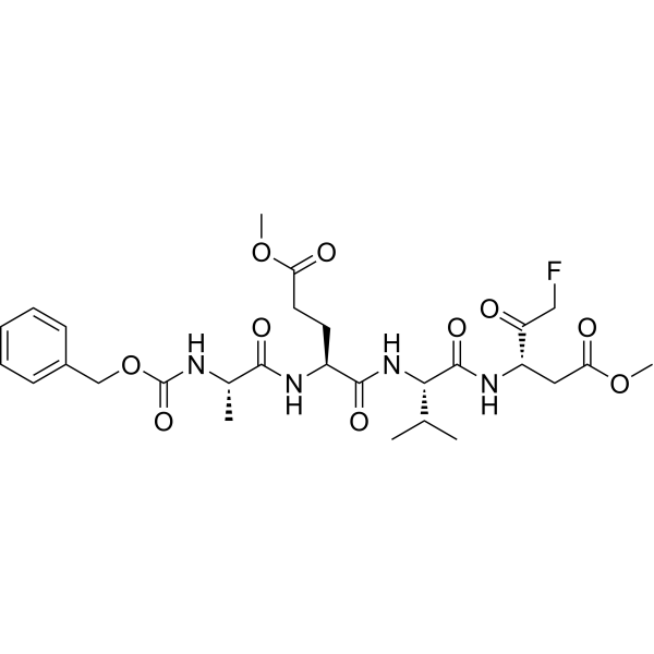
HY-125305
Z-AEVD-FMKCaspase-10 Inhibitor -
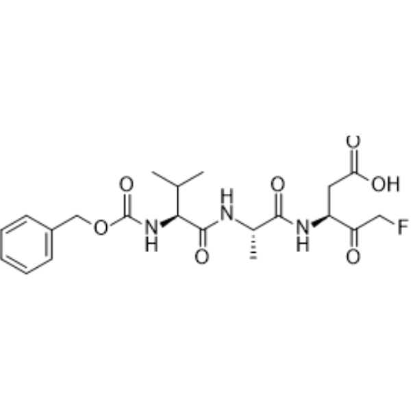
HY-16658B
Z-VAD-FMKPan-Caspase Inhibitor -
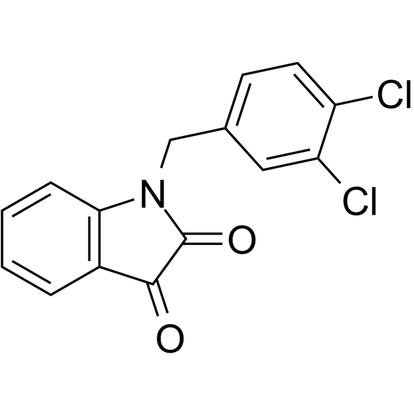
HY-18633
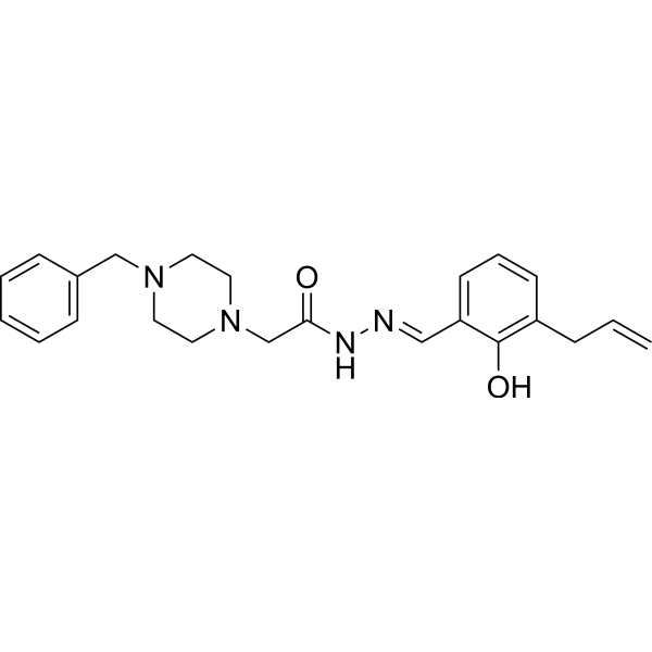
MDK83190Apoptosis Inducer -
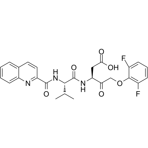
HY-12305
Q-VD-OPhPan-caspase Inhibitor -
HY-13523
PAC-1Caspase Activator -
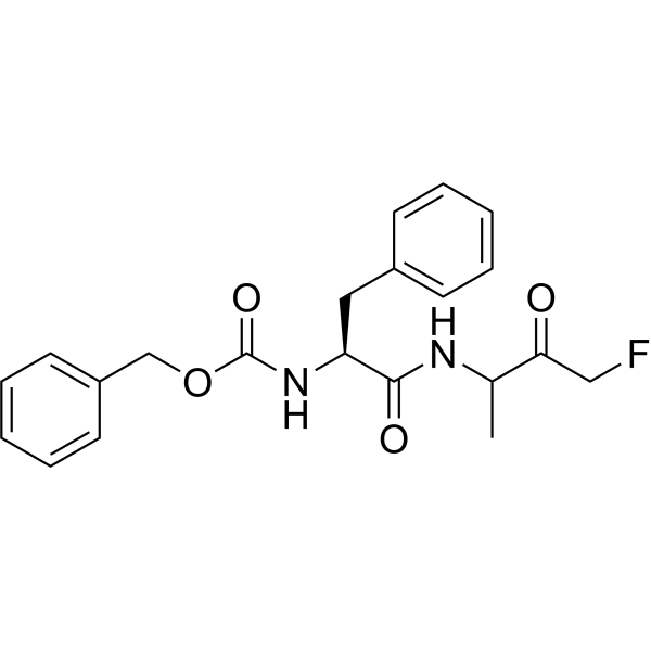
HY-P0109A
Z-FA-FMKCathepsin B/L Inhibitor -
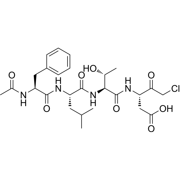
HY-111675
Ac-FLTD-CMKCaspase Inhibitor -
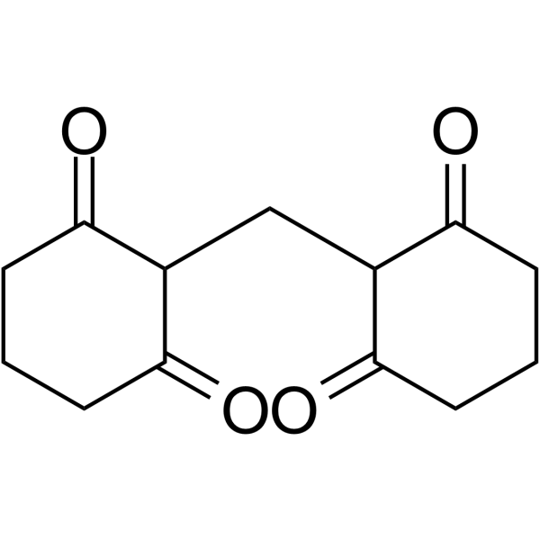
HY-103347
M50054Apoptosis Inhibitor -
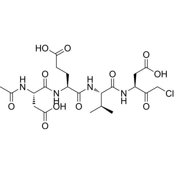
HY-P0034
Ac-DEVD-CMKCaspase-3 Inhibitor -

HY-W033813
Caspase-3/7 Inhibitor ICaspase-3/7 Inhibitor -
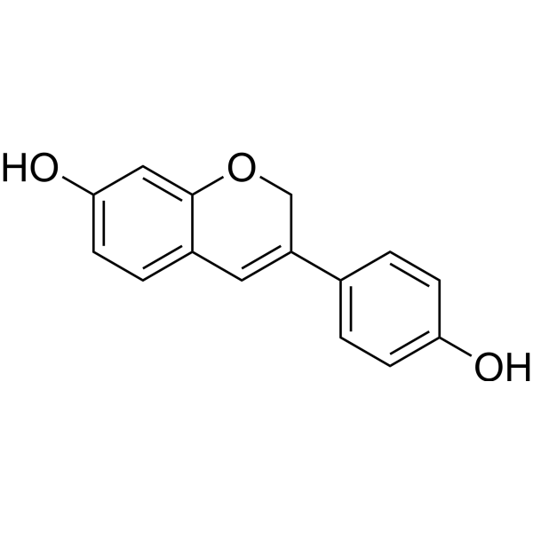
HY-13721
PhenoxodiolAnticancer Agent -
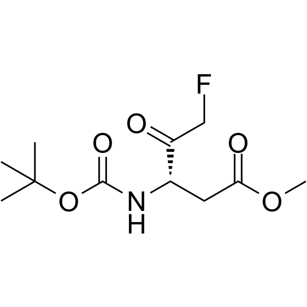
HY-103348
Boc-Asp(OMe)-fluoromethyl ketoneCaspase Inhibitor
Voir tous les produits spécifiques à Isoform Caspase
MoreVoir tous les produits spécifiques à Isoform DNA/RNA Synthesis
More

Activité biologique
Description


Chemical Information
-
CAS No. 582314-48-5
-
Appearance Solid
-
Masse moléculaire 336.21
-
Formule C17H15Cl2NO2
-
Color White to off-white
-
SMILES
O=C(O[C@H]1CCC2=C1C=CC=C2)NCC3=CC=C(Cl)C(Cl)=C3
-
Livraison
Room temperature in continental US; may vary elsewhere.
-
Stockage
Powder -20°C 3 years In solvent -80°C 6 months -20°C 1 month


Pureté et documentation
-
Fiche technique (268 KB)
-
SDS (394 KB)
- English - EN (394 KB)
- Français - FR (394 KB)
- Deutsch - DE (394 KB)
- Norwegian - NO (394 KB)
- Español - ES (394 KB)
- Swedish - SV (394 KB)
- Italian - IT (394 KB)
- Korean - KR (394 KB)
- Portuguese - PT (394 KB)
-
Instruction de manipulation (2659 KB)
Références

Calculators
Concentration (start) × Volume (start) = Concentration (final) × Volume (final)


Apoptosis inducer 34 Related Classifications
HY-116692 Related Classifications
 Powered by Bioz