



CD00509

Based on 1 Customer Validation
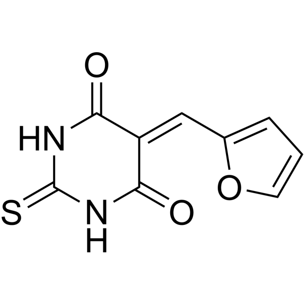
CD00509 is a tyrosyl-DNA phosphodiesterase (PDE) Tdp1 inhibitor with an IC50 of 0.71 μM. CD00509 increases DNA damage and promotes apoptosis of MCF-7 cells. CD00509 can sensitize breast cancer cells and wild-type murine embryonic fibroblasts (MEFs) to Camptothecin (CPT) (HY-16560). CD00509 can be used for the study of cancers such as breast cancer.
Nur für Forschungszwecke. Wir verkaufen nicht an Patienten.
- Reinheit : 98.0%
- CAS. Nr.: 27430-18-8
- Formel: C9H6N2O3S
- Molecular Weight:222.22
-
Speicherung:
4°C, sealed storage, away from moisture and light
* In solvent : -80°C, 6 months; -20°C, 1 month (sealed storage, away from moisture and light)

To place orders, for customer services and technical support, please contact: MedChemExpress USA
Tel: 609-228-6898 E-mail: [email protected] [email protected]


-
 Biologische Aktivität
-
Chemical Information
-
Lösungsmittel & Löslichkeit
- Protokoll
- Reinheit & Dokumentation
- Verweise
-
 Help & FAQs


Alle Caspase Isoform-spezifische Produkte anzeigen
MoreAlle DNA/RNA Synthesis Isoform-spezifische Produkte anzeigen
More

Biologische Aktivität
Beschreibung
IC50 & Target
[1]|
PDEI 0.71 μM (IC50) |
Caspase-3 |
Caspase-7 |
In Vitro
CD00509 (30 μM, 1-2 h) increases γ-H2AX Foci and DNA breaks in CPT-treated MCF-7 Cells[2].
CD00509 (10 μM, 0-96 h) reduces the number of MCF-7 cells, has almost no effect on normal breast epithelial cells, and significantly increases the caspase-3/7 activation of MCF-7 cells and co-treatment with CPT or Rucaparib (HY-10617A) is more lethal[2].
CD00509 (10 μM) effectively restores the attenuated TPI activity of PGP-inactivated MEFs to wildtype levels[3].
MedChemExpress (MCE) has not independently confirmed the accuracy of these methods. They are for reference only. Further protocols information, click here.


Chemical Information
-
CAS. Nr. 27430-18-8
-
Appearance Solid
-
Molecular Weight 222.22
-
Formel C9H6N2O3S
-
Color Light yellow to yellow
-
SMILES
S=C1NC(=O)C(C(N1)=O)=CC2=CC=CO2
-
Versand
Room temperature in continental US; may vary elsewhere.
-
Speicherung
4°C, sealed storage, away from moisture and light
* In solvent : -80°C, 6 months; -20°C, 1 month (sealed storage, away from moisture and light)


Lösungsmittel & Löslichkeit
In Vitro:
DMSO : 12.5 mg/mL (56.25 mM; Need ultrasonic; Hygroscopic DMSO has a significant impact on the solubility of product, please use newly opened DMSO)
Please refer to the solubility information to select the appropriate solvent. Once prepared, please aliquot and store the solution to prevent product inactivation from repeated freeze-thaw cycles.
Storage method and period of stock solution: -80°C, 6 months; -20°C, 1 month (sealed storage, away from moisture and light). When stored at -80°C, please use it within 6 months. When stored at -20°C, please use it within 1 month.
Please refer to the solubility information to select the appropriate solvent. Once prepared, please aliquot and store the solution to prevent product inactivation from repeated freeze-thaw cycles.
Storage method and period of stock solution: -80°C, 6 months; -20°C, 1 month (sealed storage, away from moisture and light). When stored at -80°C, please use it within 6 months. When stored at -20°C, please use it within 1 month.
Konzentration (Stammlösung) × Volumen (Stammlösung) = Konzentration (Ziellösung) × Volumen (Ziellösung)

Protokoll
-
Apoptosis
Apoptosis, also called programmed cell death, is generally characterized by distinct morphological characteristics.
-
TUNEL staining for apoptotic DNA fragmentation
TUNEL staining detects DNA strand breaks by using terminal deoxynucleotidyl transferase to add labeled nucleotides to exposed 3′-OH DNA termini, generating either microscopic staining in fixed cells or tissue sections, or fluorescence/cytometric signal in cell suspensions. TUNEL positivity reflects DNA fragmentation but should not be interpreted alone as definitive apoptosis, because TUNEL can also label necrotic, autolytic, mechanically damaged, or DNA-repair-associated DNA breaks.
-
Cell Cytotoxicity Assay
Cytotoxicity assays are usually based on the assessment of cell membrane damage, which can also be indirectly detected by measuring cell viability. Detection methods include MTT assay, CKK-8 assay, LDH assay and ATP assay, etc.
-
Annexin V plus membrane-impermeant dye apoptosis staining
Annexin V-based apoptosis assays rely on the detection of phosphatidylserine (PS) externalization from the inner leaflet of the plasma membrane to the outer leaflet, an early biochemical hallmark of apoptosis. Fluorescently labeled Annexin V binds PS in a calcium-dependent manner, enabling identification of early apoptotic cells by flow cytometry or fluorescence microscopy. When combined with a membrane-impermeant DNA-binding dye (e. g. , propidium iodide), this approach allows discrimination between viable (Annexin V−/dye−), early apoptotic (Annexin V+/dye−), and late apoptotic or necrotic (Annexin V+/dye+) cell populations by assessing membrane integrity and PS exposure.
-
Apoptosis Solutions
Apoptosis is a regulated, generally non-lytic cell-death pathway that removes unwanted, damaged, infected, or abnormal cells through coordinated morphological changes, caspase activation, DNA fragmentation, and membrane remodeling. The intrinsic apoptosis pathway is controlled mainly by mitochondrial outer membrane permeabilization, BCL-2 family proteins, cytochrome c release, apoptosome formation, caspase-9 activation, and downstream executioner caspase-3/7 activation. The extrinsic apoptosis pathway is initiated by death receptors such as Fas, TNFR, and TRAIL receptors, which recruit adaptor proteins and activate caspase-8 before engaging executioner caspases or mitochondrial amplification through BID cleavage. Apoptosis is linked to many phenotypes, including cancer cell killing, tissue homeostasis, immune regulation, neurodegeneration, infection response, and treatment-induced cytotoxicity; unresolved questions include how apoptosis interacts with necroptosis, pyroptosis, ferroptos
-
Breast Cancer Modeling
Breast cancer is a heterogeneous cancer, and it has been distinguished into four subtypes: luminal A, luminal B, HER2-positive and basal-like. Molecular mutations, epigenetic alterations, hormone exposure and immune microenvironment are related to the progression of breast cancer.
-
Genotoxicity/Mutagenicity Study
The bacterial reverse mutation assay detects point mutations that restore amino-acid prototrophy in auxotrophic Salmonella typhimurium or Escherichia coli tester strains; after exposure to a test article, mutagenic activity is read out as an increased number of revertant colonies on minimal agar compared with the vehicle control. The assay uses tester strains with different mutation targets so that base-substitution and frameshift mutagens can be detected, and testing is performed with and without exogenous mammalian metabolic activation because some chemicals require biotransformation to become mutagenic.


Reinheit & Dokumentation
-
Data Sheet (277 KB)
-
SDS (393 KB)
- English - EN (393 KB)
- Français - FR (393 KB)
- Deutsch - DE (393 KB)
- Norwegian - NO (393 KB)
- Español - ES (393 KB)
- Swedish - SV (393 KB)
- Italian - IT (393 KB)
- Korean - KR (393 KB)
- Portuguese - PT (393 KB)
-
Handling Instructions (2659 KB)
Verweise
[1]. Laev SS, et al. Tyrosyl-DNA phosphodiesterase inhibitors: Progress and potential. Bioorg Med Chem. 2016 Nov 1;24(21):5017-5027. [Content Brief]
[2]. Dean RA,et al. Identification of a putative Tdp1 inhibitor (CD00509) by in vitro and cell-based assays. J Biomol Screen. 2014 Dec;19(10):1372-82. [Content Brief]
[3]. Dean RA, et al. Identification of a putative Tdp1 inhibitor (CD00509) by in vitro and cell-based assays. J Biomol Screen. 2014 Dec;19(10):1372-82. [Content Brief]
[4]. Segerer G, et al. An essential developmental function for murine phosphoglycolate phosphatase in safeguarding cell proliferation. Sci Rep. 2016 Oct 12;6:35160. [Content Brief]


Complete Stock Solution Preparation Table
Please refer to the solubility information to select the appropriate solvent. Once prepared, please aliquot and store the solution to prevent product inactivation from repeated freeze-thaw cycles.
Storage method and period of stock solution: -80°C, 6 months; -20°C, 1 month (sealed storage, away from moisture and light). When stored at -80°C, please use it within 6 months. When stored at -20°C, please use it within 1 month.
| Optional Solvent | Concentration Solvent Mass | 1 mg | 5 mg | 10 mg | 25 mg |
|---|---|---|---|---|---|
| DMSO | 1 mM | 4.5000 mL | 22.5002 mL | 45.0004 mL | 112.5011 mL |
| 5 mM | 0.9000 mL | 4.5000 mL | 9.0001 mL | 22.5002 mL | |
| 10 mM | 0.4500 mL | 2.2500 mL | 4.5000 mL | 11.2501 mL | |
| 15 mM | 0.3000 mL | 1.5000 mL | 3.0000 mL | 7.5001 mL | |
| 20 mM | 0.2250 mL | 1.1250 mL | 2.2500 mL | 5.6251 mL | |
| 25 mM | 0.1800 mL | 0.9000 mL | 1.8000 mL | 4.5000 mL | |
| 30 mM | 0.1500 mL | 0.7500 mL | 1.5000 mL | 3.7500 mL | |
| 40 mM | 0.1125 mL | 0.5625 mL | 1.1250 mL | 2.8125 mL | |
| 50 mM | 0.0900 mL | 0.4500 mL | 0.9000 mL | 2.2500 mL |


CD00509 Related Classifications
HY-W177546 Related Classifications
 Powered by Bioz