



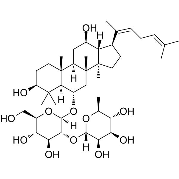
Ginsenoside Rg4

Based on 1 Customer Validation
Ginsenoside Rg4 is an orally active protopanaxatriol type ginsenoside. Ginsenoside Rg4 can activate PI3K, AKT and GSK-3β signaling. Ginsenoside Rg4 can inhibit ROS and inflammatory cytokine levels. Ginsenoside Rg4 can be used for the researches of inflammation, infection and metabolic disease, such as sepsis and lung inflammation.
Nur für Forschungszwecke. Wir verkaufen nicht an Patienten.
- Reinheit : 99.94%
- CAS. Nr.: 126223-28-7
- Formel: C42H70O12
- Molecular Weight:767.00
-
Speicherung:
-80°C, protect from light

To place orders, for customer services and technical support, please contact: MedChemExpress USA
Tel: 609-228-6898 E-mail: [email protected] [email protected]


-
 Biologische Aktivität
-
Chemical Information
-
Lösungsmittel & Löslichkeit
- Protokoll
- Reinheit & Dokumentation
- Verweise
-
 Help & FAQs


Alle TNF Receptor Isoform-spezifische Produkte anzeigen
More

Biologische Aktivität
Beschreibung
IC50 & Target
[1]|
GSK-3β |
IL-1β |
TNF-α |
In Vitro
Ginsenoside Rg4 (0.01-0.2 mg/mL, 6 h) decreases the expressions of toll-like receptor (TLR) 4 and TNF-α levels in HMGB1-activated HUVECs[1].
Ginsenoside Rg4 (0.1-0.2 mg/mL, 6 h) increases cell viability by activating PI3K/AKT signaling in HMGB1-activated HUVECs[1].
Ginsenoside Rg4 (20-50 μg/mL, 48 h) promotes the hair-inductive properties of DP cells by activating the AKT/GSK3β/β-catenin signaling pathway in dermal papilla (DP) sphere[2].
MedChemExpress (MCE) has not independently confirmed the accuracy of these methods. They are for reference only.
-
Cell Line:HMGB1-activated HUVECs
-
Concentration:0.01, 0. 1 and 0.2 mg/mL
-
Incubation Time:6 h
-
Result:Reduced TLR2, TLR4 and NF-κB expression.
In Vivo
Ginsenoside Rg4 (1-15 mg/kg, p.o., for 10 days) attenuates inflammation in PM2.5-induced pulmonary inflammation mice models[3].
MedChemExpress (MCE) has not independently confirmed the accuracy of these methods. They are for reference only.
-
Animal Model:Cecal ligation and puncture (CLP)-induced sepsis mice models[1]
-
Dosage:10 and 15 mg/kg
-
Administration:Intravenously injection, 24 h after CLP
-
Result:Reduced TNF-α and IL-1β levels.
Reduced nitric oxide (NO) levels and renal inflammation.
Increased PI3k and p-AKT levels.
-
Animal Model:PM2.5-induced pulmonary inflammation mice models[3]
-
Dosage:1, 2, 4, 7.5 and 15 mg/kg
-
Administration:Orally administration, for 10 days
-
Result:Reduced ROS levels.
Inhibited activation of p38 mitogen-activated protein kinase (MAPK) .
Activated Akt in purified pulmonary endothelial cells.
Reduced vascular protein leakage and leukocyte infiltration.
Reduced proinflammatory cytokine release in bronchoalveolar lavage fluids.


Chemical Information
-
CAS. Nr. 126223-28-7
-
Appearance Solid
-
Molecular Weight 767.00
-
Formel C42H70O12
-
Color White to off-white
-
SMILES
C[C@]12[C@@]3(C)[C@@]([H])([C@H](CC3)/C(C)=C\C/C=C(C)\C)[C@@H](C[C@@]1([C@]4(C)[C@@]([H])([C@H](C2)O[C@]5([C@H](O[C@]6([C@H](O)[C@H](O)[C@H]([C@@H](O6)C)O)[H])[C@@H](O)[C@@H]([C@H](O5)CO)O)[H])C([C@H](CC4)O)(C)C)[H])O
-
Structure Classification
-
Initial Source
-
Versand
Shipping with dry ice.
-
Speicherung
-80°C, protect from light


Lösungsmittel & Löslichkeit
In Vitro:
DMSO : 5 mg/mL (6.52 mM; Need ultrasonic; Hygroscopic DMSO has a significant impact on the solubility of product, please use newly opened DMSO)
Please refer to the solubility information to select the appropriate solvent. Once prepared, please aliquot and store the solution to prevent product inactivation from repeated freeze-thaw cycles.
Storage method and period of stock solution: -80°C, 6 months (protect from light). When stored at -80°C, please use it within 6 months.
Please refer to the solubility information to select the appropriate solvent. Once prepared, please aliquot and store the solution to prevent product inactivation from repeated freeze-thaw cycles.
Storage method and period of stock solution: -80°C, 6 months (protect from light). When stored at -80°C, please use it within 6 months.
Konzentration (Stammlösung) × Volumen (Stammlösung) = Konzentration (Ziellösung) × Volumen (Ziellösung)


Reinheit & Dokumentation
-
Data Sheet (284 KB)
-
SDS (251 KB)
- English - EN (251 KB)
- Français - FR (251 KB)
- Deutsch - DE (251 KB)
- Norwegian - NO (251 KB)
- Español - ES (251 KB)
- Swedish - SV (251 KB)
- Italian - IT (251 KB)
- Korean - KR (251 KB)
- Portuguese - PT (251 KB)
-
Handling Instructions (2659 KB)
Verweise
[1]. Kim GO, et al. Inhibitory Activities of Rare Ginsenoside Rg4 on Cecal Ligation and Puncture-Induced Sepsis. Int J Mol Sci. 2022 Sep 16;23(18):10836. [Content Brief]
[2]. Lee YH, et al. Ginsenoside Rg4 Enhances the Inductive Effects of Human Dermal Papilla Spheres on Hair Growth Via the AKT/GSK-3β/β-Catenin Signaling Pathway. J Microbiol Biotechnol. 2021 Jul 28;31(7):933-941. [Content Brief]


Complete Stock Solution Preparation Table
Please refer to the solubility information to select the appropriate solvent. Once prepared, please aliquot and store the solution to prevent product inactivation from repeated freeze-thaw cycles.
Storage method and period of stock solution: -80°C, 6 months (protect from light). When stored at -80°C, please use it within 6 months.
| Optional Solvent | Concentration Solvent Mass | 1 mg | 5 mg | 10 mg | 25 mg |
|---|---|---|---|---|---|
| DMSO | 1 mM | 1.3038 mL | 6.5189 mL | 13.0378 mL | 32.5945 mL |
| 5 mM | 0.2608 mL | 1.3038 mL | 2.6076 mL | 6.5189 mL |


Ginsenoside Rg4 Related Classifications
HY-N6580 Related Classifications
 Powered by Bioz