



Mocravimod
Based on 2 publication(s) in Google Scholar
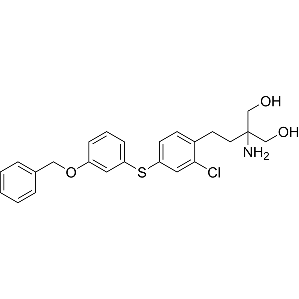
Mocravimod (KRP-203 free base) is a sphingosine-1-phosphate receptor (S1PR) modulator that blocks the signal required by T cells to egress from lymph nodes and other lymphoid organs. Mocravimod preferentially binds to S1PR1 over S1PR2 and S1PR3 in cardiomyocytes. Mocravimod significantly lowered the concentration of reactive oxygen species (ROS), prevented mitochondrial permeability transition pore opening, boosted mitochondrial membrane potential (MMP), and increased phosphorylation of AKT, EKR, GSK-3β, JAK2, and STAT3. Mocravimod retains T cell effector function. Mocravimod can be used for the study of acute myelogenous leukemia, diabetes and Myocardial Ischemia-Reperfusion Injury (MIRI).
For research use only. We do not sell to patients.
- Purity : 98.39%
- CAS No.: 509092-16-4
- Formula: C24H26ClNO3S
- Molecular Weight:443.99
-
Storage:Powder -20°C, 3 years , 4°C, 2 years ; In solvent -80°C, 6 months , -20°C, 1 month

To place orders, for customer services and technical support, please contact: MedChemExpress USA
Tel: 609-228-6898 E-mail: [email protected] [email protected]


-
 Biological Activity
-
Chemical Information
-
Solvent & Solubility
- Protocols
- Purity & Documentation
- References
-
 Help & FAQs


-
HY-139853
BMS-986278LPA1 Antagonist -
HY-16039
AM095LPA1 Antagonist -
HY-119401
CAY10444S1P3 Antagonist -
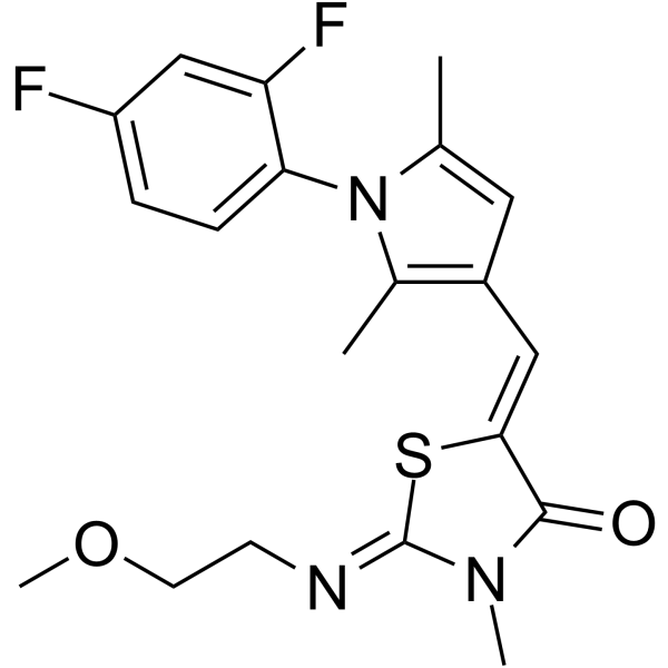
HY-108495
CYM50308S1P4 Receptor Agonist -
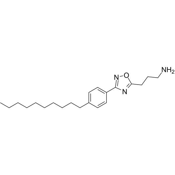
HY-149004
SLF1081851Spns2 Inhibitor -
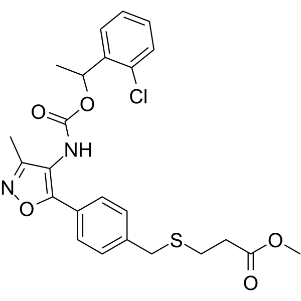
HY-18641
Ki16198LPA Receptor Antagonist -
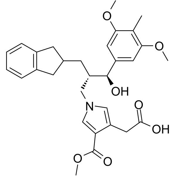
HY-117444
ONO-9780307LPA1 Antagonist -
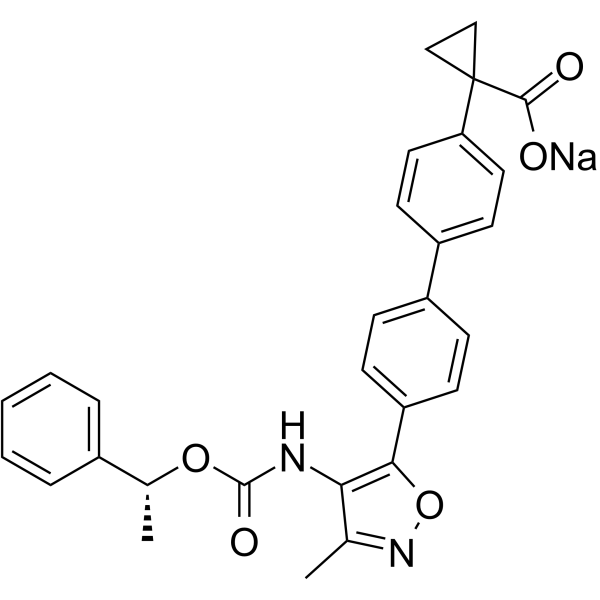
HY-100619A
BMS-986020 sodiumLPA1 Antagonist -
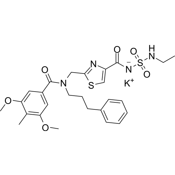
HY-120478
ASP6432LPA1 Antagonist -

HY-108495R
CYM50308 (Standard)Reference Standard -
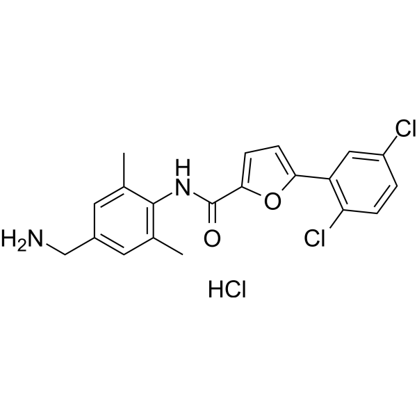
HY-110314
CYM50358 hydrochlorideS1P4 Receptor Antagonist -

HY-100619AR
BMS-986020 sodium (Standard)LPA1 Antagonist
Publications Citing Use of MedChemExpress (MCE) Mocravimod
More

Biological Activity
Description
IC50 & Target
|
S1PR1 |
GSK-3β |
STAT3 |
JAK2 |
In Vitro
Mocravimod (0.2-5 μM, 24 h) significantly improves the survival rate of the hypoxia-reoxygenation (H/R) H9c2 cells, reduces lactate dehydrogenase (LDH) release, and decreases apoptosis[4].
Mocravimod (0.2-1 μM, 24 h) significantly reduces reactive oxygen species (ROS) levels, improves mitochondrial membrane potential, and inhibits the opening of the mitochondrial permeability transition pore (mPTP) in hypoxia-reoxygenation (H/R) H9c2 cells[4].
Mocravimod (0.2-1 μM, 24 h) inhibits the expression of mitochondrial-mediated apoptosis pathway proteins such as cytochrome C, caspase-9, and caspase-3[4].
Mocravimod (0.2-1 μM, 24 h) significantly increases the phosphorylation levels of key signaling pathway proteins in hypoxia-reoxygenation (H/R) H9c2 cells, including AKT (Ser473), ERK (Thr202/Tyr204), GSK-3β (Ser9), JAK2 (Tyr1007/1008), and STAT3 (Ser727)[4].
MedChemExpress (MCE) has not independently confirmed the accuracy of these methods. They are for reference only.
-
Cell Line:Hypoxia-Reoxygenation (H/R) H9c2 Cells
-
Concentration:0.2 μM, 1 μM, 5 μM
-
Incubation Time:24 h
-
Result:Induced apoptosis.
-
Cell Line:Hypoxia-Reoxygenation (H/R) H9c2 Cells
-
Concentration:0.2 μM, 1 μM
-
Incubation Time:24 h
-
Result:Inhibited the expression of mitochondrial-mediated apoptosis pathway proteins such as cytochrome C, caspase-9, and caspase-3.
In Vivo
Mocravimod (3.0 mg/kg, i.p., thrice weekly) significantly inhibits the development of atherosclerotic lesions in mice on a high-cholesterol diet, and this effect was independent of changes in blood lipids, but rather by regulating the infiltration of inflammatory cells within plaques[3].
MedChemExpress (MCE) has not independently confirmed the accuracy of these methods. They are for reference only.
-
Animal Model:Low-density lipoprotein receptor-deficient mice (LDL-R-/-) on cholesterol-rich diet[3].
-
Dosage:3 mg/kg
-
Administration:I.p., thrice weekly for 6-16 weeks
-
Result:Significantly reduced the lesion area of the aortic root and the entire aorta.
Decreased levels of macrophages (MOMA-2 positive) and T cells (CD3 positive) in plaques.
No effect on plasma lipid levels (total cholesterol, HDL cholesterol, triglycerides), body weight, or liver and kidney function parameters (such as ALT, BUN, creatinine).
Decreased CD4+ and CD8+ T cells in the spleen and lymph nodes, with decreased expression of the activation marker CD69.
Decreased MHC-II expression (activation marker) in F4/80+ macrophages and CD11c+ dendritic cells; reduced cytokine (TNF-α, MCP-1, IL-6) production after in vitro stimulation.
- Kinase activity and phosphorylation assays
- Western Blot
- Mitochondrial membrane-potential fluorescent assay
- ROS/oxidative-stress fluorescent staining
- Fluorescent plasma-membrane potential dye assay
- Mitochondrial membrane-potential and mitochondrial mass staining
- Transepithelial/transendothelial electrical resistance assay
- Protocol for Kinase activity and phosphorylation assays
- Research Protocol for Metabolic Diseases
- Research Protocol for Cancer Immunology


Chemical Information
-
CAS No. 509092-16-4
-
Appearance Solid
-
Molecular Weight 443.99
-
Formula C24H26ClNO3S
-
Color White to off-white
-
SMILES
OCC(CCC1=CC=C(SC2=CC=CC(OCC3=CC=CC=C3)=C2)C=C1Cl)(N)CO
-
Synonyms
KRP-203 free base
-
Shipping
Room temperature in continental US; may vary elsewhere.
-
Storage
Powder -20°C 3 years 4°C 2 years In solvent -80°C 6 months -20°C 1 month


Publications (2)
-
Journal Impact Factor
-
Most Recent
-
Cell Biol Toxicol
The novel sphingosine-1-phosphate receptor modulator KRP-203 prevents myocardial ischemia-reperfusion injury by preserving mitochondrial function through activation of the RISK and SAFE signaling pathways. [Abstract]2025 Nov 27;41(1):159. PMID: 41307722 -
bioRxiv
Sphingosine-1-phosphate receptor modulators resensitize FLT3-ITD acute myeloid leukemia cells with NRAS mutations to FLT3 inhibitors. [Abstract]2025 Nov 24:2025.11.21.689510. PMID: 41341164


Solvent & Solubility
In Vitro:
DMSO : 100 mg/mL (225.23 mM; Need ultrasonic; Hygroscopic DMSO has a significant impact on the solubility of product, please use newly opened DMSO)
Please refer to the solubility information to select the appropriate solvent. Once prepared, please aliquot and store the solution to prevent product inactivation from repeated freeze-thaw cycles.
Storage method and period of stock solution: -80°C, 6 months; -20°C, 1 month. When stored at -80°C, please use it within 6 months. When stored at -20°C, please use it within 1 month.
Please refer to the solubility information to select the appropriate solvent. Once prepared, please aliquot and store the solution to prevent product inactivation from repeated freeze-thaw cycles.
Storage method and period of stock solution: -80°C, 6 months; -20°C, 1 month. When stored at -80°C, please use it within 6 months. When stored at -20°C, please use it within 1 month.
Concentration (start) × Volume (start) = Concentration (final) × Volume (final)


Purity & Documentation
-
Data Sheet (277 KB)
-
SDS (393 KB)
- English - EN (393 KB)
- Français - FR (393 KB)
- Deutsch - DE (393 KB)
- Norwegian - NO (393 KB)
- Español - ES (393 KB)
- Swedish - SV (393 KB)
- Italian - IT (393 KB)
- Korean - KR (393 KB)
- Portuguese - PT (393 KB)
-
Handling Instructions (2659 KB)
References
[1]. Dertschnig S, et al. Mocravimod, a Selective Sphingosine-1-Phosphate Receptor Modulator, in Allogeneic Hematopoietic Stem Cell Transplantation for Malignancy. Transplant Cell Ther. 2023 Jan;29(1):41.e1-41.e9. [Content Brief]
[3]. Potì F, et al. Mocravimod, sphingosine 1-phosphate receptor type 1 agonist, ameliorates atherosclerosis in LDL-R-/- mice. Arterioscler Thromb Vasc Biol. 2013 Jul;33(7):1505-12. [Content Brief]
[4]. Wu N, et al. The novel sphingosine-1-phosphate receptor modulator Mocravimod prevents myocardial ischemia-reperfusion injury by preserving mitochondrial function through activation of the RISK and SAFE signaling pathways. Cell Biol Toxicol. 2025 Nov 27;41(1):159. [Content Brief]


Complete Stock Solution Preparation Table
Please refer to the solubility information to select the appropriate solvent. Once prepared, please aliquot and store the solution to prevent product inactivation from repeated freeze-thaw cycles.
Storage method and period of stock solution: -80°C, 6 months; -20°C, 1 month. When stored at -80°C, please use it within 6 months. When stored at -20°C, please use it within 1 month.
| Optional Solvent | Concentration Solvent Mass | 1 mg | 5 mg | 10 mg | 25 mg |
|---|---|---|---|---|---|
| DMSO | 1 mM | 2.2523 mL | 11.2615 mL | 22.5230 mL | 56.3076 mL |
| 5 mM | 0.4505 mL | 2.2523 mL | 4.5046 mL | 11.2615 mL | |
| 10 mM | 0.2252 mL | 1.1262 mL | 2.2523 mL | 5.6308 mL | |
| 15 mM | 0.1502 mL | 0.7508 mL | 1.5015 mL | 3.7538 mL | |
| 20 mM | 0.1126 mL | 0.5631 mL | 1.1262 mL | 2.8154 mL | |
| 25 mM | 0.0901 mL | 0.4505 mL | 0.9009 mL | 2.2523 mL | |
| 30 mM | 0.0751 mL | 0.3754 mL | 0.7508 mL | 1.8769 mL | |
| 40 mM | 0.0563 mL | 0.2815 mL | 0.5631 mL | 1.4077 mL | |
| 50 mM | 0.0450 mL | 0.2252 mL | 0.4505 mL | 1.1262 mL | |
| 60 mM | 0.0375 mL | 0.1877 mL | 0.3754 mL | 0.9385 mL | |
| 80 mM | 0.0282 mL | 0.1408 mL | 0.2815 mL | 0.7038 mL | |
| 100 mM | 0.0225 mL | 0.1126 mL | 0.2252 mL | 0.5631 mL |


Mocravimod Related Classifications
HY-109038 Related Classifications
 Powered by Bioz
Keywords
- Mocravimod
- 509092-16-4
- KRP-203
- KRP203
- KRP 203
- LPL Receptor
- Reactive Oxygen Species (ROS)
- Mitochondrial Metabolism
- Akt
- GSK-3
- JAK
- STAT
- S1PR
- Sphingosine-1-phosphate receptor agonist
- Myocardial ischemia–reperfusion injury
- Mitochondrial function
- Reperfusion injury salvage kinase signaling pathway
- Survivor activating factor enhancement signaling pathway
- Inhibitor
- inhibitor
- inhibit