



Notoginsenoside Ft1
Based on 1 publication(s) in Google Scholar
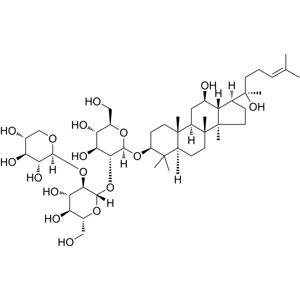
Notoginsenoside Ft1 is an orally active bioactive saponin. Notoginsenoside Ft1 inhibits the PI3K/AKT/mTOR signaling pathway, activates the p38 MAPK and ERK1/2 signaling pathways, and increases the proportion of CD8+ T cells, thereby inducing apoptosis and lysosomal cell death in various cancer cells, and promoting angiogenesis. Notoginsenoside Ft1 causes vasodilation by activating glucocorticoid receptors (GR) and estrogen receptor beta (ERβ) in endothelial cells. Notoginsenoside Ft1 increases intracellular Ca2+ accumulation, reduces cAMP levels by activating a signaling network mediated through P2Y12 receptors, and promotes platelet aggregation, thereby exerting a procoagulant effect. Notoginsenoside Ft1 inhibits ferroptosis (ferroptosis) in renal tubular epithelial cells by activating the TGR5 receptor, thereby demonstrating a renal protective effect. Notoginsenoside Ft1 acts as a TGR5 agonist and an FXR antagonist to combat obesity and insulin resistance.
Nos produits utilisent uniquement pour la recherche. Nous ne vendons pas aux patients.
- Pureté : 99.49%
- CAS No.: 155683-00-4
- Formule: C47H80O17
- Masse moléculaire:917.13
-
Stockage:
4°C, stored under nitrogen
* In solvent : -80°C, 6 months; -20°C, 1 month (stored under nitrogen)

To place orders, for customer services and technical support, please contact: MedChemExpress USA
Tel: 609-228-6898 E-mail: [email protected] [email protected]


-
 Activité biologique
-
Chemical Information
-
Solvant et solubilité
- Pureté et documentation
- Références
-
 Help & FAQs



Publications Citing Use of MedChemExpress (MCE) Notoginsenoside Ft1
MoreVoir tous les produits spécifiques à Isoform Calcium Channel
More

Activité biologique
Description
In Vitro
Notoginsenoside Ft1 (0-10 μM, 0-48 h) significantly promotes HUVECs proliferation and migration, promotes the transition of the cell cycle from G1 to S phase, significantly promotes the formation of tubular structures of HUVECs, and promotes VEGF expression and secretion by inhibiting the PI3K/AKT/mTOR pathway and the Raf/MEK/ERK pathway[1].
Notoginsenoside Ft1 (0-250 μM) promotes platelet aggregation in a dose-dependent manner in Wistar rats (EC50 = 56.42 μM) and exhibits comprehensive procoagulant effects in both human (240 μg/mL, 5 min) and rat plasma (80 μg/mL, 5 min), affecting extrinsic and common coagulation pathways[2].
Notoginsenoside Ft1 (56.4 μM) induces an increase in intracellular Ca²⁺ in platelets and P2Y12-HEK293 cells, significantly inhibits the production of cAMP and induces the phosphorylation of PI3K and Akt[2].
Notoginsenoside Ft1 (1 nM-10 mM, 30 min) stimulates endothelial GRs and ERbs with subsequent activation of the PI3K/Akt and ERK1/2 pathways in rat mesenteric arteries, resulting in phosphorylation of eNOS and the release of NO, which activates soluble guanylyl cyclase in the vascular smooth muscle cells leading to vasodilatation[3].
Notoginsenoside Ft1 (0.1-100 μM, 24 h) inhibits the proliferation of SH-SY5Y cells (IC50 = 45 μM), causes cell cycle arrest and induces apoptosis involving regulating the p38 MAPK and ERK1/2 pathways[4].
Notoginsenoside Ft1 (0.1-10 μM, 0.5-24 h) significantly activates the luciferase activity of TGR5 and increases the level of cAMP in Tgr5-HEK293 cells and promotes the secretion of GLP-1 in NCI-H716 cells[5].
Notoginsenoside Ft1 (10 μM, 24 h) significantly inhibits the FXR target genes in Caco-2 cells and inhibits the FXR activity induced by GW4064 (HY-50108) in HEK293T cells[5].
Notoginsenoside Ft1 (10 μM, 24 h) has inhibitory effects on HepG2 (IC50 = 46.3 μM), Huh7 (IC50 = 35.2 μM),
and PLC/PRF/5 (IC50 = 58.9 μM) cells, but not on THLE-2 (IC50 = 82.2 μM) cells[6].
Notoginsenoside Ft1 (12.5-50 μM, 12-24 h) induces apoptosis by inhibiting the PI3K/AKT/mTOR pathway and promotes lysosomal cell death by activating TFEB in HepG2 cells[6].
Notoginsenoside Ft1 (0-100 μM, 24 h-14 d) inhibits the growth of MC38, CT26, HT29 cell with IC50 of 32.87, 30.75 and 27.59 μM, inhibits clone formation of MC38 cells and significantly inhibits the migration ability of MC38 and CT26 cells and inhibits colony number[7].
Notoginsenoside Ft1 (5 μM, 24 h) successfully inhibits the ferroptosis model in HK2 cells, as demonstrated by increased cell viability, suppressed ROS accumulation, improved mitochondrial dysfunction, and altered expression of ferroptosis marker proteins[8].
MedChemExpress (MCE) has not independently confirmed the accuracy of these methods. They are for reference only.
-
Cell Line:HUVECs
-
Concentration:10 μM
-
Incubation Time:0, 3, 6, 12, 16, 24 h
-
Result:Increased the proportion of cells in the S phase and decreased the proportion of cells in the G1 phase.
-
Cell Line:HUVECs
-
Concentration:0, 0.5, 1, 2.5, 5 and 10 μM
-
Incubation Time:24 h
-
Result:Promoted HUVECs migration in a dose-dependent manner.
-
Cell Line:HUVECs
-
Concentration:10 μM
-
Incubation Time:0, 15, 30 min, 1, 2, 4, 8, 12 and 24 h
-
Result:Promoted PI3K, AKT, mTOR, c-Raf, MEK1/2 and ERK1/2 phosphorylation.
Promoted HIF-1α translocation from the cytoplasm to the nucleus in a time-dependent manner.
-
Cell Line:Rat mesenteric arteries
-
Concentration:100 μM
-
Incubation Time:30 min
-
Result:Significantly increased p-eNOS, but no significant change in total eNOS expression; eNOS expression was almost undetectable in the endothelial removal group.
Increased Akt phosphorylation and ERK1/2 phosphorylation.
Increased the levels of p-GR (Ser211) and p-ERβ (Ser87) in the endothelial layer.
-
Cell Line:SH-SY5Y cells
-
Concentration:22.5, 45 and 67.5 μM
-
Incubation Time:24 h
-
Result:Caused arrest at the S phase and G2/M phase22.5-45 μM.
Only causes G2/M phase arrest at 67.5 μM.
-
Cell Line:SH-SY5Y cells
-
Concentration:45 and 67.5 μM
-
Incubation Time:24 h
-
Result:Showed nuclear enrichment and fragmentation.
showed that the rate of early apoptosis increased from 13.6% to 48.9%.
-
Cell Line:SH-SY5Y cells
-
Concentration:22.5, 45 and 67.5 μM
-
Incubation Time:24 h
-
Result:Increased cleaved caspase-3 and decreased Bcl-2.
Increased p-p53, p21 and cyclin B1.
Increased p-ERK1/2, p-JNK and p-p38, decreased p-Jak2 and p-PI3K.
-
Cell Line:HepG2 and Huh7 cells
-
Concentration:12.5, 25 and 50 μM
-
Incubation Time:24 h
-
Result:Increased significantly the proportion of early and late apoptotic cells.
Increased expression of cleaved PARP, caspase-9, caspase-8, and caspase-3.
In Vivo
Notoginsenoside Ft1 (1.25 mg/kg, i.v., single dose) significantly increases thrombosis and shortens the bleeding time in Wistar rats[2].
Notoginsenoside Ft1 (50-100 mg/100 g diet, p.o., for 6 weeks) ameliorates obesity, improves glucose disorder in high fat diet (HFD) mice by activating Tgr5 and enhances hepatic bile acids (BA) synthesis by antagonizing FXR[5].
Notoginsenoside Ft1 (25-50 mg/kg, p.o., once daily for 3 weeks) shows anti-cancer effects in HepG2 xenograft tumor mice model[6].
Notoginsenoside Ft1 (10-30 mg/kg, i.p., once daily for 24 days) markedly inhibits subcutaneous tumor formation in colorectal cancer (CRC) and enhances the proportion of CD8+ T cells in tumor-bearing mice, thus restraining tumor growth[7].
Notoginsenoside Ft1 controls blood sugar in mice, reduces renal tubular damage, and intervenes in the occurrence of renal ferroptosis by activating the JUN signaling pathway[8].
MedChemExpress (MCE) has not independently confirmed the accuracy of these methods. They are for reference only.
-
Animal Model:Matrigel plug assay established in Balb/c nude mice (8 weeks old)[1]
-
Dosage:1, 5 and 25 μM with Heparin (HY-17567) in Matrigel
-
Administration:Intraperitoneal injection (i.p.), single dose
-
Result:Formed of a functional vasculature inside the Matrigel.
Increased the hemoglobin content in a dose-dependent manner.
Revealed a dose-dependent increase in staining with the blood vessel endothelial cell marker CD-31 Matrigels with more enlarged and more blood vessels.
-
Animal Model:Ear wound healing assay established in Balb/c nude mice (8 weeks old)[1]
-
Dosage:0.25 mg/kg, 2.5 mg/kg or 25 mg/kg
-
Administration:Intraperitoneal injection (i.p.), every other day for 28 days
-
Result:Significantly reduced the diameter of the wound, and more new blood vessels could be observed at the wound edge.
Significantly upregulated the expression of VEGF mRNA.
No significant difference in the weight changes of the mice in each group.
-
Animal Model:Diet-induced obesity model established in Tgr5 -/- mice, Cyp27a1-/- mice and wild-type mice[5]
-
Dosage:50 and 100 mg/100 g diet
-
Administration:Oral administration (p.o.) with diet, for 6 weeks
-
Result:Reduced the weight gain of mice, significantly decreased the diameter of fat cells, and reduced the deposition of lipid droplets in the liver.
Reduced fasting metabolic parameters, lowered blood sugar, improved insulin resistance index (HOMA-IR) and enhanced insulin sensitivity.
Increased energy digestion and improved cold tolerance.
Upregulated Cyp7a1 and Cyp27a1 mRNA in the liver and downregulated Fgf15 mRNA in the ileum.
Increased Ucp1 expression in BAT and iWAT, and upregulated genes such as Pgc1a and Prdm16.
Increased PKA substrate phosphorylation, HSL phosphorylation, and serum glycerol levels.
-
Animal Model:HepG2 xenograft tumor model established in adult male mice[6]
-
Dosage:25 and 50 mg/kg
-
Administration:Oral administration (p.o.), once daily for 3 weeks
-
Result:Significantly reduced tumor volume and weight.
No significant weight loss caused, and no obvious pathological changes found in major organs.
Reduced KRAS, ERK1/2, c-Fos, and JUND proteins levels.
-
Animal Model:MC38 or CT26 -induced CRC model established in C57BL/6J and Balb/c mice, male, SPF grade, aged 6-8 weeks, and weighing 18 g[7]
-
Dosage:10 and 30 mg/kg
-
Administration:Intraperitoneal injection (i.p.), once daily for 24 days
-
Result:Significantly inhibited tumor growth at 30 mg/kg.
No obvious weight loss, no obvious pathological changes in major organs such as liver and kidney.
Significantly increased the proportion of CD8+ T cells in tumors.


Chemical Information
-
CAS No. 155683-00-4
-
Appearance Solid
-
Masse moléculaire 917.13
-
Formule C47H80O17
-
Color White to off-white
-
SMILES
C[C@@]([C@@]12C)(CC[C@@]3([H])C4(C)C)[C@@](C[C@@H](O)[C@]1([H])[C@]([C@](C)(O)CC/C=C(C)/C)([H])CC2)([H])[C@]3(CC[C@@H]4O[C@@](O[C@H](CO)[C@@H](O)[C@@H]5O)([H])[C@@H]5O[C@@](O[C@H](CO)[C@@H](O)[C@@H]6O)([H])[C@@H]6O[C@@](OC[C@@H](O)[C@@H]7O)([H])[C@@H]7O)C
-
Structure Classification
-
Initial Source
-
Livraison
Room temperature in continental US; may vary elsewhere.
-
Stockage
4°C, stored under nitrogen
* In solvent : -80°C, 6 months; -20°C, 1 month (stored under nitrogen)


Publications (1)
-
Journal Impact Factor
-
Most Recent
-
Nat Commun
Physiological premature aging of ovarian blood vessels leads to decline in fertility in middle-aged mice. [Abstract]2025 Jan 2;16(1):72. PMID: 39747922


Solvant et solubilité
In Vitro:
DMSO : 100 mg/mL (109.04 mM; Need ultrasonic; Hygroscopic DMSO has a significant impact on the solubility of product, please use newly opened DMSO)
Please refer to the solubility information to select the appropriate solvent. Once prepared, please aliquot and store the solution to prevent product inactivation from repeated freeze-thaw cycles.
Storage method and period of stock solution: -80°C, 6 months; -20°C, 1 month (stored under nitrogen). When stored at -80°C, please use it within 6 months. When stored at -20°C, please use it within 1 month.
Please refer to the solubility information to select the appropriate solvent. Once prepared, please aliquot and store the solution to prevent product inactivation from repeated freeze-thaw cycles.
Storage method and period of stock solution: -80°C, 6 months; -20°C, 1 month (stored under nitrogen). When stored at -80°C, please use it within 6 months. When stored at -20°C, please use it within 1 month.
Concentration (start) × Volume (start) = Concentration (final) × Volume (final)
In Vivo:
Select the appropriate dissolution method based on your experimental animal and administration route.
- For the following dissolution methods, please ensure to first prepare a clear stock solution using an In Vitro approach and then sequentially add co-solvents:
- To ensure reliable experimental results, the clarified stock solution can be appropriately stored based on storage conditions. As for the working solution for In Vivo experiments, it is recommended to prepare freshly and use it on the same day.
- The percentages shown for the solvents indicate their volumetric ratio in the final prepared solution. If precipitation or phase separation occurs during preparation, heat and/or sonication can be used to aid dissolution.
Add each solvent one by one: 10% DMSO 40% PEG300 5% Tween-80 45% Saline
Solubility: ≥ 2.5 mg/mL (2.73 mM); Clear solution
This protocol yields a clear solution of ≥ 2.5 mg/mL (saturation unknown).
Taking 1 mL working solution as an example, add 100 μL DMSO stock solution (25.0 mg/mL) to 400 μL PEG300, and mix evenly; then add 50 μL Tween-80 and mix evenly; then add 450 μL Saline to adjust the volume to 1 mL.
Preparation of Saline: Dissolve 0.9 g sodium chloride in ddH₂O and dilute to 100 mL to obtain a clear Saline solution.
Add each solvent one by one: 10% DMSO 90% (20% SBE-β-CD in Saline)
Solubility: ≥ 2.5 mg/mL (2.73 mM); Clear solution
This protocol yields a clear solution of ≥ 2.5 mg/mL (saturation unknown).
Taking 1 mL working solution as an example, add 100 μL DMSO stock solution (25.0 mg/mL) to 900 μL 20% SBE-β-CD in Saline, and mix evenly.
Preparation of 20% SBE-β-CD in Saline (4°C, storage for one week): 2 g SBE-β-CD powder is dissolved in 10 mL Saline, completely dissolve until clear.
In Vivo Dissolution Calculator
Please enter the basic information of animal experiments:
-
-
-
-
Recommended: Prepare an additional quantity of animals to account for potential losses during experiments.
Please enter your animal formula composition:
-
%DMSO +
Recommended: Keep the proportion of DMSO in working solution below 2% if your animal is weak.
-
%+
-
+%Tween-80 + +
-
%Saline +
The co-solvents required include: DMSO, . All of co-solvents are available by MedChemExpress (MCE). , Tween 80. All of co-solvents are available by MedChemExpress (MCE).
Working solution concentration: 0.22 mg/mL
Method for preparing stock solution: mg drug dissolved in μL DMSO. Stock solution concentration: mg/mL. * In solvent : -80°C, 6 months; -20°C, 1 month (stored under nitrogen)
1. Take μL DMSO stock solution;
2. Add μL .
μL , mix evenly;
3. Then add μL Tween 80, mix evenly;
4. Then add μL
Please ensure that the stock solution in the first step is dissolved to a clear state, and add co-solvents in sequence. You can use ultrasonic heating (ultrasonic cleaner, recommended frequency 20-40 kHz), vortexing, etc. to assist dissolution.


Pureté et documentation
-
Fiche technique (301 KB)
-
SDS (394 KB)
- English - EN (394 KB)
- Français - FR (394 KB)
- Deutsch - DE (394 KB)
- Norwegian - NO (394 KB)
- Español - ES (394 KB)
- Swedish - SV (394 KB)
- Italian - IT (394 KB)
- Korean - KR (394 KB)
- Portuguese - PT (394 KB)
-
Instruction de manipulation (2659 KB)
Références
[1]. Shen K, et al. Notoginsenoside Ft1 promotes angiogenesis via HIF-1α mediated VEGF secretion and the regulation of PI3K/AKT and Raf/MEK/ERK signaling pathways. Biochem Pharmacol. 2012 Sep 15;84(6):784-92. [Content Brief]
[2]. Gao B, et al. Platelet P2Y12 receptors are involved in the haemostatic effect of notoginsenoside Ft1, a saponin isolated from Panax notoginseng. Br J Pharmacol. 2014 Jan;171(1):214-23. [Content Brief]
[3]. Shen K, et al. Notoginsenoside Ft1 activates both glucocorticoid and estrogen receptors to induce endothelium-dependent, nitric oxide-mediated relaxations in rat mesenteric arteries. Biochem Pharmacol. 2014 Mar 1;88(1):66-74. [Content Brief]
[4]. Gao B, et al. p38 MAPK and ERK1/2 pathways are involved in the pro-apoptotic effect of notoginsenoside Ft1 on human neuroblastoma SH-SY5Y cells. Life Sci. 2014 Jul 17;108(2):63-70. [Content Brief]
[5]. Ding L, et al. Notoginsenoside Ft1 acts as a TGR5 agonist but FXR antagonist to alleviate high fat diet-induced obesity and insulin resistance in mice. Acta Pharm Sin B. 2021 Jun;11(6):1541-1554. [Content Brief]
[6]. Jeon Y, Kwon H, Chung T, Park YN, Kim SN, Park JY, Kang KS, Woo DY, Kim T, Kim YJ. Notoginsenoside Ft1 induces lysosomal cell death and apoptosis by inhibiting the PI3K/AKT/mTOR pathway in hepatocellular carcinoma. Biomed Pharmacother. 2025 Jul;188:118181. doi: 10.1016/j.biopha.2025.118181. Epub 2025 May 31. PMID: 40451034. [Content Brief]
[7]. Feng Y, Li Y, Ma F, Wu E, Cheng Z, Zhou S, Wang Z, Yang L, Sun X, Zhang J. Notoginsenoside Ft1 inhibits colorectal cancer growth by increasing CD8+ T cell proportion in tumor-bearing mice through the USP9X signaling pathway. Chin J Nat Med. 2024 Apr;22(4):329-340. doi: 10.1016/S1875-5364(24)60623-0. PMID: 38658096. [Content Brief]
[8]. Xiao X, Zhang J, Wu Y, Yang Q, Zhou Y, Yang J, Lang Y, Cai L, Ju X, Liu F. Mechanism of TGR5 in Ferroptosis of the Renal Tubular Epithelial Cells in Diabetes Mellitus and the Effect of Notoginsenoside Ft1. FASEB J. 2025 Jun 30;39(12):e70686. doi: 10.1096/fj.202402534R. PMID: 40549482. [Content Brief]


Complete Stock Solution Preparation Table
Please refer to the solubility information to select the appropriate solvent. Once prepared, please aliquot and store the solution to prevent product inactivation from repeated freeze-thaw cycles.
Storage method and period of stock solution: -80°C, 6 months; -20°C, 1 month (stored under nitrogen). When stored at -80°C, please use it within 6 months. When stored at -20°C, please use it within 1 month.
| Optional Solvent | Concentration Solvent Mass | 1 mg | 5 mg | 10 mg | 25 mg |
|---|---|---|---|---|---|
| DMSO | 1 mM | 1.0904 mL | 5.4518 mL | 10.9036 mL | 27.2589 mL |
| 5 mM | 0.2181 mL | 1.0904 mL | 2.1807 mL | 5.4518 mL | |
| 10 mM | 0.1090 mL | 0.5452 mL | 1.0904 mL | 2.7259 mL | |
| 15 mM | 0.0727 mL | 0.3635 mL | 0.7269 mL | 1.8173 mL | |
| 20 mM | 0.0545 mL | 0.2726 mL | 0.5452 mL | 1.3629 mL | |
| 25 mM | 0.0436 mL | 0.2181 mL | 0.4361 mL | 1.0904 mL | |
| 30 mM | 0.0363 mL | 0.1817 mL | 0.3635 mL | 0.9086 mL | |
| 40 mM | 0.0273 mL | 0.1363 mL | 0.2726 mL | 0.6815 mL | |
| 50 mM | 0.0218 mL | 0.1090 mL | 0.2181 mL | 0.5452 mL | |
| 60 mM | 0.0182 mL | 0.0909 mL | 0.1817 mL | 0.4543 mL | |
| 80 mM | 0.0136 mL | 0.0681 mL | 0.1363 mL | 0.3407 mL | |
| 100 mM | 0.0109 mL | 0.0545 mL | 0.1090 mL | 0.2726 mL |


Notoginsenoside Ft1 Related Classifications
HY-N0910 Related Classifications
 Powered by Bioz
Keywords
- Notoginsenoside Ft1
- 155683-00-4
- Notoginsenoside Ft 1
- Notoginsenoside Ft-1
- PI3K
- mTOR
- Akt
- Apoptosis
- p38 MAPK
- ERK
- Transmembrane Glycoprotein
- Glutathione Reductase (GR)
- Estrogen Receptor/ERR
- Calcium Channel
- Ferroptosis
- G protein-coupled Bile Acid Receptor 1
- FXR
- Notoginsenoside Ft1 (Ft1)
- Angiogenesis
- Wound healing
- Vascular endothelial growth factor (VEGF)
- Hypoxia-inducible factor-1a (HIF-1a)
- Obesity
- Insulin resistance
- Bile acids
- TGR5
- Inhibitor
- inhibitor
- inhibit