



CDK9-IN-41

Based on 1 Customer Validation
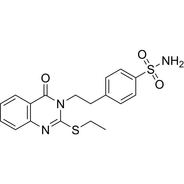
CDK9-IN-41 is a potent CDK9, EGFR, HER2, and COX-2 kinases inhibitor with IC50s of 192.81 nM, 254.03 nM, 238.81 nM, and 775 nM. COX-2-IN-18 exhibits remarkable antitumor activity against leukemia cancer cells, colon cancer cells, melanoma cells, ovarian cells and breast cells.
For research use only. We do not sell to patients.
- Purity : 98.84%
- CAS No.: 1038061-96-9
- Formula: C18H19N3O3S2
- Molecular Weight:389.49
-
Storage:Powder -20°C, 3 years , 4°C, 2 years ; In solvent -80°C, 6 months , -20°C, 1 month

To place orders, for customer services and technical support, please contact: MedChemExpress USA
Tel: 609-228-6898 E-mail: [email protected] [email protected]


-
 Biological Activity
-
Chemical Information
- Protocols
- Purity & Documentation
- References
-
 Help & FAQs


All EGFR Isoforms
More

Biological Activity
Description
IC50 & Target
[1]|
CDK9 192.81 nM (IC50) |
HER2 238.81 nM (IC50) |
EGFR 254.03 nM (IC50) |
COX-2 775 nM (IC50) |
In Vitro
CDK9-IN-41 (Compound 3) has antitumor activity presented as growth inhibition percentages (GI%) for 59 subpanel tumor cell lines. Leukemia (CCRF-CEM, 20%; HL-60(TB), 30%; MOLT-4, 20%; RPMI-8226, 20%; SR, 19%), NSC Lung (A549/ATCC, 20%; HOP-92, 14%; NCI-H322M, 19%; NCI-H23, 12%; NCI-H522, 24%), Colon (HCT-116, 21%; HCT-15, 12%), CNS (SF-539, 12%; SNB-19, 12%; U251, 12%), Melanoma (LOX IMVI, 23%; MALME-3 M, 25%; SK-MEL-5, 11%; UACC-62, 24%), Ovarian (OVCAR-8, 12%; A-498, 12%; RXF-393, 13%; UO-31, 35%), Prostate (PC-3, 13%), Breast (MDA-MB-231/ATCC, 29%; HS-578 T, 18%; T-47D, 15%; MDA-MB-468, 12%)[1].
MedChemExpress (MCE) has not independently confirmed the accuracy of these methods. They are for reference only.


Chemical Information
-
CAS No. 1038061-96-9
-
Appearance Solid
-
Molecular Weight 389.49
-
Formula C18H19N3O3S2
-
Color White to off-white
-
SMILES
O=S(C1=CC=C(CCN2C(SCC)=NC3=C(C=CC=C3)C2=O)C=C1)(N)=O
-
Shipping
Room temperature in continental US; may vary elsewhere.
-
Storage
Powder -20°C 3 years 4°C 2 years In solvent -80°C 6 months -20°C 1 month


Purity & Documentation
-
Data Sheet (269 KB)
-
SDS (251 KB)
- English - EN (251 KB)
- Français - FR (251 KB)
- Deutsch - DE (251 KB)
- Norwegian - NO (251 KB)
- Español - ES (251 KB)
- Swedish - SV (251 KB)
- Italian - IT (251 KB)
- Korean - KR (251 KB)
- Portuguese - PT (251 KB)
-
Handling Instructions (2659 KB)
References

Calculators
Concentration (start) × Volume (start) = Concentration (final) × Volume (final)


CDK9-IN-41 Related Classifications
HY-147794 Related Classifications
 Powered by Bioz