



Glycochenodeoxycholic acid sodium salt
Based on 9 publication(s) in Google Scholar
Glycochenodeoxycholic acid sodium salt (Sodium glycochenodeoxycholate) is a relatively toxic bile salt generated in the liver from chenodeoxycholic acid and glycine. Glycochenodeoxycholic acid sodium salt inhibits Autophagosome formation and impairs lysosomal function by inhibiting lysosomal proteolysis and increasing lysosomal pH in human normal liver cells, leading to the Apoptosis of human hepatocyte cells. Glycochenodeoxycholic acid sodium salt induces stemness and chemoresistance via activating STAT3 signaling pathway in hepatocellular carcinoma cells (HCC). Glycochenodeoxycholic acid sodium salt is promising for research in the field of cholestasis desease, hepatocellular carcinoma and primary sclerosing cholangitis (PSC).
For research use only. We do not sell to patients.
- Purity : 98.51%
- CAS No.: 16564-43-5
- Formula: C26H42NNaO5
- Molecular Weight:471.61
-
Storage:
4°C, sealed storage, away from moisture
* In solvent : -80°C, 6 months; -20°C, 1 month (sealed storage, away from moisture)

To place orders, for customer services and technical support, please contact: MedChemExpress USA
Tel: 609-228-6898 E-mail: [email protected] [email protected]


-
 Biological Activity
-
Chemical Information
-
Solvent & Solubility
- Protocols
- Purity & Documentation
- References
-
 Help & FAQs


-
HY-N0545
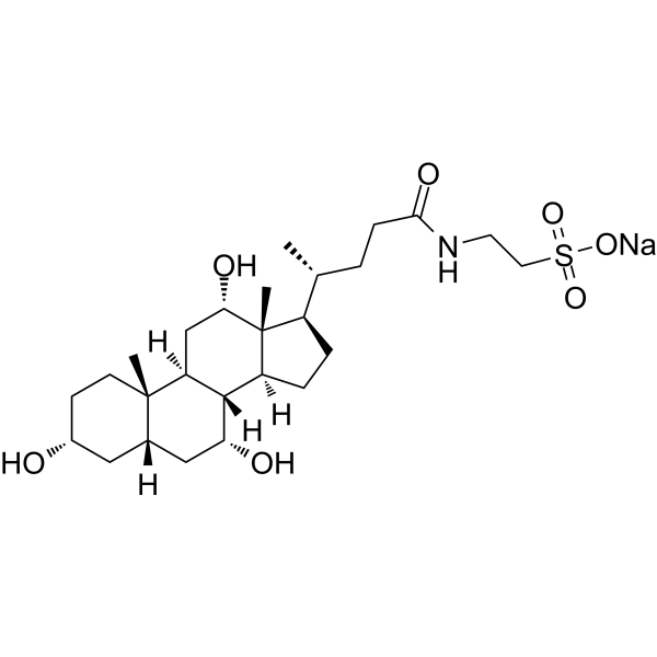
Taurocholic acid sodiumImmunoregulation Agent -
HY-N1424
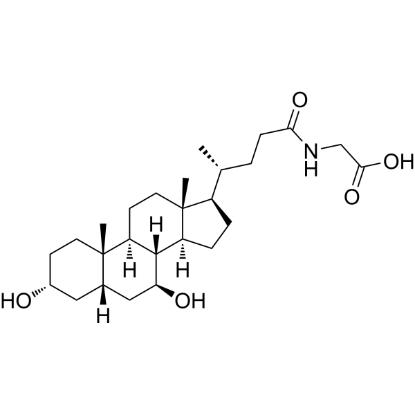
Glycoursodeoxycholic acidEndogenous Metabolite -
HY-N1429
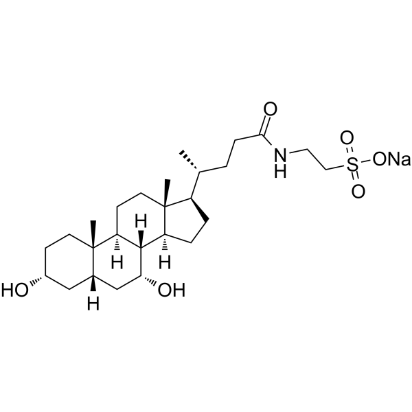
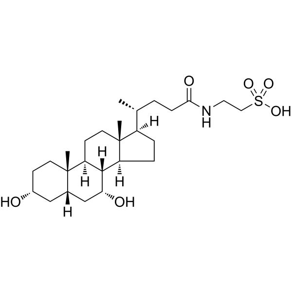
Taurochenodeoxycholic acid sodiumAnti-inflammatory Drug -
HY-N2027
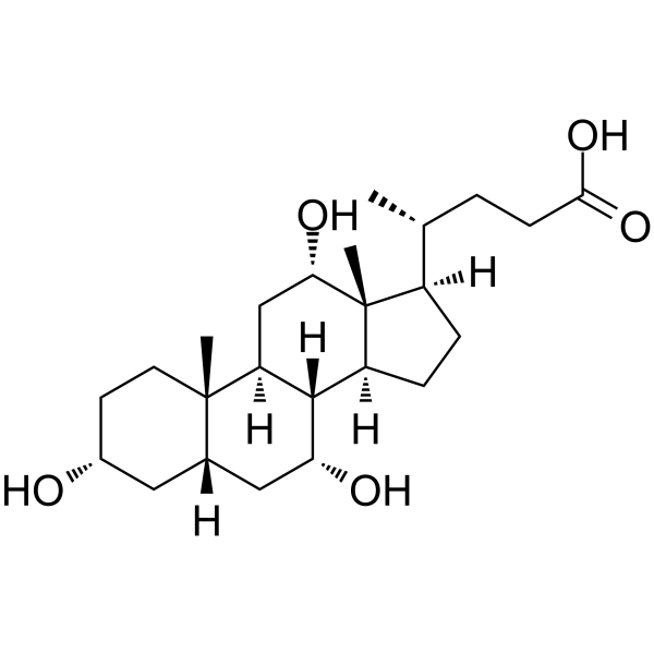
Taurochenodeoxycholic acidEndogenous Metabolite -
HY-N0324
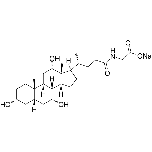
Cholic acidEndogenous Metabolite -
HY-N1423A
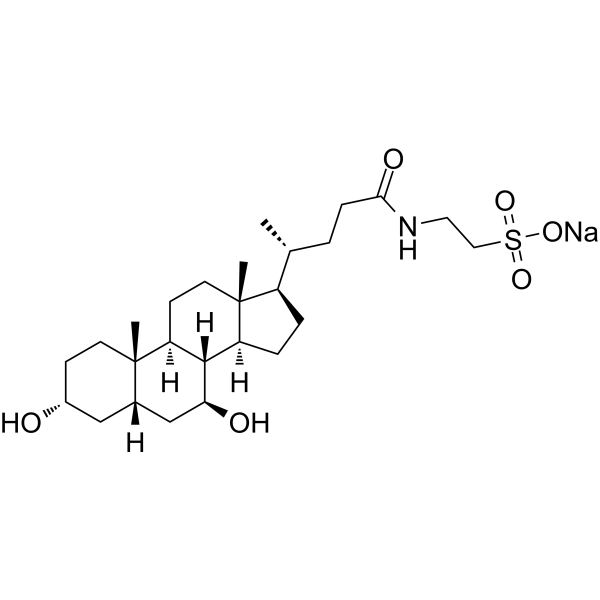
Glycocholic acid sodiumBile Acid Derivative -
HY-19696A
Tauroursodeoxycholate sodiumEndoplasmic Reticulum Stress Inhibitor -
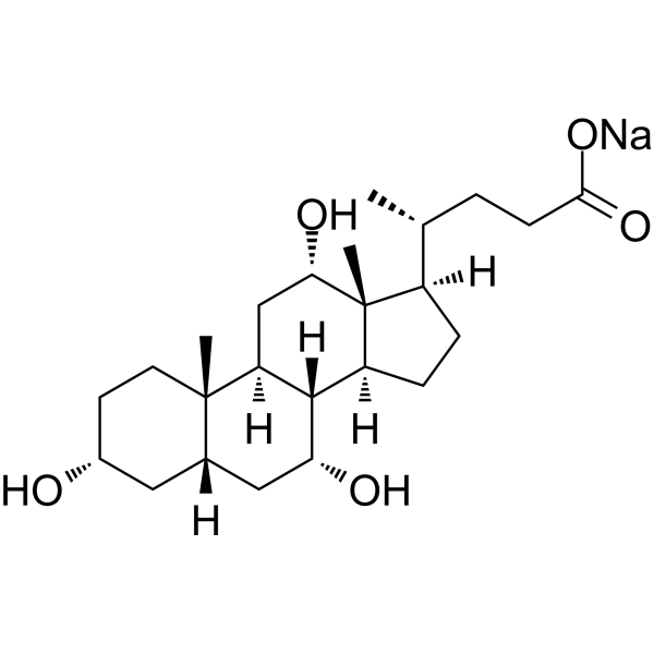
HY-N0324A
Cholic acid sodiumEndogenous Metabolite -
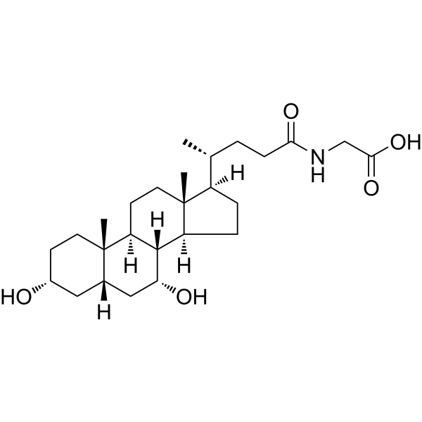
HY-N2334
Glycochenodeoxycholic acidEndogenous Metabolite -
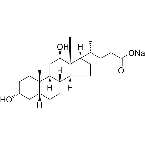
HY-N0593A
Deoxycholic acid sodium saltBile Acid Receptor Activator -
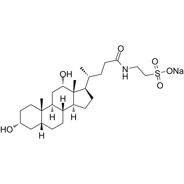
HY-128853
Taurodeoxycholate sodiumAnti-inflammatory Agent -

HY-W013105
Sodium glycocholate hydrate, 98%Bile Acid Derivative
Publications Citing Use of MedChemExpress (MCE) Glycochenodeoxycholic acid sodium salt
More- Adv Sci (Weinh). 2025 Mar;12(12):e2411719. [Abstract]
- ACS Appl Mater Interfaces. 2026 Jun 3;18(21):29739-29755. [Abstract]
- Hepatol Int. 2024 Jun;18(3):1040-1052. [Abstract]
- J Agric Food Chem. 2026 Jun 26. [Abstract]
- Food Funct. 2026 Jun 22. [Abstract]
- Biomolecules. 2025 Jun 28;15(7):943. [Abstract]
- Discov Oncol. 2023 Jan 11;14(1):4. [Abstract]
- bioRxiv. 2026 May 5:2026.04.30.721772. [Abstract]
- SSRN. 2022 Jan 26.
 Customer Validation & Images

-
RT-PCR
-
RT-PCR
All Endogenous Metabolite Isoforms
MoreAll Caspase Isoforms
More

Biological Activity
Description
IC50 & Target
|
Microbial Metabolite |
Human Endogenous Metabolite |
Cellular Effect
|
Cell Line

|
Type | Value | Description | References |
|---|---|---|---|---|
| CHO | EC50 |
3.88 μM
Compound: 4b, glyco
|
Agonist activity at human TGR5 expressed in CHO cells by luciferase assay
Agonist activity at human TGR5 expressed in CHO cells by luciferase assay
|
[PMID: 18307294] |
| HET-1A | CC50 |
729 μM
Compound: 3, TCDCA
|
Cytotoxicity against human HET-1A cells assessed as cell viability after 24 hrs by MTT assay
Cytotoxicity against human HET-1A cells assessed as cell viability after 24 hrs by MTT assay
|
[PMID: 20713311] |
In Vitro
Glycochenodeoxycholic acid sodium salt (0-100 μM, 6 h) significantly increases the amount of dead cells and decreases in LC3, ATG5 and BECN1 expression in human normal liver cells, leading to Autophagosome formation inhibition[1].
Glycochenodeoxycholic acid sodium salt (5–500 μM, 24 h) exerts no induction or reduction of TGF-β mRNA expression in KMBC cells and LX-2 cells[2].
Glycochenodeoxycholic acid sodium salt (200 μM, 24 h and 48 h) enhances stemness and chemoresistance of hepatocellular carcinoma cells (HCC) by activating the STAT3 signaling pathway, suppressing the expression of apoptotic genes (Bcl10, Caspase 3, Caspase 4, Tp53, BAD) and increasing the expression of anti-apoptotic genes (Bcl2, Bcl-xl and IL10)[4].
MedChemExpress (MCE) has not independently confirmed the accuracy of these methods. They are for reference only. Further protocols information, click here.
-
Cell Line:Huh7 and LM3 cell lines
-
Concentration:200 μM
-
Incubation Time:24 h and 48 h
-
Result:Increased the cell viability and promoted chemoresistance treated with 5-FU (HY-90006) (120 µg/mL) and cisplatinin (10 µg/mL) in Huh7 and LM3 cell lines
-
Cell Line:L02 cells
-
Concentration:100 μM
-
Incubation Time:6 h
-
Result:Significantly reduced GFP-LC3 puncta and decreased microtubule-associated protein 1 light chain 3 (LC3), autophagy related 5 (ATG5) and beclin 1 (BECN1) expression in L02 cells.
-
Cell Line:Human normal liver cells
-
Concentration:0-100 μM
-
Incubation Time:6 h
-
Result:Decreased the TFE3 levels in a dose-dependent manner in human normal liver cells.
-
Cell Line:KMBC cells
-
Concentration:5–500 μM
-
Incubation Time:24 h
-
Result:Unaltered TGF-β mRNA expression in KMBC cells and LX-2 cells.
-
Cell Line:Huh7 and LM3 cell lines
-
Concentration:200 μM
-
Incubation Time:24 h and 48 h
-
Result:Suppressed the expression of apoptotic genes (Bcl10, Caspase 3, Caspase 4, Tp53, BAD) and increased the expression of anti-apoptotic genes (Bcl2, Bcl-xl and IL10) in Huh7 and LM3 cell lines.


Chemical Information
-
CAS No. 16564-43-5
-
Appearance Solid
-
Molecular Weight 471.61
-
Formula C26H42NNaO5
-
Color White to off-white
-
SMILES
C[C@@]12[C@](CC[C@]2([H])[C@H](C)CCC(NCC(O[Na])=O)=O)([H])[C@@]3([H])[C@]([C@@]4([C@](C[C@H](O)CC4)([H])C[C@H]3O)C)([H])CC1
-
Synonyms
Chenodeoxycholylglycine sodium salt; Sodium glycochenodeoxycholate
-
Structure Classification
-
Initial Source
-
Shipping
Room temperature in continental US; may vary elsewhere.
-
Storage
4°C, sealed storage, away from moisture
* In solvent : -80°C, 6 months; -20°C, 1 month (sealed storage, away from moisture)


Publications (9)
-
Journal Impact Factor
-
Most Recent
-
Adv Sci (Weinh)
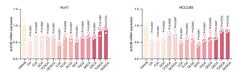
Targeting FDFT1 Reduces Cholesterol and Bile Acid Production and Delays Hepatocellular Carcinoma Progression Through the HNF4A/ALDOB/AKT1 Axis. [Abstract]2025 Mar;12(12):e2411719. PMID: 39899681
Glycochenodeoxycholic acid sodium salt purchased from MedChemExpress. Usage Cited in: Adv Sci (Weinh). 2025 Mar;12(12):e2411719. [Abstract]
The mRNA levels of ALDOB in Huh7 or HCCLM3 cells treated with various bile acids (100 μM, 24 h). All data are presented as mean ± SD. Data were analyzed by one-way ANOVA with Bonferroni multiple-comparison correction. CA, cholic acid; TCA, taurocholic acid; GCA, glycocholic acid; TCDCA, taurochenodeoxycholic acid; GCDCA, glycochenodeoxycholic acid; LCA, lithocholic acid; TLCA, taurolithocholic acid; GLCA, glycolithocholic acid; DCA, deoxycholic acid; TDCA, taurodeoxycholic acid; GDCA, glycodeoxycholic acid; UDCA, ursodeoxycholic acid; TUDCA, tauroursodeoxycholic acid; GUDCA, glycoursodeoxycholic acid.
-
ACS Appl Mater Interfaces
Glutathione-Chitosan Modified Methyl Eugenol Liposomes Promote Spinal Cord Injury Repair by Alleviating Neuroinflammation. [Abstract]2026 Jun 3;18(21):29739-29755. PMID: 42176032 -
Hepatol Int
Bile acids induce liver fibrosis through the NLRP3 inflammasome pathway and the mechanism of FXR inhibition of NLRP3 activation. [Abstract]2024 Jun;18(3):1040-1052. PMID: 38172440 -
J Agric Food Chem
Protein-Free Diet Aggravates Food Allergy Response via the Consumption of Glycochenodeoxycholic Acid in a Murine Model. [Abstract]2026 Jun 26. PMID: 42360286 -
Food Funct
Association between daidzein metabolism to equol and fecal bile acid profiles in young women. [Abstract]2026 Jun 22. PMID: 42329218 -
Biomolecules
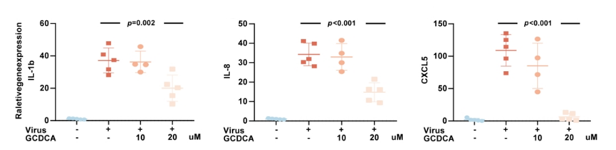
Significant Reduction of Chenodeoxycholic Acid and Glycochenodeoxycholic Acid in the Elderly with Severe COVID-19. [Abstract]2025 Jun 28;15(7):943. PMID: 40723815
Glycochenodeoxycholic acid sodium salt purchased from MedChemExpress. Usage Cited in: Biomolecules. 2025 Jun 28;15(7):943. [Abstract]
SARS-CoV-2-stimulated THP-1 cells were treated with 10 μM or 20 μM Glycochenodeoxycholic acid (GCDCA) for 24 h. The intracellular mRNA expression levels of IL-1β, IL-8, and CXCL5 were measured by RT–qPCR.
-
Discov Oncol
Sphingosine 1-phosphate receptor 2 promotes the onset and progression of non-alcoholic fatty liver disease-related hepatocellular carcinoma through the PI3K/AKT/mTOR pathway. [Abstract]2023 Jan 11;14(1):4. PMID: 36631680 -
bioRxiv
2026 May 5:2026.04.30.721772. PMID: 42146479 -


Solvent & Solubility
In Vitro:
DMSO : 250 mg/mL (530.10 mM; Need ultrasonic; Hygroscopic DMSO has a significant impact on the solubility of product, please use newly opened DMSO)
H2O : ≥ 100 mg/mL (212.04 mM)
* "≥" means soluble, but saturation unknown.
Please refer to the solubility information to select the appropriate solvent. Once prepared, please aliquot and store the solution to prevent product inactivation from repeated freeze-thaw cycles.
Storage method and period of stock solution: -80°C, 6 months; -20°C, 1 month (sealed storage, away from moisture). When stored at -80°C, please use it within 6 months. When stored at -20°C, please use it within 1 month.
* Note: If you choose water as the stock solution, please dilute it to the working solution, then filter and sterilize it with a 0.22 μm filter before use.
Please refer to the solubility information to select the appropriate solvent. Once prepared, please aliquot and store the solution to prevent product inactivation from repeated freeze-thaw cycles.
Storage method and period of stock solution: -80°C, 6 months; -20°C, 1 month (sealed storage, away from moisture). When stored at -80°C, please use it within 6 months. When stored at -20°C, please use it within 1 month.
* Note: If you choose water as the stock solution, please dilute it to the working solution, then filter and sterilize it with a 0.22 μm filter before use.
Concentration (start) × Volume (start) = Concentration (final) × Volume (final)
In Vivo:
Select the appropriate dissolution method based on your experimental animal and administration route.
- For the following dissolution methods, please ensure to first prepare a clear stock solution using an In Vitro approach and then sequentially add co-solvents:
- To ensure reliable experimental results, the clarified stock solution can be appropriately stored based on storage conditions. As for the working solution for In Vivo experiments, it is recommended to prepare freshly and use it on the same day.
- The percentages shown for the solvents indicate their volumetric ratio in the final prepared solution. If precipitation or phase separation occurs during preparation, heat and/or sonication can be used to aid dissolution.
Add each solvent one by one: 10% DMSO 40% PEG300 5% Tween-80 45% Saline
Solubility: ≥ 2.08 mg/mL (4.41 mM); Clear solution
This protocol yields a clear solution of ≥ 2.08 mg/mL (saturation unknown).
Taking 1 mL working solution as an example, add 100 μL DMSO stock solution (20.8 mg/mL) to 400 μL PEG300, and mix evenly; then add 50 μL Tween-80 and mix evenly; then add 450 μL Saline to adjust the volume to 1 mL.
Preparation of Saline: Dissolve 0.9 g sodium chloride in ddH₂O and dilute to 100 mL to obtain a clear Saline solution.
Add each solvent one by one: 10% DMSO 90% (20% SBE-β-CD in Saline)
Solubility: ≥ 2.08 mg/mL (4.41 mM); Clear solution
This protocol yields a clear solution of ≥ 2.08 mg/mL (saturation unknown).
Taking 1 mL working solution as an example, add 100 μL DMSO stock solution (20.8 mg/mL) to 900 μL 20% SBE-β-CD in Saline, and mix evenly.
Preparation of 20% SBE-β-CD in Saline (4°C, storage for one week): 2 g SBE-β-CD powder is dissolved in 10 mL Saline, completely dissolve until clear.
For the following dissolution methods, please prepare the working solution directly:
It is recommended to prepare fresh solutions and use them promptly within a short period of time.
The percentages shown for the solvents indicate their volumetric ratio in the final prepared solution. If precipitation or phase separation occurs during preparation, heat and/or sonication can be used to aid dissolution.
Add each solvent one by one: PBS
Solubility: 50 mg/mL (106.02 mM); Clear solution; Need ultrasonic
In Vivo Dissolution Calculator
Please enter the basic information of animal experiments:
-
-
-
-
Recommended: Prepare an additional quantity of animals to account for potential losses during experiments.
Working solution concentration: 0.22 mg/mL
This product has good water solubility, please refer to the measured solubility data in water/PBS/Saline for details.

Protocols
-
Apoptosis
Apoptosis, also called programmed cell death, is generally characterized by distinct morphological characteristics.
-
TUNEL staining for apoptotic DNA fragmentation
TUNEL staining detects DNA strand breaks by using terminal deoxynucleotidyl transferase to add labeled nucleotides to exposed 3′-OH DNA termini, generating either microscopic staining in fixed cells or tissue sections, or fluorescence/cytometric signal in cell suspensions. TUNEL positivity reflects DNA fragmentation but should not be interpreted alone as definitive apoptosis, because TUNEL can also label necrotic, autolytic, mechanically damaged, or DNA-repair-associated DNA breaks.
-
Annexin V plus membrane-impermeant dye apoptosis staining
Annexin V-based apoptosis assays rely on the detection of phosphatidylserine (PS) externalization from the inner leaflet of the plasma membrane to the outer leaflet, an early biochemical hallmark of apoptosis. Fluorescently labeled Annexin V binds PS in a calcium-dependent manner, enabling identification of early apoptotic cells by flow cytometry or fluorescence microscopy. When combined with a membrane-impermeant DNA-binding dye (e. g. , propidium iodide), this approach allows discrimination between viable (Annexin V−/dye−), early apoptotic (Annexin V+/dye−), and late apoptotic or necrotic (Annexin V+/dye+) cell populations by assessing membrane integrity and PS exposure.
-
Autophagy
Autophagy is a process in which eukaryotic cells use lysosomes to degrade their own cytoplasmic proteins and damaged organelles under the regulation of autophagy related gene (Atg). Microtubule-associated proteins light chain 3 (LC3) is recognized as autophagy marker, which transfers from cytoplasmic LC3 (LC3-I) to membrane type (LC3-II). LC3-II/I ratio could be detected by Western Blot and fluorescence microscopy.
-
Apoptosis Solutions
Apoptosis is a regulated, generally non-lytic cell-death pathway that removes unwanted, damaged, infected, or abnormal cells through coordinated morphological changes, caspase activation, DNA fragmentation, and membrane remodeling. The intrinsic apoptosis pathway is controlled mainly by mitochondrial outer membrane permeabilization, BCL-2 family proteins, cytochrome c release, apoptosome formation, caspase-9 activation, and downstream executioner caspase-3/7 activation. The extrinsic apoptosis pathway is initiated by death receptors such as Fas, TNFR, and TRAIL receptors, which recruit adaptor proteins and activate caspase-8 before engaging executioner caspases or mitochondrial amplification through BID cleavage. Apoptosis is linked to many phenotypes, including cancer cell killing, tissue homeostasis, immune regulation, neurodegeneration, infection response, and treatment-induced cytotoxicity; unresolved questions include how apoptosis interacts with necroptosis, pyroptosis, ferroptos
-
Lysosome and acidic-vesicle live-cell staining
Lysosome and acidic-vesicle live-cell staining detects acidic intracellular compartments by using membrane-permeant acidotropic probes that accumulate in low-pH vesicles, including lysosomes, late endosomes, autolysosomes, and acidic phagosomes. LysoTracker staining is commonly used as an intensity-based readout of acidic lysosomal compartment abundance or enlargement, while acridine orange produces green fluorescence in less concentrated compartments and red fluorescence after concentration-dependent accumulation in acidic vesicular organelles. Loss or reduction of acridine-orange red signal can be used as a readout of lysosomal membrane permeabilization or reduced acidic-vesicle integrity. This protocol is designed for live cultured cells and can be adapted for fluorescence microscopy, high-content imaging, plate-reader readout, or flow cytometry when the selected literature supports the readout. Because these dyes report acidotropic accumulation rather than lysosome identity alone,
-
Liver Cancer Modeling
Liver cancer can be classified into primary liver cancer and secondary liver cancer. Secondary liver cancer is the metastatic liver cancer. Primary liver cancer includes hepatocellular carcinoma (HCC), intrahepatic cholangiocarcinoma (ICC) and fibrolamellar HCC, of which HCC is the most common form, accounting for approximately 90% of primary liver cancers[1]. HCC mouse models include chemical agent-induced models, transplanted tumor models, and genetic engineered models.
-
Macroautophagy Solutions
Macroautophagy is a conserved lysosome-dependent degradation pathway in which cytoplasmic material is sequestered into double-membrane autophagosomes and delivered to lysosomes for degradation and recycling. The pathway supports cellular homeostasis during nutrient limitation, organelle stress, protein-aggregate accumulation, infection, differentiation, and tissue remodeling by coupling cargo sequestration, autophagosome maturation, lysosomal fusion, and degradation of cargo-derived macromolecules. The core molecular sequence includes initiation by nutrient- and stress-regulated autophagy machinery, autophagosome nucleation, LC3/ATG8-family conjugation to autophagosomal membranes, cargo selection through receptors such as SQSTM1/p62, autophagosome-lysosome fusion, and lysosomal degradation. LC3 was identified as a mammalian homolog of yeast Atg8 that localizes to autophagosomal membranes after processing, and p62/SQSTM1 was shown to connect ubiquitinated cargo with autophagic degradati


Purity & Documentation
-
Data Sheet (279 KB)
-
SDS (393 KB)
- English - EN (393 KB)
- Français - FR (393 KB)
- Deutsch - DE (393 KB)
- Norwegian - NO (393 KB)
- Español - ES (393 KB)
- Swedish - SV (393 KB)
- Italian - IT (393 KB)
- Korean - KR (393 KB)
- Portuguese - PT (393 KB)
-
Handling Instructions (2659 KB)
References
[1]. Lan W, et al. Glycochenodeoxycholic acid sodium salt impairs transcription factor E3 -dependent autophagy-lysosome machinery by disrupting reactive oxygen species homeostasis in L02 cells[J]. Toxicol Lett. 2020 Oct 1;331:11-21. [Content Brief]
[2]. Wang A, et al. Glycochenodeoxycholic acid sodium salt Does Not Increase Transforming Growth Factor-Beta Expression in Bile Duct Epithelial Cells or Collagen Synthesis in Myofibroblasts[J]. J Clin Exp Hepatol. 2017 Dec;7(4):316-320. [Content Brief]
[3]. Gonzalez B, et al. Glycochenodeoxycholic acid (GCDC) induced hepatocyte apoptosis is associated with early modulation of intracellular PKC activity. Mol Cell Biochem. 2000 Apr;207(1-2):19-27. [Content Brief]
[4]. Shi C, et al. Glycochenodeoxycholic acid sodium salt induces stemness and chemoresistance via the STAT3 signaling pathway in hepatocellular carcinoma cells[J]. Aging (Albany NY). 2020 Aug 3;12(15):15546-15555. [Content Brief]


Complete Stock Solution Preparation Table
Please refer to the solubility information to select the appropriate solvent. Once prepared, please aliquot and store the solution to prevent product inactivation from repeated freeze-thaw cycles.
Storage method and period of stock solution: -80°C, 6 months; -20°C, 1 month (sealed storage, away from moisture). When stored at -80°C, please use it within 6 months. When stored at -20°C, please use it within 1 month.
| Optional Solvent | Concentration Solvent Mass | 1 mg | 5 mg | 10 mg | 25 mg |
|---|---|---|---|---|---|
| H2O / DMSO | 1 mM | 2.1204 mL | 10.6020 mL | 21.2040 mL | 53.0099 mL |
| 5 mM | 0.4241 mL | 2.1204 mL | 4.2408 mL | 10.6020 mL | |
| 10 mM | 0.2120 mL | 1.0602 mL | 2.1204 mL | 5.3010 mL | |
| 15 mM | 0.1414 mL | 0.7068 mL | 1.4136 mL | 3.5340 mL | |
| 20 mM | 0.1060 mL | 0.5301 mL | 1.0602 mL | 2.6505 mL | |
| 25 mM | 0.0848 mL | 0.4241 mL | 0.8482 mL | 2.1204 mL | |
| 30 mM | 0.0707 mL | 0.3534 mL | 0.7068 mL | 1.7670 mL | |
| 40 mM | 0.0530 mL | 0.2650 mL | 0.5301 mL | 1.3252 mL | |
| 50 mM | 0.0424 mL | 0.2120 mL | 0.4241 mL | 1.0602 mL | |
| 60 mM | 0.0353 mL | 0.1767 mL | 0.3534 mL | 0.8835 mL | |
| 80 mM | 0.0265 mL | 0.1325 mL | 0.2650 mL | 0.6626 mL | |
| 100 mM | 0.0212 mL | 0.1060 mL | 0.2120 mL | 0.5301 mL |
* Note: If you choose water as the stock solution, please dilute it to the working solution, then filter and sterilize it with a 0.22 μm filter before use.


Glycochenodeoxycholic acid sodium salt Related Classifications
HY-N2334A Related Classifications
 Powered by Bioz
Keywords
- Glycochenodeoxycholic acid
- 16564-43-5
- Chenodeoxycholylglycine
- Sodium glycochenodeoxycholate
- Endogenous Metabolite
- Apoptosis
- STAT
- BCL6
- Interleukin Related
- Caspase
- bile salt
- hepatocellular carcinoma cells
- autophagosome inhibition
- STAT3 signaling pathway
- cholestasis desease
- primary sclerosing cholangitis
- Inhibitor
- inhibitor
- inhibit