



Notoginsenoside Ft1
Based on 1 publication(s) in Google Scholar
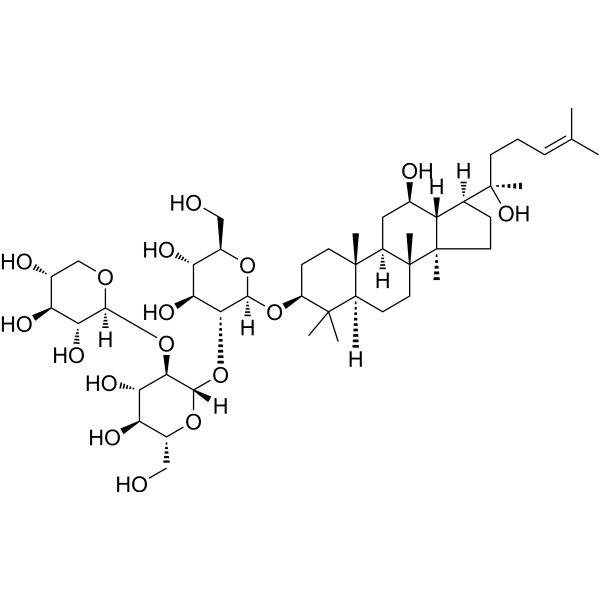
Notoginsenoside Ft1 is an orally active bioactive saponin. Notoginsenoside Ft1 inhibits the PI3K/AKT/mTOR signaling pathway, activates the p38 MAPK and ERK1/2 signaling pathways, and increases the proportion of CD8+ T cells, thereby inducing apoptosis and lysosomal cell death in various cancer cells, and promoting angiogenesis. Notoginsenoside Ft1 causes vasodilation by activating glucocorticoid receptors (GR) and estrogen receptor beta (ERβ) in endothelial cells. Notoginsenoside Ft1 increases intracellular Ca2+ accumulation, reduces cAMP levels by activating a signaling network mediated through P2Y12 receptors, and promotes platelet aggregation, thereby exerting a procoagulant effect. Notoginsenoside Ft1 inhibits ferroptosis (ferroptosis) in renal tubular epithelial cells by activating the TGR5 receptor, thereby demonstrating a renal protective effect. Notoginsenoside Ft1 acts as a TGR5 agonist and an FXR antagonist to combat obesity and insulin resistance.
商品は「研究用試薬」です。人や動物の医療用・臨床診断用・食品用の製品ではありません。
研究用途以外に使用した場合、当社は一切の責任を負いかねます。
- 純度 : 99.49%
- CAS 番号: 155683-00-4
- 分子式: C47H80O17
- 分子量:917.13
-
保管条件:
4°C, stored under nitrogen
* In solvent : -80°C, 6 months; -20°C, 1 month (stored under nitrogen)

To place orders, for customer services and technical support, please contact: MedChemExpress USA
Tel: 609-228-6898 E-mail: [email protected] [email protected]


-
 生物活性
生物活性
-
化学情報
-
溶剤 & 溶解度
- プロトコル
- 純度とドキュメンテーション
- 参考文献
-
 Help & FAQs



MedChemExpress(MCE)の使用を引用している文献 Notoginsenoside Ft1
MoreCalcium Channel アイソフォーム固有の製品をすべて表示
More

生物活性
製品説明
体外実験
Notoginsenoside Ft1 (0-10 μM, 0-48 h) significantly promotes HUVECs proliferation and migration, promotes the transition of the cell cycle from G1 to S phase, significantly promotes the formation of tubular structures of HUVECs, and promotes VEGF expression and secretion by inhibiting the PI3K/AKT/mTOR pathway and the Raf/MEK/ERK pathway[1].
Notoginsenoside Ft1 (0-250 μM) promotes platelet aggregation in a dose-dependent manner in Wistar rats (EC50 = 56.42 μM) and exhibits comprehensive procoagulant effects in both human (240 μg/mL, 5 min) and rat plasma (80 μg/mL, 5 min), affecting extrinsic and common coagulation pathways[2].
Notoginsenoside Ft1 (56.4 μM) induces an increase in intracellular Ca²⁺ in platelets and P2Y12-HEK293 cells, significantly inhibits the production of cAMP and induces the phosphorylation of PI3K and Akt[2].
Notoginsenoside Ft1 (1 nM-10 mM, 30 min) stimulates endothelial GRs and ERbs with subsequent activation of the PI3K/Akt and ERK1/2 pathways in rat mesenteric arteries, resulting in phosphorylation of eNOS and the release of NO, which activates soluble guanylyl cyclase in the vascular smooth muscle cells leading to vasodilatation[3].
Notoginsenoside Ft1 (0.1-100 μM, 24 h) inhibits the proliferation of SH-SY5Y cells (IC50 = 45 μM), causes cell cycle arrest and induces apoptosis involving regulating the p38 MAPK and ERK1/2 pathways[4].
Notoginsenoside Ft1 (0.1-10 μM, 0.5-24 h) significantly activates the luciferase activity of TGR5 and increases the level of cAMP in Tgr5-HEK293 cells and promotes the secretion of GLP-1 in NCI-H716 cells[5].
Notoginsenoside Ft1 (10 μM, 24 h) significantly inhibits the FXR target genes in Caco-2 cells and inhibits the FXR activity induced by GW4064 (HY-50108) in HEK293T cells[5].
Notoginsenoside Ft1 (10 μM, 24 h) has inhibitory effects on HepG2 (IC50 = 46.3 μM), Huh7 (IC50 = 35.2 μM),
and PLC/PRF/5 (IC50 = 58.9 μM) cells, but not on THLE-2 (IC50 = 82.2 μM) cells[6].
Notoginsenoside Ft1 (12.5-50 μM, 12-24 h) induces apoptosis by inhibiting the PI3K/AKT/mTOR pathway and promotes lysosomal cell death by activating TFEB in HepG2 cells[6].
Notoginsenoside Ft1 (0-100 μM, 24 h-14 d) inhibits the growth of MC38, CT26, HT29 cell with IC50 of 32.87, 30.75 and 27.59 μM, inhibits clone formation of MC38 cells and significantly inhibits the migration ability of MC38 and CT26 cells and inhibits colony number[7].
Notoginsenoside Ft1 (5 μM, 24 h) successfully inhibits the ferroptosis model in HK2 cells, as demonstrated by increased cell viability, suppressed ROS accumulation, improved mitochondrial dysfunction, and altered expression of ferroptosis marker proteins[8].
MedChemExpress (MCE) has not independently confirmed the accuracy of these methods. They are for reference only. Further protocols information, click here.
-
Cell Line:HUVECs
-
Concentration:10 μM
-
Incubation Time:0, 3, 6, 12, 16, 24 h
-
Result:Increased the proportion of cells in the S phase and decreased the proportion of cells in the G1 phase.
-
Cell Line:HUVECs
-
Concentration:0, 0.5, 1, 2.5, 5 and 10 μM
-
Incubation Time:24 h
-
Result:Promoted HUVECs migration in a dose-dependent manner.
-
Cell Line:HUVECs
-
Concentration:10 μM
-
Incubation Time:0, 15, 30 min, 1, 2, 4, 8, 12 and 24 h
-
Result:Promoted PI3K, AKT, mTOR, c-Raf, MEK1/2 and ERK1/2 phosphorylation.
Promoted HIF-1α translocation from the cytoplasm to the nucleus in a time-dependent manner.
-
Cell Line:Rat mesenteric arteries
-
Concentration:100 μM
-
Incubation Time:30 min
-
Result:Significantly increased p-eNOS, but no significant change in total eNOS expression; eNOS expression was almost undetectable in the endothelial removal group.
Increased Akt phosphorylation and ERK1/2 phosphorylation.
Increased the levels of p-GR (Ser211) and p-ERβ (Ser87) in the endothelial layer.
-
Cell Line:SH-SY5Y cells
-
Concentration:22.5, 45 and 67.5 μM
-
Incubation Time:24 h
-
Result:Caused arrest at the S phase and G2/M phase22.5-45 μM.
Only causes G2/M phase arrest at 67.5 μM.
-
Cell Line:SH-SY5Y cells
-
Concentration:45 and 67.5 μM
-
Incubation Time:24 h
-
Result:Showed nuclear enrichment and fragmentation.
showed that the rate of early apoptosis increased from 13.6% to 48.9%.
-
Cell Line:SH-SY5Y cells
-
Concentration:22.5, 45 and 67.5 μM
-
Incubation Time:24 h
-
Result:Increased cleaved caspase-3 and decreased Bcl-2.
Increased p-p53, p21 and cyclin B1.
Increased p-ERK1/2, p-JNK and p-p38, decreased p-Jak2 and p-PI3K.
-
Cell Line:HepG2 and Huh7 cells
-
Concentration:12.5, 25 and 50 μM
-
Incubation Time:24 h
-
Result:Increased significantly the proportion of early and late apoptotic cells.
Increased expression of cleaved PARP, caspase-9, caspase-8, and caspase-3.
体内実験
Notoginsenoside Ft1 (1.25 mg/kg, i.v., single dose) significantly increases thrombosis and shortens the bleeding time in Wistar rats[2].
Notoginsenoside Ft1 (50-100 mg/100 g diet, p.o., for 6 weeks) ameliorates obesity, improves glucose disorder in high fat diet (HFD) mice by activating Tgr5 and enhances hepatic bile acids (BA) synthesis by antagonizing FXR[5].
Notoginsenoside Ft1 (25-50 mg/kg, p.o., once daily for 3 weeks) shows anti-cancer effects in HepG2 xenograft tumor mice model[6].
Notoginsenoside Ft1 (10-30 mg/kg, i.p., once daily for 24 days) markedly inhibits subcutaneous tumor formation in colorectal cancer (CRC) and enhances the proportion of CD8+ T cells in tumor-bearing mice, thus restraining tumor growth[7].
Notoginsenoside Ft1 controls blood sugar in mice, reduces renal tubular damage, and intervenes in the occurrence of renal ferroptosis by activating the JUN signaling pathway[8].
MedChemExpress (MCE) has not independently confirmed the accuracy of these methods. They are for reference only.
-
Animal Model:Matrigel plug assay established in Balb/c nude mice (8 weeks old)[1]
-
Dosage:1, 5 and 25 μM with Heparin (HY-17567) in Matrigel
-
Administration:Intraperitoneal injection (i.p.), single dose
-
Result:Formed of a functional vasculature inside the Matrigel.
Increased the hemoglobin content in a dose-dependent manner.
Revealed a dose-dependent increase in staining with the blood vessel endothelial cell marker CD-31 Matrigels with more enlarged and more blood vessels.
-
Animal Model:Ear wound healing assay established in Balb/c nude mice (8 weeks old)[1]
-
Dosage:0.25 mg/kg, 2.5 mg/kg or 25 mg/kg
-
Administration:Intraperitoneal injection (i.p.), every other day for 28 days
-
Result:Significantly reduced the diameter of the wound, and more new blood vessels could be observed at the wound edge.
Significantly upregulated the expression of VEGF mRNA.
No significant difference in the weight changes of the mice in each group.
-
Animal Model:Diet-induced obesity model established in Tgr5 -/- mice, Cyp27a1-/- mice and wild-type mice[5]
-
Dosage:50 and 100 mg/100 g diet
-
Administration:Oral administration (p.o.) with diet, for 6 weeks
-
Result:Reduced the weight gain of mice, significantly decreased the diameter of fat cells, and reduced the deposition of lipid droplets in the liver.
Reduced fasting metabolic parameters, lowered blood sugar, improved insulin resistance index (HOMA-IR) and enhanced insulin sensitivity.
Increased energy digestion and improved cold tolerance.
Upregulated Cyp7a1 and Cyp27a1 mRNA in the liver and downregulated Fgf15 mRNA in the ileum.
Increased Ucp1 expression in BAT and iWAT, and upregulated genes such as Pgc1a and Prdm16.
Increased PKA substrate phosphorylation, HSL phosphorylation, and serum glycerol levels.
-
Animal Model:HepG2 xenograft tumor model established in adult male mice[6]
-
Dosage:25 and 50 mg/kg
-
Administration:Oral administration (p.o.), once daily for 3 weeks
-
Result:Significantly reduced tumor volume and weight.
No significant weight loss caused, and no obvious pathological changes found in major organs.
Reduced KRAS, ERK1/2, c-Fos, and JUND proteins levels.
-
Animal Model:MC38 or CT26 -induced CRC model established in C57BL/6J and Balb/c mice, male, SPF grade, aged 6-8 weeks, and weighing 18 g[7]
-
Dosage:10 and 30 mg/kg
-
Administration:Intraperitoneal injection (i.p.), once daily for 24 days
-
Result:Significantly inhibited tumor growth at 30 mg/kg.
No obvious weight loss, no obvious pathological changes in major organs such as liver and kidney.
Significantly increased the proportion of CD8+ T cells in tumors.


化学情報
-
CAS 番号 155683-00-4
-
性状 Solid
-
分子量 917.13
-
分子式 C47H80O17
-
Color White to off-white
-
SMILES
C[C@@]([C@@]12C)(CC[C@@]3([H])C4(C)C)[C@@](C[C@@H](O)[C@]1([H])[C@]([C@](C)(O)CC/C=C(C)/C)([H])CC2)([H])[C@]3(CC[C@@H]4O[C@@](O[C@H](CO)[C@@H](O)[C@@H]5O)([H])[C@@H]5O[C@@](O[C@H](CO)[C@@H](O)[C@@H]6O)([H])[C@@H]6O[C@@](OC[C@@H](O)[C@@H]7O)([H])[C@@H]7O)C
-
Structure Classification
-
Initial Source
-
輸送条件
Room temperature in continental US; may vary elsewhere.
-
保管条件
4°C, stored under nitrogen
* In solvent : -80°C, 6 months; -20°C, 1 month (stored under nitrogen)


Publications (1)
-
Journal Impact Factor
-
Most Recent
-
Nat Commun
Physiological premature aging of ovarian blood vessels leads to decline in fertility in middle-aged mice. [Abstract]2025 Jan 2;16(1):72. PMID: 39747922


溶剤 & 溶解度
体外:
DMSO : 100 mg/mL (109.04 mM; Need ultrasonic; Hygroscopic DMSO has a significant impact on the solubility of product, please use newly opened DMSO)
Please refer to the solubility information to select the appropriate solvent. Once prepared, please aliquot and store the solution to prevent product inactivation from repeated freeze-thaw cycles.
Storage method and period of stock solution: -80°C, 6 months; -20°C, 1 month (stored under nitrogen). When stored at -80°C, please use it within 6 months. When stored at -20°C, please use it within 1 month.
Please refer to the solubility information to select the appropriate solvent. Once prepared, please aliquot and store the solution to prevent product inactivation from repeated freeze-thaw cycles.
Storage method and period of stock solution: -80°C, 6 months; -20°C, 1 month (stored under nitrogen). When stored at -80°C, please use it within 6 months. When stored at -20°C, please use it within 1 month.
濃度 (開始) × 体積 (開始) = 濃度 (終了) × 体積 (終了)
体内:
Select the appropriate dissolution method based on your experimental animal and administration route.
- For the following dissolution methods, please ensure to first prepare a clear stock solution using an In Vitro approach and then sequentially add co-solvents:
- To ensure reliable experimental results, the clarified stock solution can be appropriately stored based on storage conditions. As for the working solution for In Vivo experiments, it is recommended to prepare freshly and use it on the same day.
- The percentages shown for the solvents indicate their volumetric ratio in the final prepared solution. If precipitation or phase separation occurs during preparation, heat and/or sonication can be used to aid dissolution.
Add each solvent one by one: 10% DMSO 40% PEG300 5% Tween-80 45% Saline
Solubility: ≥ 2.5 mg/mL (2.73 mM); Clear solution
This protocol yields a clear solution of ≥ 2.5 mg/mL (saturation unknown).
Taking 1 mL working solution as an example, add 100 μL DMSO stock solution (25.0 mg/mL) to 400 μL PEG300, and mix evenly; then add 50 μL Tween-80 and mix evenly; then add 450 μL Saline to adjust the volume to 1 mL.
Preparation of Saline: Dissolve 0.9 g sodium chloride in ddH₂O and dilute to 100 mL to obtain a clear Saline solution.
Add each solvent one by one: 10% DMSO 90% (20% SBE-β-CD in Saline)
Solubility: ≥ 2.5 mg/mL (2.73 mM); Clear solution
This protocol yields a clear solution of ≥ 2.5 mg/mL (saturation unknown).
Taking 1 mL working solution as an example, add 100 μL DMSO stock solution (25.0 mg/mL) to 900 μL 20% SBE-β-CD in Saline, and mix evenly.
Preparation of 20% SBE-β-CD in Saline (4°C, storage for one week): 2 g SBE-β-CD powder is dissolved in 10 mL Saline, completely dissolve until clear.
In Vivo Dissolution Calculator
Please enter the basic information of animal experiments:
-
-
-
-
Recommended: Prepare an additional quantity of animals to account for potential losses during experiments.
Please enter your animal formula composition:
-
%DMSO +
Recommended: Keep the proportion of DMSO in working solution below 2% if your animal is weak.
-
%+
-
+%Tween-80 + +
-
%Saline +
The co-solvents required include: DMSO, . All of co-solvents are available by MedChemExpress (MCE). , Tween 80. All of co-solvents are available by MedChemExpress (MCE).
Working solution concentration: 0.22 mg/mL
Method for preparing stock solution: mg drug dissolved in μL DMSO. Stock solution concentration: mg/mL. * In solvent : -80°C, 6 months; -20°C, 1 month (stored under nitrogen)
1. Take μL DMSO stock solution;
2. Add μL .
μL , mix evenly;
3. Then add μL Tween 80, mix evenly;
4. Then add μL
Please ensure that the stock solution in the first step is dissolved to a clear state, and add co-solvents in sequence. You can use ultrasonic heating (ultrasonic cleaner, recommended frequency 20-40 kHz), vortexing, etc. to assist dissolution.

プロトコル
-
Human pluripotent stem cell endothelial-cell differentiation
Human pluripotent stem cell endothelial differentiation is based on stepwise developmental patterning: early activation of WNT/GSK3β inhibition promotes mesodermal or vascular progenitor entry, followed by endothelial specification using VEGF-related signaling, BMP4, FGF2, Notch modulation, or cAMP depending on the published protocol. Endothelial differentiation is read out by acquisition of CD31, CD34, VE-cadherin/CD144, KDR/VEGFR2, vWF, Tie2, NOS3, acetylated LDL uptake, tube/network formation, barrier function, and in vivo vessel-forming capacity where tested.
-
Mammalian live/dead viability and cytotoxicity staining
Live/dead viability and cytotoxicity staining assays are based on the simultaneous detection of intracellular esterase activity in metabolically active (viable) cells and membrane integrity loss in non-viable cells. In commonly used dual-staining approaches, membrane-permeant fluorogenic substrates are converted by intracellular esterases into fluorescent products in live cells, while impermeant DNA-binding dyes selectively enter cells with compromised plasma membranes and label nucleic acids in dead or dying cells, enabling discrimination between viable and non-viable populations by fluorescence microscopy or flow cytometry.
-
Vascular/Branching Fractal Analysis
Vascular/branching fractal analysis quantifies the geometric complexity of vessel trees or vascular networks from segmented 2D images, commonly by converting vessels into binary and/or skeletonized maps and estimating fractal dimension using box-counting or related approaches. Fractal dimension is interpreted as an image-derived readout of vascular branching complexity, space filling, or density, and has been applied to retinal photographs, fluorescein angiography, OCT angiography, capillary perfusion maps, and in vitro Matrigel angiogenesis networks. The assay readout is generated from vessel-positive pixels after image preprocessing, vessel segmentation, binarization, and optional skeletonization; reported outputs include fractal dimension, vessel density, branchpoint density, endpoint density, vessel length density, tortuosity, and generation-based branching metrics when VESGEN-style analysis is used. The biological interpretation is limited to quantitative vascular patterning and s
-
Annexin V plus membrane-impermeant dye apoptosis staining
Annexin V-based apoptosis assays rely on the detection of phosphatidylserine (PS) externalization from the inner leaflet of the plasma membrane to the outer leaflet, an early biochemical hallmark of apoptosis. Fluorescently labeled Annexin V binds PS in a calcium-dependent manner, enabling identification of early apoptotic cells by flow cytometry or fluorescence microscopy. When combined with a membrane-impermeant DNA-binding dye (e. g. , propidium iodide), this approach allows discrimination between viable (Annexin V−/dye−), early apoptotic (Annexin V+/dye−), and late apoptotic or necrotic (Annexin V+/dye+) cell populations by assessing membrane integrity and PS exposure.
-
Ca2+ Staining Technique
Ca2+ staining is an experimental technique that utilizes specific fluorescent probes (such as Fluo-4 AM, Fura-2, etc.) to qualitatively or quantitatively detect dynamic changes in intracellular Ca2+ concentrations; this is achieved by monitoring the changes in fluorescent signals generated when these probes bind to free intracellular calcium ions. The underlying principle relies primarily on the presence of chelating groups within the probe's molecular structure that possess high affinity for calcium ions.
-
Research Protocol for Metabolic Diseases
AMP-activated protein kinase, AMPK, is a conserved cellular energy sensor that responds to reduced cellular energy status and coordinates metabolism by increasing ATP-generating catabolic pathways while suppressing ATP-consuming anabolic processes. In metabolic disease research, the AMPK pathway is experimentally relevant because it regulates hepatic lipid synthesis, fatty acid oxidation, glucose production, skeletal-muscle glucose disposal, mTORC1-linked biosynthesis, autophagy, mitochondrial homeostasis, and whole-body energy balance. The central pathway logic is that energy stress, metformin, exercise-like stimulation, or direct AMPK activators increase AMPKα Thr172 phosphorylation and downstream substrate phosphorylation, including ACC and RAPTOR. Phosphorylation of ACC suppresses lipogenesis and supports fatty acid oxidation, whereas phosphorylation of RAPTOR suppresses mTORC1 signaling and links cellular energy status to growth and protein synthesis control. The pathway is linked
-
Apoptosis
Apoptosis, also called programmed cell death, is generally characterized by distinct morphological characteristics.
-
TUNEL staining for apoptotic DNA fragmentation
TUNEL staining detects DNA strand breaks by using terminal deoxynucleotidyl transferase to add labeled nucleotides to exposed 3′-OH DNA termini, generating either microscopic staining in fixed cells or tissue sections, or fluorescence/cytometric signal in cell suspensions. TUNEL positivity reflects DNA fragmentation but should not be interpreted alone as definitive apoptosis, because TUNEL can also label necrotic, autolytic, mechanically damaged, or DNA-repair-associated DNA breaks.
-
Cell Cytotoxicity Assay
Cytotoxicity assays are usually based on the assessment of cell membrane damage, which can also be indirectly detected by measuring cell viability. Detection methods include MTT assay, CKK-8 assay, LDH assay and ATP assay, etc.
-
Research Protocol for Endocrine Diseases
Endocrine diseases often arise from disrupted hormone production, hormone signaling, or target-tissue responsiveness; for diabetes-focused endocrine disease models, insulin signaling regulates glucose uptake, hepatic glucose output, lipid metabolism, and β-cell compensation. Type 2 diabetes develops through interacting defects in insulin resistance, β-cell dysfunction, adipose inflammation, hepatic glucose overproduction, altered incretin signaling, and ectopic lipid metabolism. A major unresolved question is whether endocrine dysfunction is driven primarily by target-tissue insulin resistance, intrinsic β-cell failure, immune/inflammatory stress, or combined multi-organ failure that differs by disease stage.
-
Endothelial Tube Formation Assay
Endothelial tube formation assay evaluates the ability of endothelial cells to attach, migrate, align, and organize into capillary-like networks when cultured on gelled basement membrane extract or Matrigel; the readout is the morphology and quantity of tube-like networks, which reflects an in vitro endothelial morphogenesis step related to angiogenesis. Basement membrane extract/Matrigel provides laminin-rich extracellular matrix cues that support endothelial differentiation into capillary-like structures, but it can contain biologically active growth factors, so growth-factor-reduced matrix is preferred when testing defined angiogenic stimulators or inhibitors.
-
Apoptosis Solutions
Apoptosis is a regulated, generally non-lytic cell-death pathway that removes unwanted, damaged, infected, or abnormal cells through coordinated morphological changes, caspase activation, DNA fragmentation, and membrane remodeling. The intrinsic apoptosis pathway is controlled mainly by mitochondrial outer membrane permeabilization, BCL-2 family proteins, cytochrome c release, apoptosome formation, caspase-9 activation, and downstream executioner caspase-3/7 activation. The extrinsic apoptosis pathway is initiated by death receptors such as Fas, TNFR, and TRAIL receptors, which recruit adaptor proteins and activate caspase-8 before engaging executioner caspases or mitochondrial amplification through BID cleavage. Apoptosis is linked to many phenotypes, including cancer cell killing, tissue homeostasis, immune regulation, neurodegeneration, infection response, and treatment-induced cytotoxicity; unresolved questions include how apoptosis interacts with necroptosis, pyroptosis, ferroptos
-
Lysosome and acidic-vesicle live-cell staining
Lysosome and acidic-vesicle live-cell staining detects acidic intracellular compartments by using membrane-permeant acidotropic probes that accumulate in low-pH vesicles, including lysosomes, late endosomes, autolysosomes, and acidic phagosomes. LysoTracker staining is commonly used as an intensity-based readout of acidic lysosomal compartment abundance or enlargement, while acridine orange produces green fluorescence in less concentrated compartments and red fluorescence after concentration-dependent accumulation in acidic vesicular organelles. Loss or reduction of acridine-orange red signal can be used as a readout of lysosomal membrane permeabilization or reduced acidic-vesicle integrity. This protocol is designed for live cultured cells and can be adapted for fluorescence microscopy, high-content imaging, plate-reader readout, or flow cytometry when the selected literature supports the readout. Because these dyes report acidotropic accumulation rather than lysosome identity alone,
-
Ferroptosis Solutions
Ferroptosis is an iron-dependent, non-apoptotic form of regulated cell death characterized by lethal lipid peroxidation and sensitivity to suppression by iron chelators or lipophilic radical-trapping antioxidants. The core pathway links cystine uptake through system Xc−, glutathione availability, GPX4-dependent detoxification of phospholipid hydroperoxides, iron-dependent oxidative reactions, and polyunsaturated-phospholipid metabolism into a cell-death program that is biochemically and morphologically distinct from apoptosis, necrosis, and autophagy. The ferroptosis pathway is experimentally linked to phenotype through chemical and genetic perturbation. Erastin induces ferroptosis by inhibiting cystine uptake through system Xc− and weakening antioxidant defenses, while GPX4 inhibition or depletion causes lipid peroxide accumulation and ferroptotic cancer-cell death. ACSL4 and oxidizable arachidonoyl- or adrenoyl-containing phosphatidylethanolamines shape ferroptosis sensitivity by con
-
Endothelial Cell Migration/Angiogenic Sprouting Assay
Endothelial cell migration and angiogenic sprouting assays are in vitro (and partially ex vivo-adapted) functional models that quantify the ability of endothelial cells to undergo coordinated migration, extracellular matrix invasion, and multicellular organization into capillary-like sprouts in response to pro-angiogenic stimuli such as VEGF, bFGF, or conditioned microenvironments. These assays are used to model early angiogenic events including tip-cell formation, directional migration, and lumen-like sprout extension, which collectively reflect angiogenic activation and vascular morphogenesis processes observed in vivo.


純度とドキュメンテーション
-
データシート (301 KB)
-
SDS (394 KB)
- English - EN (394 KB)
- Français - FR (394 KB)
- Deutsch - DE (394 KB)
- Norwegian - NO (394 KB)
- Español - ES (394 KB)
- Swedish - SV (394 KB)
- Italian - IT (394 KB)
- Korean - KR (394 KB)
- Portuguese - PT (394 KB)
-
取扱説明書 (2659 KB)
参考文献
[1]. Shen K, et al. Notoginsenoside Ft1 promotes angiogenesis via HIF-1α mediated VEGF secretion and the regulation of PI3K/AKT and Raf/MEK/ERK signaling pathways. Biochem Pharmacol. 2012 Sep 15;84(6):784-92. [Content Brief]
[2]. Gao B, et al. Platelet P2Y12 receptors are involved in the haemostatic effect of notoginsenoside Ft1, a saponin isolated from Panax notoginseng. Br J Pharmacol. 2014 Jan;171(1):214-23. [Content Brief]
[3]. Shen K, et al. Notoginsenoside Ft1 activates both glucocorticoid and estrogen receptors to induce endothelium-dependent, nitric oxide-mediated relaxations in rat mesenteric arteries. Biochem Pharmacol. 2014 Mar 1;88(1):66-74. [Content Brief]
[4]. Gao B, et al. p38 MAPK and ERK1/2 pathways are involved in the pro-apoptotic effect of notoginsenoside Ft1 on human neuroblastoma SH-SY5Y cells. Life Sci. 2014 Jul 17;108(2):63-70. [Content Brief]
[5]. Ding L, et al. Notoginsenoside Ft1 acts as a TGR5 agonist but FXR antagonist to alleviate high fat diet-induced obesity and insulin resistance in mice. Acta Pharm Sin B. 2021 Jun;11(6):1541-1554. [Content Brief]
[6]. Jeon Y, Kwon H, Chung T, Park YN, Kim SN, Park JY, Kang KS, Woo DY, Kim T, Kim YJ. Notoginsenoside Ft1 induces lysosomal cell death and apoptosis by inhibiting the PI3K/AKT/mTOR pathway in hepatocellular carcinoma. Biomed Pharmacother. 2025 Jul;188:118181. doi: 10.1016/j.biopha.2025.118181. Epub 2025 May 31. PMID: 40451034. [Content Brief]
[7]. Feng Y, Li Y, Ma F, Wu E, Cheng Z, Zhou S, Wang Z, Yang L, Sun X, Zhang J. Notoginsenoside Ft1 inhibits colorectal cancer growth by increasing CD8+ T cell proportion in tumor-bearing mice through the USP9X signaling pathway. Chin J Nat Med. 2024 Apr;22(4):329-340. doi: 10.1016/S1875-5364(24)60623-0. PMID: 38658096. [Content Brief]
[8]. Xiao X, Zhang J, Wu Y, Yang Q, Zhou Y, Yang J, Lang Y, Cai L, Ju X, Liu F. Mechanism of TGR5 in Ferroptosis of the Renal Tubular Epithelial Cells in Diabetes Mellitus and the Effect of Notoginsenoside Ft1. FASEB J. 2025 Jun 30;39(12):e70686. doi: 10.1096/fj.202402534R. PMID: 40549482. [Content Brief]


Complete Stock Solution Preparation Table
Please refer to the solubility information to select the appropriate solvent. Once prepared, please aliquot and store the solution to prevent product inactivation from repeated freeze-thaw cycles.
Storage method and period of stock solution: -80°C, 6 months; -20°C, 1 month (stored under nitrogen). When stored at -80°C, please use it within 6 months. When stored at -20°C, please use it within 1 month.
| Optional Solvent | Concentration Solvent Mass | 1 mg | 5 mg | 10 mg | 25 mg |
|---|---|---|---|---|---|
| DMSO | 1 mM | 1.0904 mL | 5.4518 mL | 10.9036 mL | 27.2589 mL |
| 5 mM | 0.2181 mL | 1.0904 mL | 2.1807 mL | 5.4518 mL | |
| 10 mM | 0.1090 mL | 0.5452 mL | 1.0904 mL | 2.7259 mL | |
| 15 mM | 0.0727 mL | 0.3635 mL | 0.7269 mL | 1.8173 mL | |
| 20 mM | 0.0545 mL | 0.2726 mL | 0.5452 mL | 1.3629 mL | |
| 25 mM | 0.0436 mL | 0.2181 mL | 0.4361 mL | 1.0904 mL | |
| 30 mM | 0.0363 mL | 0.1817 mL | 0.3635 mL | 0.9086 mL | |
| 40 mM | 0.0273 mL | 0.1363 mL | 0.2726 mL | 0.6815 mL | |
| 50 mM | 0.0218 mL | 0.1090 mL | 0.2181 mL | 0.5452 mL | |
| 60 mM | 0.0182 mL | 0.0909 mL | 0.1817 mL | 0.4543 mL | |
| 80 mM | 0.0136 mL | 0.0681 mL | 0.1363 mL | 0.3407 mL | |
| 100 mM | 0.0109 mL | 0.0545 mL | 0.1090 mL | 0.2726 mL |


Notoginsenoside Ft1 関連分類
HY-N0910 関連分類
 Powered by Bioz
Keywords
- Notoginsenoside Ft1
- 155683-00-4
- Notoginsenoside Ft 1
- Notoginsenoside Ft-1
- PI3K
- mTOR
- Akt
- Apoptosis
- p38 MAPK
- ERK
- Transmembrane Glycoprotein
- Glutathione Reductase (GR)
- Estrogen Receptor/ERR
- Calcium Channel
- Ferroptosis
- G protein-coupled Bile Acid Receptor 1
- FXR
- Notoginsenoside Ft1 (Ft1)
- Angiogenesis
- Wound healing
- Vascular endothelial growth factor (VEGF)
- Hypoxia-inducible factor-1a (HIF-1a)
- Obesity
- Insulin resistance
- Bile acids
- TGR5
- Inhibitor
- inhibitor
- inhibit