



Pizotifen
Based on 2 publication(s) in Google Scholar
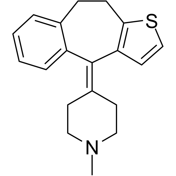
Pizotifen (Pizotyline) is a 5-HT2 receptor antagonist. Pizotifen inhibits the Wnt/β-catenin-EMT signaling pathway and induces mitochondria-mediated Apoptosis. Pizotifen causes transient ERK1/2 phosphorylation and restores ATP. Pizotifen blocks Serotonin (HY-B1473A)-mediated platelet activation, inhibits Serotonin-enhanced ADP-induced platelet aggregation, and prolongs carotid artery occlusion and tail bleeding time. Pizotifen exhibits anticancer activity against gastric cancer and colon cancer. Pizotifen exerts neuroprotective effects in the striatum of R6/2 mice. Pizotifen can be used for research on gastric cancer, colon cancer, Huntington's disease, metastasis, and thromboembolic diseases.
商品は「研究用試薬」です。人や動物の医療用・臨床診断用・食品用の製品ではありません。
研究用途以外に使用した場合、当社は一切の責任を負いかねます。
- 純度 : 99.63%
- CAS 番号: 15574-96-6
- 分子式: C19H21NS
- 分子量:295.44
-
保管条件:Powder -20°C, 3 years , 4°C, 2 years ; In solvent -80°C, 2 years , -20°C, 1 year

To place orders, for customer services and technical support, please contact: MedChemExpress USA
Tel: 609-228-6898 E-mail: [email protected] [email protected]


-
 生物活性
生物活性
-
化学情報
-
溶剤 & 溶解度
- プロトコル
- 純度とドキュメンテーション
- 参考文献
-
 Help & FAQs


-

HY-B0115R
Pizotifen (Standard)Reference Standard -
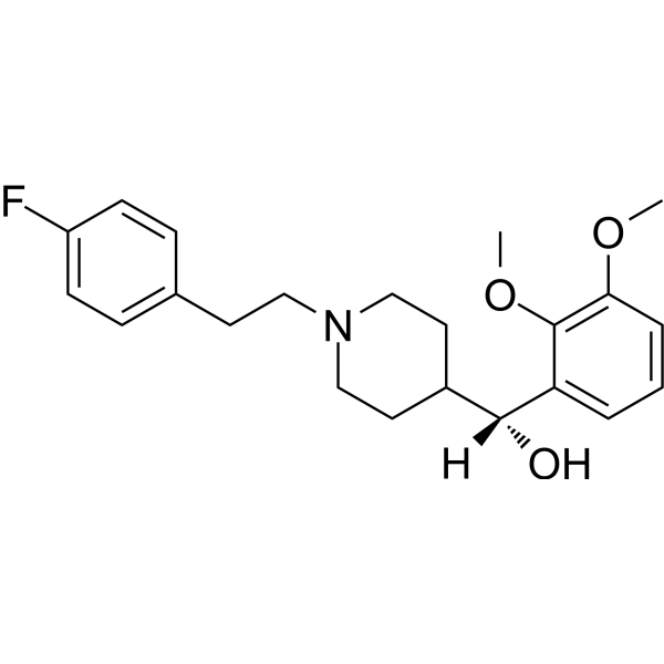
HY-14940
Volinanserin5-HT2 Receptor Antagonist -
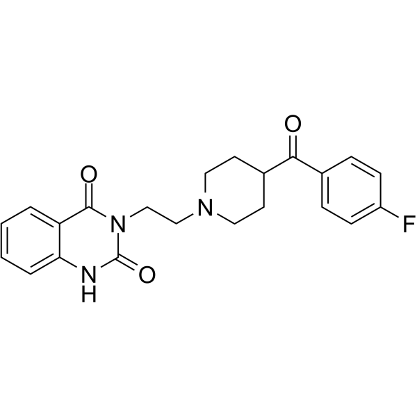
HY-10562
Ketanserin -
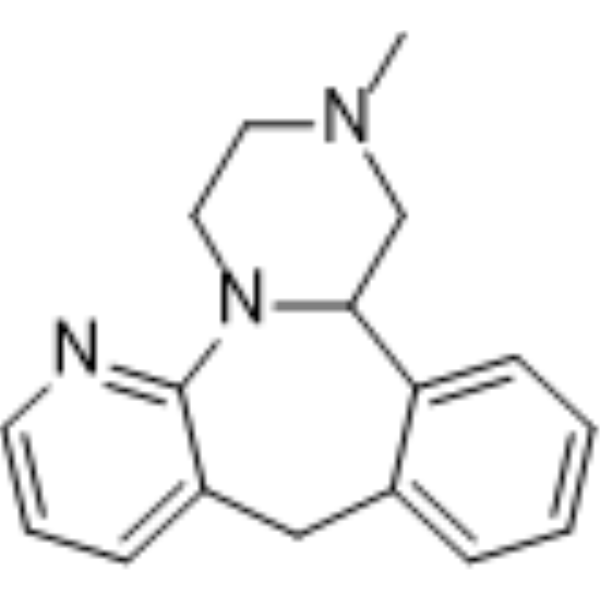
HY-B0352
MirtazapineNaSSA Agent -
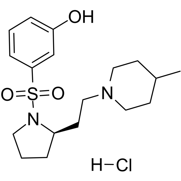
HY-15370A
SB-269970 hydrochloride5-HT7 Receptor Antagonist -
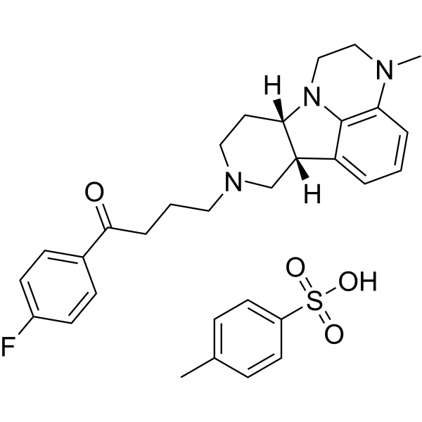
HY-19733
Lumateperone tosylate5-HT2A Receptor Antagonist -
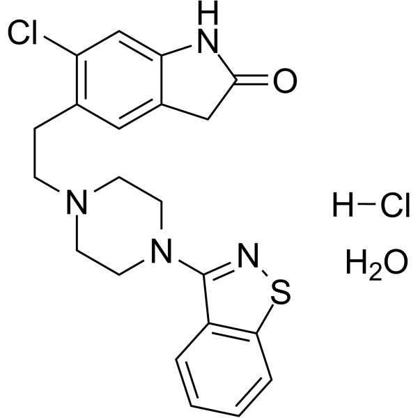
HY-17407
Ziprasidone hydrochloride monohydrate5-HT/DA Receptor Antagonist -
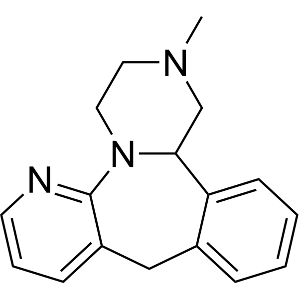
HY-B0352R
Mirtazapine (Standard) -
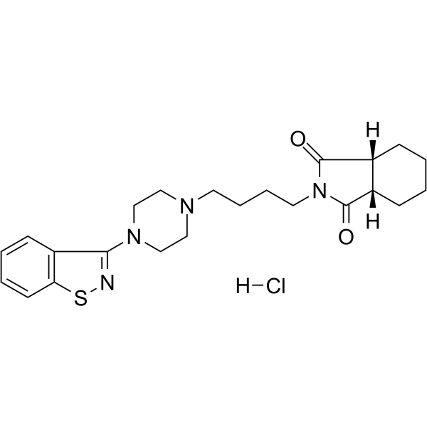
HY-B0731
Perospirone hydrochlorideAntipsychotic Agent -
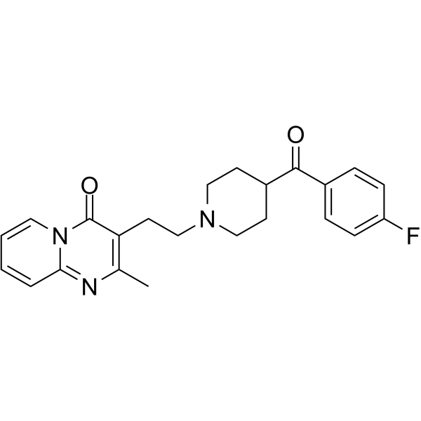
HY-B1737
Pirenperone5-HT2 Serotonin Receptor Antagonist -
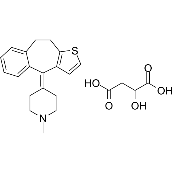
HY-B0115A
Pizotifen malate5-HT2R Antagonist -

HY-10562R
Ketanserin (Standard)5-HT2A Receptor Antagonist
MedChemExpress(MCE)の使用を引用している文献 Pizotifen
More5-HT Receptor アイソフォーム固有の製品をすべて表示
More

生物活性
製品説明
|
ERK1 |
ERK2 |
5-HT2 Receptor |
体外実験
Pizotifen (10-40 µM; 24-72 h) inhibits the viability of MNK45 and AGS gastric cancer cells in a dose-dependent manner[1].
Pizotifen (10-20 µM; 24 h) inhibits the migration and invasion of MNK45 and AGS cells in Transwell assays[1].
Pizotifen (20 µM; 24 h) induces apoptosis in MNK45 and AGS cells[1].
Pizotifen (20 µM; 48 h) decreases Bcl-2 and increases Bax and active caspase-3 in MNK45 and AGS cells[1].
Pizotifen (5-25 μM; 48 h) inhibits HCT116 cell proliferation in a dose-dependent manner, with significant effects at 15-25 μM after 48 h and at 20 μM at 48 and 72 h[2].
Pizotifen (20 μM; 24 h) inhibits HCT116 cell migration and invasion, reducing migrated cells to 45 and invaded cells to 21[2].
Pizotifen (20 μM; 24 h) promotes apoptosis in HCT116 cells, increasing the apoptosis rate to 18.01%[2].
Pizotifen blocks serum withdrawal-induced Caspase-3/7 activation in STHdhQ111/Q111 cells[3].
Pizotifen (0.01-30 nM; 1 min) dose-dependently inhibits Serotonin (HY-B1473A)-enhanced ADP- and U46619 (HY-108566)-induced platelet aggregation[5].
MedChemExpress (MCE) has not independently confirmed the accuracy of these methods. They are for reference only.
-
Cell Line:MNK45 and AGS cell lines
-
Concentration:10, 20, 40 µM
-
Incubation Time:24, 48, 72 h (viability measurement); 3 days (culture)
-
Result:Inhibited viability of MNK45 cells in a dose-dependent manner at 48 h or 72 h.
Inhibited viability of AGS cells.
-
Cell Line:MNK45 and AGS cell lines
-
Concentration:10, 20 µM
-
Incubation Time:24 h
-
Result:Decreased migratory abilities of MNK45 cells in a dose-dependent manner at 10 and 20 µM.
Decreased migratory abilities of AGS cells at 10 and 20 µM.
-
Cell Line:MNK45 and AGS cell lines
-
Concentration:20 µM
-
Incubation Time:48 h
-
Result:Reduced Bcl-2 expression in MNK45 and AGS cells at 20 µM for 48 h.
Increased Bax and active caspase-3 expression in MNK45 and AGS cells at 20 µM for 48 h.
-
Cell Line:HCT116 cells
-
Concentration:5, 10, 15, 20, 25 μM (48 h); 20 μM (time-course)
-
Incubation Time:48 h (dose-response); 0, 24, 48, 72 h (time-course)
-
Result:Inhibited HCT116 cell proliferation dose-dependently.
Reduced proliferation significantly at 15, 20, and 25 μM after 48 h.
Decreased OD values significantly at 20 μM after 48 h and 72 h compared with negative control.
-
Cell Line:HCT116 cells
-
Concentration:20 μM
-
Incubation Time:24 h (serum-free)
-
Result:Increased apoptosis rate to 18.01% compared with 7.8% in negative control.
体内実験
Pizotifen (10 mg/kg; i.p.; once daily; starting on day 2 after inoculation) inhibits metastatic progression without affecting primary tumor growth in the 4T1 mouse breast cancer model[4].
Pizotifen (3 mg/kg; i.p.; once daily; 5 days) prolongs the time to vascular occlusion in the FeCl3-induced carotid artery thrombosis model in mice[5].
Pizotifen (3 mg/kg; i.p.; once daily; 5 days) increases tail bleeding time in mice[5].
MedChemExpress (MCE) has not independently confirmed the accuracy of these methods. They are for reference only.
-
Animal Model:BALB/c (female)[4]
-
Dosage:10 mg/kg
-
Administration:i.p.; once daily; starting on day 2 post inoculation
-
Result:Showed primary tumors equal in size to vehicle group at day 10.
Showed Ki67-positive cells in resected primary tumors same as vehicle group.
Showed less than 1% cleaved caspase 3-positive cells.
Showed no anti-tumor effect on primary tumor.
Detected no light emission in lungs, livers, or lymph nodes by bioluminescence imaging at 70 days.
Formed 0-5 metastatic nodules per lung in all 10 mice.
Detected lung metastatic lesions in only 2 of 10 mice; remaining mice showed metastatic colony formations around the bronchiole.
Showed no liver metastases in 10 mice; half showed metastatic colony formation around the portal tract.
-
Animal Model:C57BL/6 (8-10 weeks old)[5]
-
Dosage:3 mg/kg
-
Administration:i.p.; once daily; 5 days
-
Result:Prolonged time to vessel occlusion to 1199 sec.
-
Animal Model:C57BL/6[5]
-
Dosage:3 mg/kg
-
Administration:i.p.; once daily; 5 days
-
Result:Increased tail bleeding time to 714.4 sec.


化学情報
-
CAS 番号 15574-96-6
-
性状 Solid
-
分子量 295.44
-
分子式 C19H21NS
-
Color White to off-white
-
SMILES
CN1CC/C(CC1)=C2C3=CC=CC=C3CCC4=C\2C=CS4
-
別名
Pizotyline; BC-105
-
輸送条件
Room temperature in continental US; may vary elsewhere.
-
保管条件
Powder -20°C 3 years 4°C 2 years In solvent -80°C 2 years -20°C 1 year


Publications (2)
-
Journal Impact Factor
-
Most Recent
-
PLoS Negl Trop Dis
Identification of anti-flaviviral drugs with mosquitocidal and anti-Zika virus activity in Aedes aegypti. [Abstract]2019 Aug 20;13(8):e0007681. PMID: 31430351 -
Exp Ther Med
2020 Feb;19(2):817-824. PMID: 32010241


溶剤 & 溶解度
体外:
DMSO : 20 mg/mL (67.70 mM; Need ultrasonic; Hygroscopic DMSO has a significant impact on the solubility of product, please use newly opened DMSO)
H2O : < 0.1 mg/mL (insoluble)
Please refer to the solubility information to select the appropriate solvent. Once prepared, please aliquot and store the solution to prevent product inactivation from repeated freeze-thaw cycles.
Storage method and period of stock solution: -80°C, 2 years; -20°C, 1 year. When stored at -80°C, please use it within 2 years. When stored at -20°C, please use it within 1 year.
Please refer to the solubility information to select the appropriate solvent. Once prepared, please aliquot and store the solution to prevent product inactivation from repeated freeze-thaw cycles.
Storage method and period of stock solution: -80°C, 2 years; -20°C, 1 year. When stored at -80°C, please use it within 2 years. When stored at -20°C, please use it within 1 year.
濃度 (開始) × 体積 (開始) = 濃度 (終了) × 体積 (終了)
体内:
Select the appropriate dissolution method based on your experimental animal and administration route.
- For the following dissolution methods, please ensure to first prepare a clear stock solution using an In Vitro approach and then sequentially add co-solvents:
- To ensure reliable experimental results, the clarified stock solution can be appropriately stored based on storage conditions. As for the working solution for In Vivo experiments, it is recommended to prepare freshly and use it on the same day.
- The percentages shown for the solvents indicate their volumetric ratio in the final prepared solution. If precipitation or phase separation occurs during preparation, heat and/or sonication can be used to aid dissolution.
Add each solvent one by one: 10% DMSO 40% PEG300 5% Tween-80 45% Saline
Solubility: ≥ 2 mg/mL (6.77 mM); Clear solution
This protocol yields a clear solution of ≥ 2 mg/mL (saturation unknown).
Taking 1 mL working solution as an example, add 100 μL DMSO stock solution (20.0 mg/mL) to 400 μL PEG300, and mix evenly; then add 50 μL Tween-80 and mix evenly; then add 450 μL Saline to adjust the volume to 1 mL.
Preparation of Saline: Dissolve 0.9 g sodium chloride in ddH₂O and dilute to 100 mL to obtain a clear Saline solution.
Add each solvent one by one: 10% DMSO 90% (20% SBE-β-CD in Saline)
Solubility: ≥ 2 mg/mL (6.77 mM); Clear solution
This protocol yields a clear solution of ≥ 2 mg/mL (saturation unknown).
Taking 1 mL working solution as an example, add 100 μL DMSO stock solution (20.0 mg/mL) to 900 μL 20% SBE-β-CD in Saline, and mix evenly.
Preparation of 20% SBE-β-CD in Saline (4°C, storage for one week): 2 g SBE-β-CD powder is dissolved in 10 mL Saline, completely dissolve until clear.
In Vivo Dissolution Calculator
Please enter the basic information of animal experiments:
-
-
-
-
Recommended: Prepare an additional quantity of animals to account for potential losses during experiments.
Please enter your animal formula composition:
-
%DMSO +
Recommended: Keep the proportion of DMSO in working solution below 2% if your animal is weak.
-
%+
-
+%Tween-80 + +
-
%Saline +
The co-solvents required include: DMSO, . All of co-solvents are available by MedChemExpress (MCE). , Tween 80. All of co-solvents are available by MedChemExpress (MCE).
Working solution concentration: 0.22 mg/mL
Method for preparing stock solution: mg drug dissolved in μL DMSO. Stock solution concentration: mg/mL.
1. Take μL DMSO stock solution;
2. Add μL .
μL , mix evenly;
3. Then add μL Tween 80, mix evenly;
4. Then add μL
Please ensure that the stock solution in the first step is dissolved to a clear state, and add co-solvents in sequence. You can use ultrasonic heating (ultrasonic cleaner, recommended frequency 20-40 kHz), vortexing, etc. to assist dissolution.


純度とドキュメンテーション
-
データシート (290 KB)
-
SDS (418 KB)
- English - EN (418 KB)
- Français - FR (418 KB)
- Deutsch - DE (418 KB)
- Norwegian - NO (418 KB)
- Español - ES (418 KB)
- Swedish - SV (418 KB)
- Italian - IT (418 KB)
- Korean - KR (418 KB)
- Portuguese - PT (418 KB)
-
取扱説明書 (2659 KB)
参考文献
[1]. Jiang Y, et al. Pizotifen inhibits the proliferation and invasion of gastric cancer cells. Experimental and therapeutic medicine. 2020 Feb;19(2):817-824. [Content Brief]
[5]. Lin OA, et al. The antidepressant 5-HT2A receptor antagonists pizotifen and cyproheptadine inhibit serotonin-enhanced platelet function. PloS one. 2014;9(1):e87026. [Content Brief]


Complete Stock Solution Preparation Table
Please refer to the solubility information to select the appropriate solvent. Once prepared, please aliquot and store the solution to prevent product inactivation from repeated freeze-thaw cycles.
Storage method and period of stock solution: -80°C, 2 years; -20°C, 1 year. When stored at -80°C, please use it within 2 years. When stored at -20°C, please use it within 1 year.
| Optional Solvent | Concentration Solvent Mass | 1 mg | 5 mg | 10 mg | 25 mg |
|---|---|---|---|---|---|
| DMSO | 1 mM | 3.3848 mL | 16.9239 mL | 33.8478 mL | 84.6195 mL |
| 5 mM | 0.6770 mL | 3.3848 mL | 6.7696 mL | 16.9239 mL | |
| 10 mM | 0.3385 mL | 1.6924 mL | 3.3848 mL | 8.4620 mL | |
| 15 mM | 0.2257 mL | 1.1283 mL | 2.2565 mL | 5.6413 mL | |
| 20 mM | 0.1692 mL | 0.8462 mL | 1.6924 mL | 4.2310 mL | |
| 25 mM | 0.1354 mL | 0.6770 mL | 1.3539 mL | 3.3848 mL | |
| 30 mM | 0.1128 mL | 0.5641 mL | 1.1283 mL | 2.8207 mL | |
| 40 mM | 0.0846 mL | 0.4231 mL | 0.8462 mL | 2.1155 mL | |
| 50 mM | 0.0677 mL | 0.3385 mL | 0.6770 mL | 1.6924 mL | |
| 60 mM | 0.0564 mL | 0.2821 mL | 0.5641 mL | 1.4103 mL |


Pizotifen 関連分類
HY-B0115 関連分類
 Powered by Bioz
Keywords
- Pizotifen
- 15574-96-6
- Pizotyline
- BC-105
- BC105
- BC 105
- BC-105
- 5-HT Receptor
- Wnt
- β-catenin
- Apoptosis
- ERK
- ATP Synthase
- Mitochondrial Metabolism
- ERK1/2 phosphorylation
- Huntington's disease
- Wnt/β-catenin-EMT signaling
- colon cancer
- gastric cancer
- metastasis
- mitochondrial-mediated apoptosis
- platelet activation
- serotonin 5-HT2A/HTR2C receptor
- thromboembolic disorders
- Inhibitor
- inhibitor
- inhibit