



Apoptosis inducer 34

Based on 1 Customer Validation
Apoptosis inducer 34 (Compound 4) is a small molecule compound that induces apoptosis by directly activating the intrinsic apoptotic pathway. Apoptosis inducer 34 promotes Apaf-1 oligomerization to form mature apoptosomes, thereby activating caspase-9 and caspase-3. It significantly activates the apoptotic pathway in Jurkat cells by enhancing the cytochrome c-dependent apoptotic signaling pathway, inducing PARP cleavage and chromosomal DNA fragmentation. Furthermore, Apoptosis inducer 34 exhibits low toxicity to normal cells, demonstrating potential for selective targeting of cancer cells. Apoptosis inducer 34 is a promising candidate for studying cancer related to apoptotic pathways.
商品は「研究用試薬」です。人や動物の医療用・臨床診断用・食品用の製品ではありません。
研究用途以外に使用した場合、当社は一切の責任を負いかねます。
- 純度 : 99.9%
- CAS 番号: 582314-48-5
- 分子式: C17H15Cl2NO2
- 分子量:336.21
-
保管条件:Powder -20°C, 3 years ; In solvent -80°C, 6 months , -20°C, 1 month


To place orders, for customer services and technical support, please contact: MedChemExpress USA
Tel: 609-228-6898 E-mail: [email protected] [email protected]


-
 生物活性
生物活性
-
化学情報
- プロトコル
- 純度とドキュメンテーション
- 参考文献
-
 Help & FAQs


-
HY-125305
Z-AEVD-FMKCaspase-10 Inhibitor -
HY-16658B
Z-VAD-FMKPan-Caspase Inhibitor -
HY-18633
MDK83190Apoptosis Inducer -
HY-12305
Q-VD-OPhPan-caspase Inhibitor -
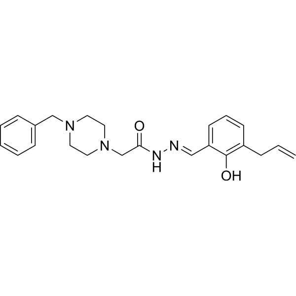
HY-13523
PAC-1Caspase Activator -
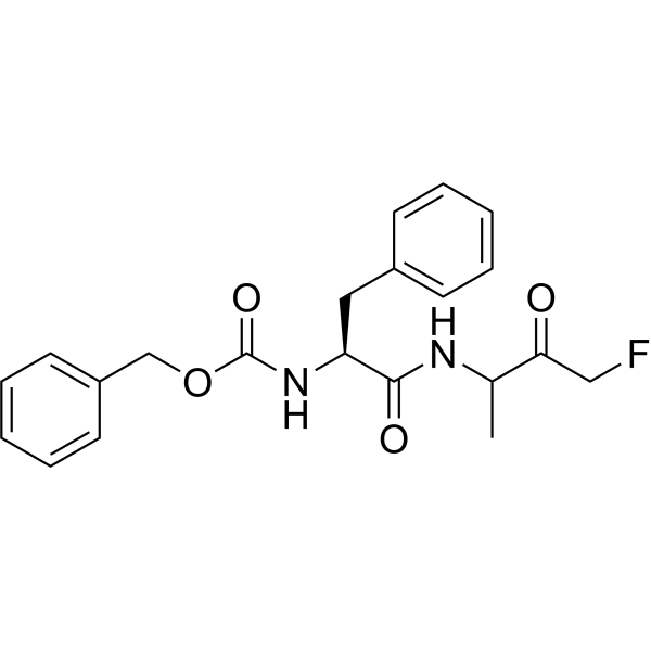
HY-P0109A
Z-FA-FMKCathepsin B/L Inhibitor -
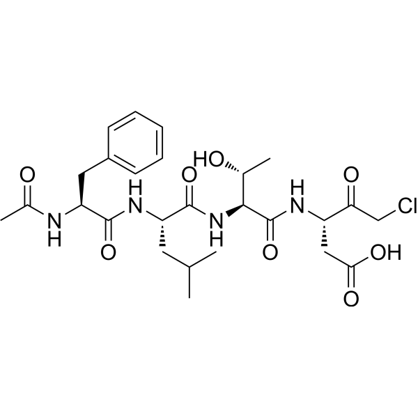
HY-111675
Ac-FLTD-CMKCaspase Inhibitor -
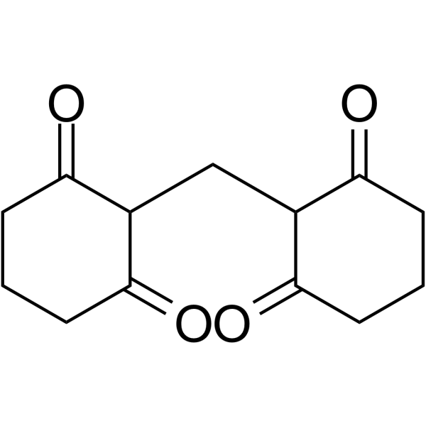
HY-103347
M50054Apoptosis Inhibitor -
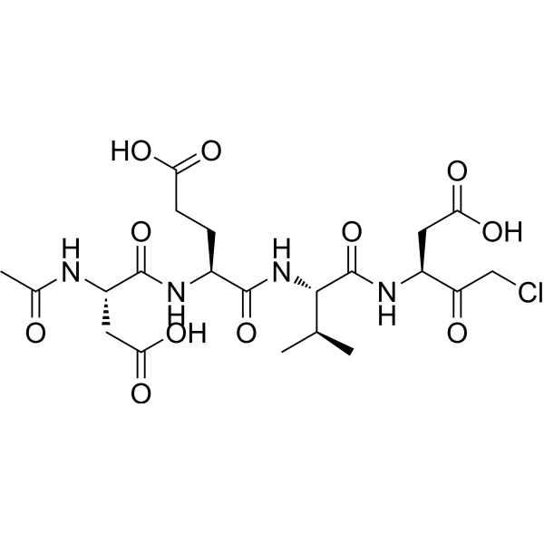
HY-P0034
Ac-DEVD-CMKCaspase-3 Inhibitor -

HY-W033813
Caspase-3/7 Inhibitor ICaspase-3/7 Inhibitor -
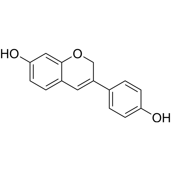
HY-13721
PhenoxodiolAnticancer Agent -
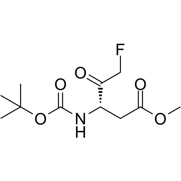
HY-103348
Boc-Asp(OMe)-fluoromethyl ketoneCaspase Inhibitor


生物活性
製品説明


化学情報
-
CAS 番号 582314-48-5
-
性状 Solid
-
分子量 336.21
-
分子式 C17H15Cl2NO2
-
Color White to off-white
-
SMILES
O=C(O[C@H]1CCC2=C1C=CC=C2)NCC3=CC=C(Cl)C(Cl)=C3
-
輸送条件
Room temperature in continental US; may vary elsewhere.
-
保管条件
Powder -20°C 3 years In solvent -80°C 6 months -20°C 1 month


純度とドキュメンテーション
-
データシート (268 KB)
-
SDS (394 KB)
- English - EN (394 KB)
- Français - FR (394 KB)
- Deutsch - DE (394 KB)
- Norwegian - NO (394 KB)
- Español - ES (394 KB)
- Swedish - SV (394 KB)
- Italian - IT (394 KB)
- Korean - KR (394 KB)
- Portuguese - PT (394 KB)
-
取扱説明書 (2659 KB)
参考文献

Calculators
濃度 (開始) × 体積 (開始) = 濃度 (終了) × 体積 (終了)


Apoptosis inducer 34 関連分類
HY-116692 関連分類
 Powered by Bioz