



Based on 1 Customer Validation
Polyinosinic-polycytidylic acid (Poly(I:C)) (GMP Like) is the GMP Like class Polyinosinic-polycytidylic acid (HY-107202), and can be used as pharmaceutical excipients. Polyinosinic-polycytidylic acid (Poly(I:C)) is a synthetic analog of double-stranded RNA and an agonist of toll-like receptor 3 (TLR3) and retinoic acid inducible gene I (RIG-I)-like receptors (RIG-I and MDA5). Polyinosinic-polycytidylic acid can be used as a vaccine adjuvant to enhance innate and adaptive immune responses, and to alter the tumor microenvironment. Polyinosinic-polycytidylic acid can directly trigger cancer cells to undergo apoptosis.
For research use only. We do not sell to patients.
- Assay : 90.5%
- CAS No.: 24939-03-5
- Formula: (C10H13N4O8P)x.(C9H14N3O8P)x
-
Storage:
Please store the product under the recommended conditions in the Certificate of Analysis.


To place orders, for customer services and technical support, please contact: MedChemExpress USA
Tel: 609-228-6898 E-mail: [email protected] [email protected]


-
 Biological Activity
-
Chemical Information
- Protocols
- Purity & Documentation
- References
-
 Help & FAQs


-
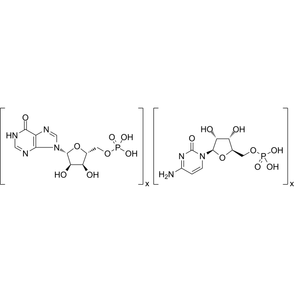
HY-107202
Polyinosinic-polycytidylic acidTLR-3 Agonist -
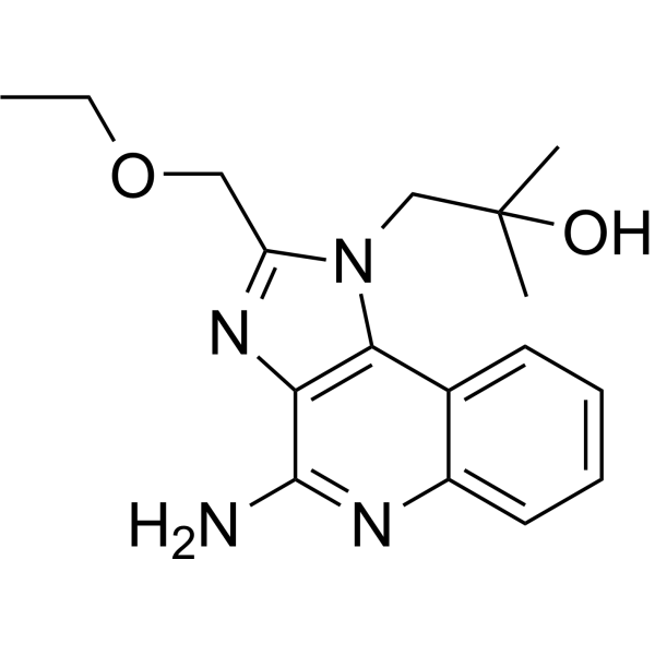
HY-13740G
Resiquimod (GMP)TLR7/TLR8 Agonist (GMP) -
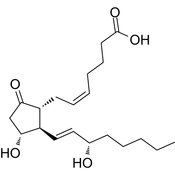
HY-101952G
Prostaglandin E2 (GMP)Inflammatory Mediator (GMP) -
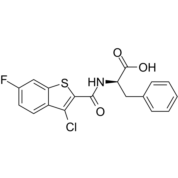
HY-108473
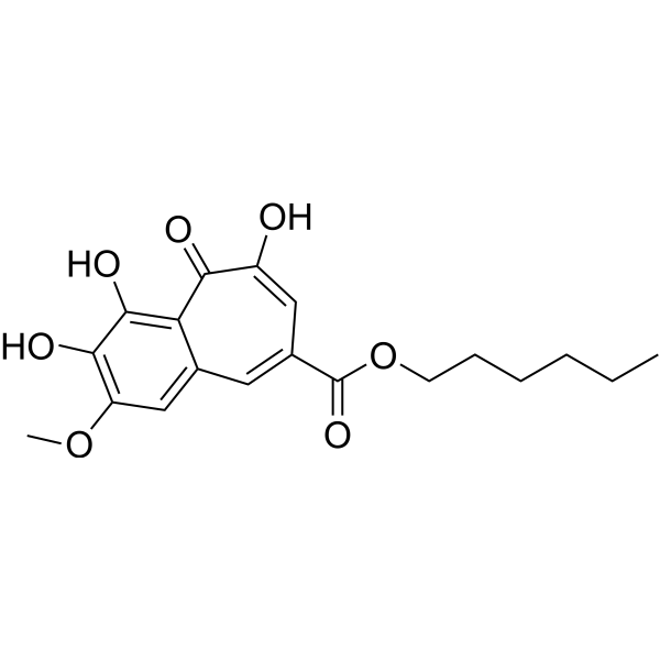
CU-CPT 4aTLR3 Inhibitor -
HY-108471
CU-CPT22TLR1/TLR2 Antagonist -
HY-11109
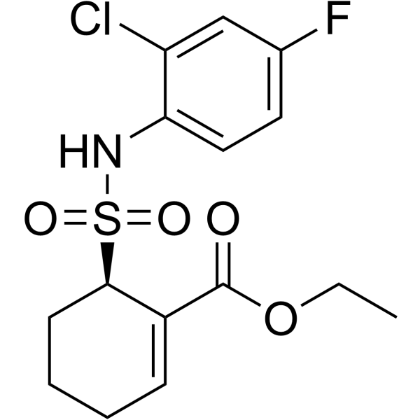
ResatorvidTLR4 Inhibitor -
HY-B0180A
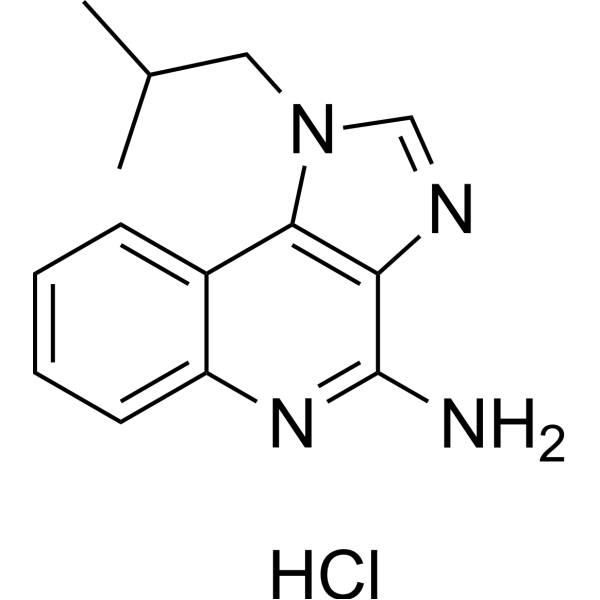
Imiquimod hydrochlorideTLR7 Agonist -
HY-15601
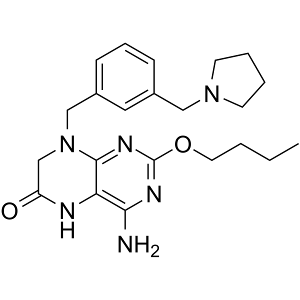
VesatolimodTLR7 Agonist -
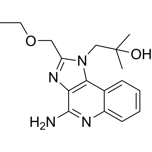
HY-13740
ResiquimodTLR7/8 Agonist -
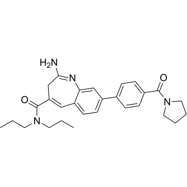
HY-13773
MotolimodTLR8 Agonist -
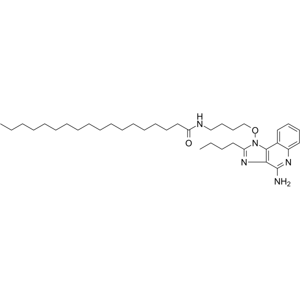
HY-109104
TelratolimodTLR7/8 Agonist -
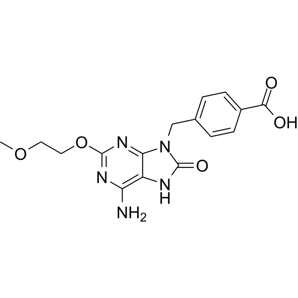
HY-115400
1V209TLR7 Agonist


Biological Activity
Description
In Vitro
Polyinosinic-polycytidylic acid (0.5-5 μg/mL, 3-24 h) (GMP Like) induces a dose- and time-dependent increase in paracellular permeability of immortalized airway epithelial cells[4].
Polyinosinic-polycytidylic acid (5 μg/mL, 24 h) (GMP Like) does not have cytotoxicity to 16HBE14o- cells[4].
Polyinosinic-polycytidylic acid (5 μg/mL, 6 h) (GMP Like) induces disruption of epithelial apical junctional complexes
(AJCs) and tight junctions (TJs) in 16HBE14o- cells[4].
MedChemExpress (MCE) has not independently confirmed the accuracy of these methods. They are for reference only. Further protocols information, click here.
-
Cell Line:16HBE14o- cells
-
Concentration:5 μg/mL
-
Incubation Time:24 h
-
Result:Did not lead to significant accumulation of LDH in cell-culture medium.
In Vivo
Polyinosinic-polycytidylic acid (10 μg/mouse, Intraperitoneal injection, single dose) (GMP Like) decreases lung tumor growth in mice[3].
Polyinosinic-polycytidylic acid (1.25 mg/kg, Intraperitoneal injection, single dose) (GMP Like) exerts therapeutic effects against cerebral I/R injury through the downregulation of TLR4/MyD88 signaling via TLR3 in MCAO model mice[5].
MedChemExpress (MCE) has not independently confirmed the accuracy of these methods. They are for reference only.
-
Animal Model:Middle cerebral artery occlusion (MCAO) model mice[5]
-
Dosage:1.25 mg/kg
-
Administration:Intraperitoneal injection (i.p.)
-
Result:Reduced focal cerebral I/R injury. Increased the expression of Bcl2, Hsp27, and Hsp70, decreased Bax expression, and reduced cellular degeneration and apoptosis. Protected against cerebral ischemia and conferred protection against cerebral I/R injury through the downregulation of TLR4 signaling via TLR3.
-
Animal Model:Lung tumor-bearing mice[3]
-
Dosage:10 μg/mouse
-
Administration:Intraperitoneal injection (i.p.)
-
Result:Induced a significant decrease in the growth of pulmonary metastases in tumor-bearing mice. Reduced the amount of lung foci to ≈ 40%. Significantly increased BAL fluid cell numbers. Increased the level of INF-γ and IL-17A, decreased the levels of IL-13. Increased TLR3 expression.


Chemical Information
-
CAS No. 24939-03-5
-
Appearance Solid
-
Formula (C10H13N4O8P)x.(C9H14N3O8P)x
-
Color White to off-white
-
SMILES
O[C@H]1[C@@H](O[C@H](COP(O)(O)=O)[C@H]1O)N2C3=C(C(NC=N3)=O)N=C2.O[C@H]4[C@H](N5C(N=C(N)C=C5)=O)O[C@H](COP(O)(O)=O)[C@H]4O.[x].[x]
-
Synonyms
Poly(I:C) (GMP Like)
-
Shipping
Room temperature in continental US; may vary elsewhere.
-
Storage
Please store the product under the recommended conditions in the Certificate of Analysis.

Protocols
-
Multiplex immunofluorescence IHC
Multiplex immunofluorescence IHC detects multiple protein biomarkers in one tissue section by sequential antibody staining, HRP-mediated tyramide fluorophore deposition, heat-mediated antibody stripping, nuclear counterstaining, multispectral imaging, spectral unmixing, and digital cell phenotyping; TSA deposits fluorophore near the antigen so the fluorescence signal remains after primary and secondary antibodies are removed, enabling repeated staining cycles, including with antibodies from the same host species. Classic FFPE tumor immune-profiling applications use panels such as CD3, CD8, CD68/CD163, FOXP3, PD-1, PD-L1, pancytokeratin, Ki67, and DAPI to identify tumor cells, immune-cell subsets, checkpoint-marker expression, co-expression phenotypes, cell density, and spatial relationships in the tumor microenvironment.
-
Apoptosis
Apoptosis, also called programmed cell death, is generally characterized by distinct morphological characteristics.
-
TUNEL staining for apoptotic DNA fragmentation
TUNEL staining detects DNA strand breaks by using terminal deoxynucleotidyl transferase to add labeled nucleotides to exposed 3′-OH DNA termini, generating either microscopic staining in fixed cells or tissue sections, or fluorescence/cytometric signal in cell suspensions. TUNEL positivity reflects DNA fragmentation but should not be interpreted alone as definitive apoptosis, because TUNEL can also label necrotic, autolytic, mechanically damaged, or DNA-repair-associated DNA breaks.
-
Annexin V plus membrane-impermeant dye apoptosis staining
Annexin V-based apoptosis assays rely on the detection of phosphatidylserine (PS) externalization from the inner leaflet of the plasma membrane to the outer leaflet, an early biochemical hallmark of apoptosis. Fluorescently labeled Annexin V binds PS in a calcium-dependent manner, enabling identification of early apoptotic cells by flow cytometry or fluorescence microscopy. When combined with a membrane-impermeant DNA-binding dye (e. g. , propidium iodide), this approach allows discrimination between viable (Annexin V−/dye−), early apoptotic (Annexin V+/dye−), and late apoptotic or necrotic (Annexin V+/dye+) cell populations by assessing membrane integrity and PS exposure.
-
Multiplex immunohistochemistry
Multiplex immunohistochemistry (mIHC), also known as tyramide dignal amplification (TSA), is an enzymatic detection method that uses horseradish peroxidase (HRP) to perform high-density in-situ labeling of target proteins or nucleic acids.
-
Apoptosis Solutions
Apoptosis is a regulated, generally non-lytic cell-death pathway that removes unwanted, damaged, infected, or abnormal cells through coordinated morphological changes, caspase activation, DNA fragmentation, and membrane remodeling. The intrinsic apoptosis pathway is controlled mainly by mitochondrial outer membrane permeabilization, BCL-2 family proteins, cytochrome c release, apoptosome formation, caspase-9 activation, and downstream executioner caspase-3/7 activation. The extrinsic apoptosis pathway is initiated by death receptors such as Fas, TNFR, and TRAIL receptors, which recruit adaptor proteins and activate caspase-8 before engaging executioner caspases or mitochondrial amplification through BID cleavage. Apoptosis is linked to many phenotypes, including cancer cell killing, tissue homeostasis, immune regulation, neurodegeneration, infection response, and treatment-induced cytotoxicity; unresolved questions include how apoptosis interacts with necroptosis, pyroptosis, ferroptos
-
Research Protocol for Cancer Immunology
Cancer immunology studies how the immune system recognizes, suppresses, edits, or fails to eliminate malignant cells through tumor antigen release, antigen presentation, T-cell priming, immune trafficking, tumor-cell killing, and feedback inhibition in the tumor microenvironment. The cancer-immunity cycle links tumor antigenicity, dendritic-cell priming, CD8+ T-cell infiltration, cytotoxic function, and immune-checkpoint regulation to tumor rejection or immune escape. Immune-checkpoint pathways such as PD-1/PD-L1 and CTLA-4 suppress antitumor T-cell activity and can be therapeutically blocked, but many tumors remain resistant because of poor antigen presentation, weak T-cell infiltration, suppressive myeloid cells, regulatory T cells, and tumor-intrinsic immune-exclusion programs. Unresolved questions include which immune-cell states predict response, how tumor-intrinsic pathways exclude immune cells, how myeloid suppression limits checkpoint blockade, and which combination strategies


Purity & Documentation
-
Data Sheet (277 KB)
-
SDS (393 KB)
- English - EN (393 KB)
- Français - FR (393 KB)
- Deutsch - DE (393 KB)
- Norwegian - NO (393 KB)
- Español - ES (393 KB)
- Swedish - SV (393 KB)
- Italian - IT (393 KB)
- Korean - KR (393 KB)
- Portuguese - PT (393 KB)
-
Handling Instructions (2659 KB)
References
[1]. Cheng Y, Xu F. Anticancer function of polyinosinic-polycytidylic acid [J]. Cancer biology & therapy, 2010, 10(12): 1219-1223. [Content Brief]
[2]. Deleidi M, Hallett P J, Koprich J B, et al. The Toll-like receptor-3 agonist polyinosinic: polycytidylic acid triggers nigrostriatal dopaminergic degeneration [J]. Journal of Neuroscience, 2010, 30(48): 16091-16101. [Content Brief]
[3]. Forte G, Rega A, Morello S, et al. Polyinosinic-polycytidylic acid limits tumor outgrowth in a mouse model of metastatic lung cancer [J]. The Journal of Immunology, 2012, 188(11): 5357-5364. [Content Brief]
[4]. Rezaee F, Meednu N, Emo J A, et al. Polyinosinic: polycytidylic acid induces protein kinase D–dependent disassembly of apical junctions and barrier dysfunction in airway epithelial cells [J]. Journal of Allergy and Clinical Immunology, 2011, 128(6): 1216-1224. e11. [Content Brief]
[5]. Wang P F, Fang H, Chen J, et al. Polyinosinic-polycytidylic acid has therapeutic effects against cerebral ischemia/reperfusion injury through the downregulation of TLR4 signaling via TLR3 [J]. The Journal of Immunology, 2014, 192(10): 4783-4794. [Content Brief]
[6]. Alexopoulou L, Holt AC, Medzhitov R, Flavell RA. Recognition of double-stranded RNA and activation of NF-kappaB by Toll-like receptor 3. Nature. 2001;413(6857):732-738. [Content Brief]
[7]. Matsumoto M, Kikkawa S, Kohase M, Miyake K, Seya T. Establishment of a monoclonal antibody against human Toll-like receptor 3 that blocks double-stranded RNA-mediated signaling. Biochem Biophys Res Commun. 2002;293(5):1364-1369. [Content Brief]

Calculators
Concentration (start) × Volume (start) = Concentration (final) × Volume (final)


Polyinosinic-polycytidylic acid (GMP Like) Related Classifications
HY-107202GL Related Classifications
 Powered by Bioz