



IKK 16
Based on 32 publication(s) in Google Scholar
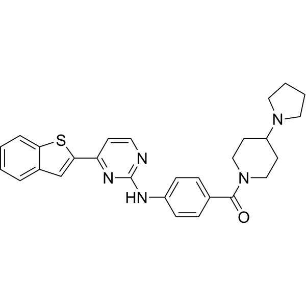
IKK 16 is an orally active IKK inhibitor. IKK 16 shows IC50s of 40 nM, 70 nM, 200 nM, and 50 nM for IKK2, IKK complex, IKK1, and LRRK 2, respectively. IKK 16 is also a pan-PKD inhibitor, inhibiting PKD1, PKD2, and PKD3 with IC50s of 153.9, 115, and 99.7 nM, respectively. IKK 16 is also an ABCB1 inhibitor, interfering with the binding of ABCB1 to its substrates. IKK 16 protects against LPS (HY-D1056)-induced multiple organ dysfunction by reducing the acute inflammatory response induced by endotoxin exposure. IKK 16 can restore renal function and alleviate fibrosis in acute kidney injury. IKK 16 attenuates cardiac dysfunction associated with polymicrobial sepsis in mice with type 2 diabetes mellitus (T2DM) by inhibiting the NF-κB pathway.
For research use only. We do not sell to patients.
- Purity : 99.88%
- CAS No.: 873225-46-8
- Formula: C28H29N5OS
- Molecular Weight:483.63
-
Storage:Powder -20°C, 3 years , 4°C, 2 years ; In solvent -80°C, 2 years , -20°C, 1 year

To place orders, for customer services and technical support, please contact: MedChemExpress USA
Tel: 609-228-6898 E-mail: [email protected] [email protected]


-
 Biological Activity
-
Chemical Information
-
Solvent & Solubility
- Protocols
- Purity & Documentation
- References
-
 Help & FAQs


-
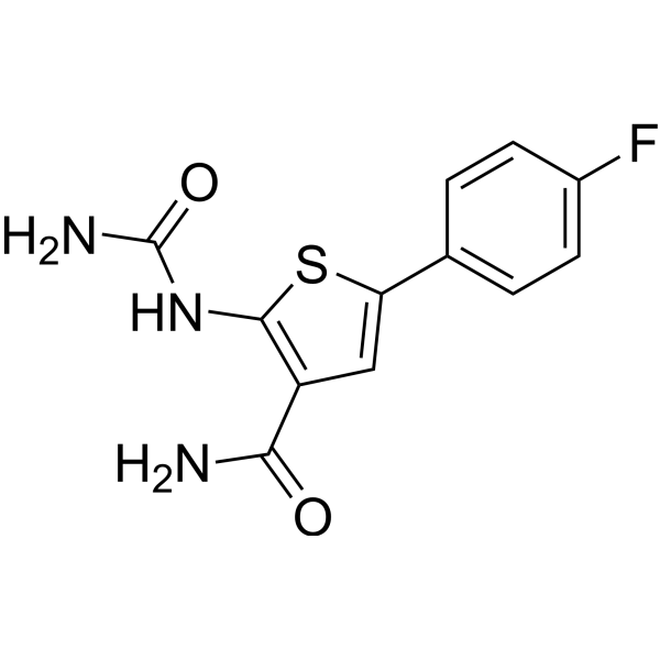
HY-10074
TPCA-1IKK-2 Inhibitor -
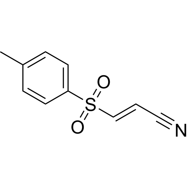
HY-13453
BAY 11-7082IκBα/NF-κB Inhibitor -
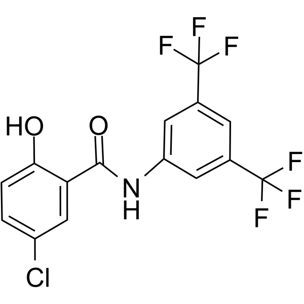
HY-10172
IMD-0354IKKβ Inhibitor -
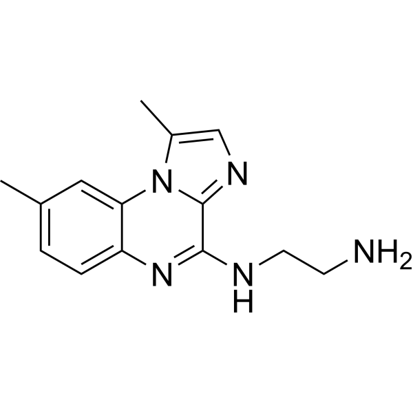
HY-10519
BMS-345541IKK-1/IKK-2 Inhibitor -
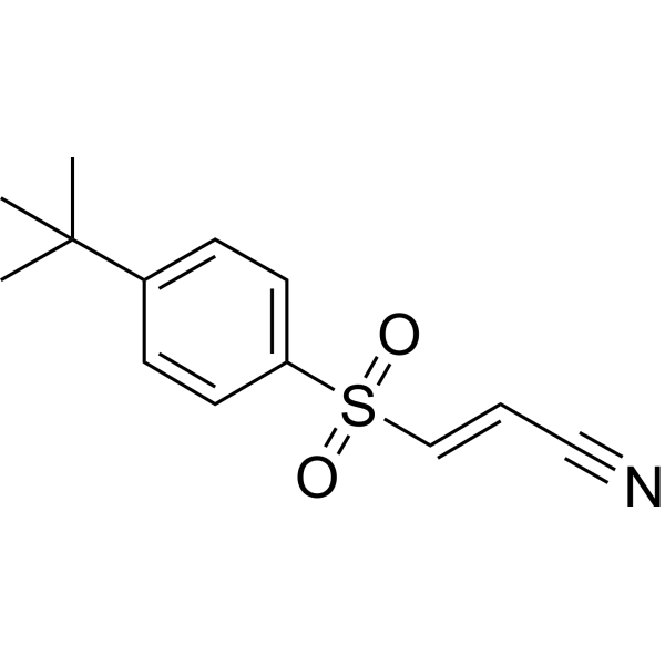
HY-10257
BAY 11-7085NF-κB Activation Inhibitor -
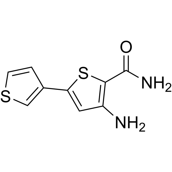
HY-13802
SC-514IKK Inhibitor -
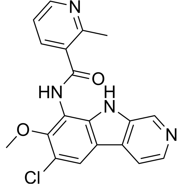
HY-15473
MLN120BIKKβ Inhibitor -

HY-13259
MG-132Proteasome Inhibitor -
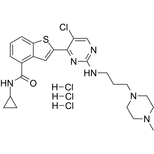
HY-B0788A
LY2409881 trihydrochlorideIKK2 Inhibitor -
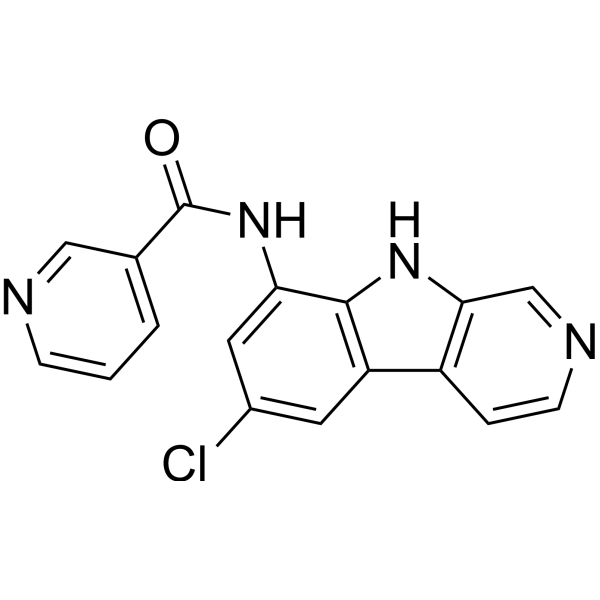
HY-18008
PS-1145Apoptosis Inducer -
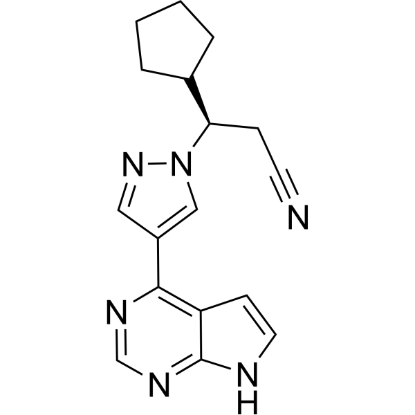
HY-50856
RuxolitinibJAK1/2 Inhibitor -
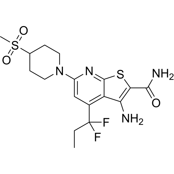
HY-13019
BI605906IKK Inhibitor
Publications Citing Use of MedChemExpress (MCE) IKK 16
More- Cancer Cell. 2026 Jul 24:S1535-6108(26)00305-3.
- Nature. 2026 May;653(8113):212-220. [Abstract]
- Mol Cancer. 2025 Jul 5;24(1):189. [Abstract]
- J Hepatol. 2021 Aug;75(2):363-376. [Abstract]
- Nat Commun. 2022 Mar 31;13(1):1700. [Abstract]
- Mol Cell. 2024 Feb 15;84(4):744-759.e6. [Abstract]
- Theranostics. 2020 Feb 18;10(8):3579-3593. [Abstract]
- J Exp Clin Cancer Res. 2023 Jul 13;42(1):166. [Abstract]
- J Exp Clin Cancer Res. 2021 Aug 27;40(1):273. [Abstract]
- Cell Death Dis. 2026 Mar 25;17(1):377. [Abstract]
- Cell Death Dis. 2019 Apr 3;10(4):304. [Abstract]
- J Immunother Cancer. 2020 Sep;8(2):e000517. [Abstract]
- Phytomedicine. 2026 Apr:153:157988. [Abstract]
- Oncogene. 2024 Dec;43(50):3633-3644. [Abstract]
- J Invest Dermatol. 2022 Nov;142(11):3041-3051.e10. [Abstract]
- Cancer Cell Int. 2021 Jun 5;21(1):291. [Abstract]
- Respir Res. 2017 Sep 20;18(1):174. [Abstract]
- Int Immunopharmacol. 2022 Oct:111:109179. [Abstract]
- Inflammation. 2023 Aug;46(4):1445-1457. [Abstract]
- FASEB J. 2025 Oct 31;39(20):e71164. [Abstract]
- mSphere. 2025 Nov 25;10(11):e0068125. [Abstract]
- J Immunol. 2025 Jul 1;214(7):1714-1732. [Abstract]
- J Immunol. 2018 Apr 15;200(8):2826-2834. [Abstract]
- Mol Immunol. 2018 Dec:104:69-78. [Abstract]
- Biol Pharm Bull. 2023;46(6):803-810. [Abstract]
- bioRxiv. 2026 Jun 25.
- bioRxiv. 2026 Apr 30:2026.04.27.720940. [Abstract]
- bioRxiv. 2026 Feb 14.
- bioRxiv. 2026 Jan 24.
- University of Bonn. 2025.
- Ribeirao Preto Medical School. 2025.
- SSRN. 2023 Dec 13.
 Customer Validation & Images

-
WB
-
WB
-
IF


Biological Activity
Description
|
IKK2 40 nM (IC50) |
IKK1 200 nM (IC50) |
IKK 70 nM (IC50) |
LRRK2 50 nM (IC50) |
PKD1/PKCμ 153.9 nM (IC50) |
PKD2 115 nM (IC50) |
PKD3 99.7 nM (IC50) |
Cellular Effect
|
Cell Line

|
Type | Value | Description | References |
|---|---|---|---|---|
| HEK293 | EC50 |
0.003 μM
Compound: IKK-1
|
Inhibition of atypical PKCzeta in HEK293 cells assessed as TNF-induced NFkappaB activation incubated for 3 hrs prior to TNF induction measured after 5 hrs by bright-glo luciferase reporter gene assay
Inhibition of atypical PKCzeta in HEK293 cells assessed as TNF-induced NFkappaB activation incubated for 3 hrs prior to TNF induction measured after 5 hrs by bright-glo luciferase reporter gene assay
|
[PMID: 23566515] |
| HeLa | IC50 |
0.07 μM
Compound: 16
|
Inhibitory activity against IKK complex isolated from HeLa cells
Inhibitory activity against IKK complex isolated from HeLa cells
|
[PMID: 16236504] |
| HUVEC | IC50 |
0.3 μM
Compound: 16
|
Inhibition of TNF-alpha-stimulated expression of ICAM1 in HUVEC cells
Inhibition of TNF-alpha-stimulated expression of ICAM1 in HUVEC cells
|
[PMID: 16236504] |
| HUVEC | IC50 |
0.3 μM
Compound: 16
|
Inhibition of TNF-alpha-stimulated expression of VCAM1 in HUVEC cells
Inhibition of TNF-alpha-stimulated expression of VCAM1 in HUVEC cells
|
[PMID: 16236504] |
| HUVEC | IC50 |
0.5 μM
Compound: 16
|
Inhibition of TNF-alpha-stimulated expression of E-selectin in HUVEC cells
Inhibition of TNF-alpha-stimulated expression of E-selectin in HUVEC cells
|
[PMID: 16236504] |
In Vitro
IKK 16 inhibits IκB degradation and inhibits TNFα-stimulated E-Sel, ICAM, VCAM, β2M, and HLA-DR expression in HUVEC cells, with IC50s of 1, 0.5, 0.3, 0.3, 2, 2 μM, respectively[3].
IKK 16 (20 μM) inhibits phosphorylation of Ser910, Ser935, Ser955 and Ser973 in Flp-In T-REx™ HEK293 GFP-LRRK2 G2019S cells[4].
IKK 16 (20 μM) rescues phosphorylation of Ser910, Ser935, Ser955 and Ser973 in Flp-In T-REx™ HEK293 LRRK2 A2016T/G2019S mutant[4].
IKK 16 (1 μM) inhibits PKD1 activity by 67%,is ATP-competitive inhibitors[6].
IKK 16 (1-100 μM) inhibits PKCα and PKCδ[6].
MedChemExpress (MCE) has not independently confirmed the accuracy of these methods. They are for reference only.
In Vivo
IKK 16 (1 mg/kg, i.v., once) improves recovery of renal glomerular and tubular function, prevents fibrosis at 28 days after the AKI insult in male Wistar rats model[2].
IKK 16 (30 mg/kg, s.c., once) suppresses systemic TNFα levels in LPS-induced rats model[3].
IKK 16 (10 mg/kg, s.c., once) inhibits neutrophil extravasation in thioglycollate-induced peritonitis mice model[3].
IKK 16 reduces CLP-induced cardiac dysfunction, NF-κB pathway activation and iNOS expression in high fat (HFD)-caecal ligation and puncture (CLP) mice model[5].
MedChemExpress (MCE) has not independently confirmed the accuracy of these methods. They are for reference only.
-
Animal Model:Male Wistar rats (240-450 g) model[1]
-
Dosage:1 mg/kg
-
Administration:i.v., once
-
Result:Increased the serum concentration amylase and lipase, decreased serum concentration creatine kinase.
Prevented the elevation of urea, creatinine, AST, ALT and CK serum levels, had no significant alterations in serum pancreatic enzymes (amylase and lipase) levels induced by LPS treatment.
Decreased the mRNA gene expression of TNF-α and IL-1β, increased the gene expression of IL-6 in the heart, overexpressed the mRNA gene expression of TNF-α, IL-6 and IL-1β in kidney and liver.
-
Animal Model:Male Wistar rats (240-290 g) model[2]
-
Dosage:0.1 mg/kg, 0.3 mg/kg, 1 mg/kg
-
Administration:i.v., once
-
Result:Attenuated serum urea, SCr, and eCCL, reduced signs of histological injury with 1 mg/kg at 24 h into reperfusion. Attenuated the increase in the phosphorylation of IκBα on Ser32/36, phosphorylation (Ser176/180 of IKKα/β) and activation of IKK, and the subsequent nuclear translocation of the p65 NF‐κB subunit. Attenuated the increase in Sirius red staining for collagen I and III and, therefore, fibrosis. Attenuated myofibroblast formation and macrophage infiltration, and decreased TGF-β and Smad2/3 phosphorylation. prevented the expression of fibronectin caused by unilateral IRI and 28 days of reperfusion. Reduced Sirius red, α-smooth muscle actin and CD68 staining, reduced macrophage accumulation in the renal tissue.
-
Animal Model:LPS-induced rats model[3]
-
Dosage:30 mg/kg
-
Administration:s.c., once
-
Result:Suppressed systemic TNFα levels.
-
Animal Model:Thioglycollate-induced peritonitis mice model[3]
-
Dosage:10 mg/kg
-
Administration:s.c., once
-
Result:Inhibited neutrophil extravasation.


Chemical Information
-
CAS No. 873225-46-8
-
Appearance Solid
-
Molecular Weight 483.63
-
Formula C28H29N5OS
-
Color White to yellow
-
SMILES
O=C(C1=CC=C(NC2=NC=CC(C3=CC4=CC=CC=C4S3)=N2)C=C1)N5CCC(N6CCCC6)CC5
-
Shipping
Room temperature in continental US; may vary elsewhere.
-
Storage
Powder -20°C 3 years 4°C 2 years In solvent -80°C 2 years -20°C 1 year


Publications (32)
-
Journal Impact Factor
-
Most Recent
-
-
Nature
2026 May;653(8113):212-220. PMID: 41813898 -
Mol Cancer
Targeting CD30L in B-cell non-Hodgkin lymphoma: novel peptide conjugates and their therapeutic potential. [Abstract]2025 Jul 5;24(1):189. PMID: 40615839 -
J Hepatol
FOSL1 promotes cholangiocarcinoma via transcriptional effectors that could be therapeutically targeted. [Abstract]2021 Aug;75(2):363-376. PMID: 33887357 -
Nat Commun
USP8 inhibition reshapes an inflamed tumor microenvironment that potentiates the immunotherapy. [Abstract]2022 Mar 31;13(1):1700. PMID: 35361799 -
Mol Cell
Serine synthesis sustains macrophage IL-1β production via NAD+-dependent protein acetylation. [Abstract]2024 Feb 15;84(4):744-759.e6. PMID: 38266638 -
Theranostics
NIK links inflammation to hepatic steatosis by suppressing PPARα in alcoholic liver disease. [Abstract]2020 Feb 18;10(8):3579-3593. PMID: 32206109 -
J Exp Clin Cancer Res
Pulmonary interleukin 1 beta/serum amyloid A3 axis promotes lung metastasis of hepatocellular carcinoma by facilitating the pre-metastatic niche formation. [Abstract]2023 Jul 13;42(1):166. PMID: 37443052 -
J Exp Clin Cancer Res
Cyclin G2 reverses immunosuppressive tumor microenvironment and potentiates PD-1 blockade in glioma. [Abstract]2021 Aug 27;40(1):273. PMID: 34452627 -
Cell Death Dis
cGAS-STING/HMGB1-mediated senescence induced by LRRK2 accelerates cartilage degeneration in osteoarthritis. [Abstract]2026 Mar 25;17(1):377. PMID: 41881967 -
Cell Death Dis
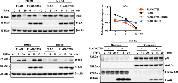
Cancer testis antigen 55 deficiency attenuates colitis-associated colorectal cancer by inhibiting NF-κB signaling. [Abstract]2019 Apr 3;10(4):304. PMID: 30944312
IKK 16 purchased from MedChemExpress. Usage Cited in: Cell Death Dis. 2019 Apr 3;10(4):304. [Abstract]
IKK 16 treatment overwhelms the function of CT55 to promote the activation of p65 and IκBα degradation. HCT116 cells transfected with Flag-CT55 were treated with IKK 16 (20 mM) or DMSO for 2 h before TNF-α treatment. Western blotting is performed to detect the protein levels of IκBα, p-p65, and p65.
-
J Immunother Cancer
Targeted delivery of miR-99b reprograms tumor-associated macrophage phenotype leading to tumor regression. [Abstract]2020 Sep;8(2):e000517. PMID: 32948650 -
Phytomedicine
Celastrol disrupts the inflammatory feedback loop driven by Tenascin-C⁺ fibroblast subtype in psoriasis: Insights from single-cell RNA sequencing and experimental validation. [Abstract]2026 Apr:153:157988. PMID: 41763138 -
Oncogene
PTPRZ1 dephosphorylates and stabilizes RNF26 to reduce the efficacy of TKIs and PD-1 blockade in ccRCC. [Abstract]2024 Dec;43(50):3633-3644. PMID: 39443724 -
J Invest Dermatol
M1 Macrophages Induce Protumor Inflammation in Melanoma Cells through TNFR-NF-κB Signaling. [Abstract]2022 Nov;142(11):3041-3051.e10. PMID: 35580697 -
Cancer Cell Int
Construction of a prognostic model with histone modification-related genes and identification of potential drugs in pancreatic cancer. [Abstract]2021 Jun 5;21(1):291. PMID: 34090418 -
Respir Res
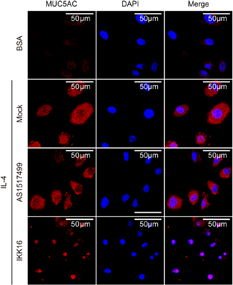
IL-4-induced caveolin-1-containing lipid rafts aggregation contributes to MUC5AC synthesis in bronchial epithelial cells. [Abstract]2017 Sep 20;18(1):174. PMID: 28931396
IKK 16 purchased from MedChemExpress. Usage Cited in: Respir Res. 2017 Sep 20;18(1):174. [Abstract]
BECs are pretreated with specific STAT6 inhibitor (AS1517499) or a selective IκB kinase inhibitor (IKK 16) for half an hour, then treated for 24 h in the presence or absence of 20 ng/mL IL-4. Immunofluorescence staining of MUC5AC protein is performed according to the Methods.
-
Int Immunopharmacol
Extracellular matrix of early pulmonary fibrosis modifies the polarization of alveolar macrophage. [Abstract]2022 Oct:111:109179. PMID: 36029666 -
Inflammation
2023 Aug;46(4):1445-1457. PMID: 37171694 -
FASEB J
Integrated Multi-Omics and Experimental Validation Unveil the GZMK/NF-κB Axis Driving Inflammation and Fibroblast Proliferation in Rosacea: A Novel Drug Target Selection Strategy. [Abstract]2025 Oct 31;39(20):e71164. PMID: 41139274 -
mSphere
The Pseudomonas aeruginosa effector protein TesG regulates alternative activation of macrophages through NLRC5. [Abstract]2025 Nov 25;10(11):e0068125. PMID: 41170846 -
J Immunol
MBL regulates phagocytosis and bactericidal activity of macrophages by triggering AKT/NF-κB/Rab5A axis occurred early in vertebrate evolution. [Abstract]2025 Jul 1;214(7):1714-1732. PMID: 40280188 -
J Immunol
2018 Apr 15;200(8):2826-2834. PMID: 29563176 -
Mol Immunol
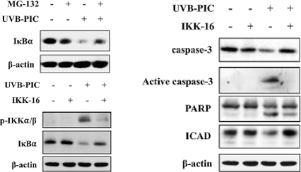
Persistent IKKα phosphorylation induced apoptosis in UVB and Poly I:C co-treated HaCaT cells plausibly through pro-apoptotic p73 and abrogation of IκBα. [Abstract]2018 Dec:104:69-78. PMID: 30445257
IKK 16 purchased from MedChemExpress. Usage Cited in: Mol Immunol. 2018 Dec:104:69-78. [Abstract]
Western analysis of proteins expression with treatment of IKK-16 or MG-132.
-
Biol Pharm Bull
Ponicidin Induces Apoptosis of Murine Melanoma by Inhibiting the NF-κB Signaling Pathway. [Abstract]2023;46(6):803-810. PMID: 37258145 -
-
bioRxiv
Transient efferocytosis-induced activation of IKKβ reprograms macrophages to promote tissue resolution. [Abstract]2026 Apr 30:2026.04.27.720940. PMID: 42094405 -
-
-
-
-


Solvent & Solubility
In Vitro:
DMSO : ≥ 27 mg/mL (55.83 mM; Hygroscopic DMSO has a significant impact on the solubility of product, please use newly opened DMSO)
* "≥" means soluble, but saturation unknown.
Please refer to the solubility information to select the appropriate solvent. Once prepared, please aliquot and store the solution to prevent product inactivation from repeated freeze-thaw cycles.
Storage method and period of stock solution: -80°C, 2 years; -20°C, 1 year. When stored at -80°C, please use it within 2 years. When stored at -20°C, please use it within 1 year.
Please refer to the solubility information to select the appropriate solvent. Once prepared, please aliquot and store the solution to prevent product inactivation from repeated freeze-thaw cycles.
Storage method and period of stock solution: -80°C, 2 years; -20°C, 1 year. When stored at -80°C, please use it within 2 years. When stored at -20°C, please use it within 1 year.
Concentration (start) × Volume (start) = Concentration (final) × Volume (final)
In Vivo:
Select the appropriate dissolution method based on your experimental animal and administration route.
- For the following dissolution methods, please ensure to first prepare a clear stock solution using an In Vitro approach and then sequentially add co-solvents:
- To ensure reliable experimental results, the clarified stock solution can be appropriately stored based on storage conditions. As for the working solution for In Vivo experiments, it is recommended to prepare freshly and use it on the same day.
- The percentages shown for the solvents indicate their volumetric ratio in the final prepared solution. If precipitation or phase separation occurs during preparation, heat and/or sonication can be used to aid dissolution.
Add each solvent one by one: 10% DMSO 40% PEG300 5% Tween-80 45% Saline
Solubility: ≥ 2.5 mg/mL (5.17 mM); Clear solution
This protocol yields a clear solution of ≥ 2.5 mg/mL (saturation unknown).
Taking 1 mL working solution as an example, add 100 μL DMSO stock solution (25.0 mg/mL) to 400 μL PEG300, and mix evenly; then add 50 μL Tween-80 and mix evenly; then add 450 μL Saline to adjust the volume to 1 mL.
Preparation of Saline: Dissolve 0.9 g sodium chloride in ddH₂O and dilute to 100 mL to obtain a clear Saline solution.
Add each solvent one by one: 10% DMSO 90% (20% SBE-β-CD in Saline)
Solubility: ≥ 2.5 mg/mL (5.17 mM); Clear solution
This protocol yields a clear solution of ≥ 2.5 mg/mL (saturation unknown).
Taking 1 mL working solution as an example, add 100 μL DMSO stock solution (25.0 mg/mL) to 900 μL 20% SBE-β-CD in Saline, and mix evenly.
Preparation of 20% SBE-β-CD in Saline (4°C, storage for one week): 2 g SBE-β-CD powder is dissolved in 10 mL Saline, completely dissolve until clear.
In Vivo Dissolution Calculator
Please enter the basic information of animal experiments:
-
-
-
-
Recommended: Prepare an additional quantity of animals to account for potential losses during experiments.
Please enter your animal formula composition:
-
%DMSO +
Recommended: Keep the proportion of DMSO in working solution below 2% if your animal is weak.
-
%+
-
+%Tween-80 + +
-
%Saline +
The co-solvents required include: DMSO, . All of co-solvents are available by MedChemExpress (MCE). , Tween 80. All of co-solvents are available by MedChemExpress (MCE).
Working solution concentration: 0.22 mg/mL
Method for preparing stock solution: mg drug dissolved in μL DMSO. Stock solution concentration: mg/mL.
1. Take μL DMSO stock solution;
2. Add μL .
μL , mix evenly;
3. Then add μL Tween 80, mix evenly;
4. Then add μL
Please ensure that the stock solution in the first step is dissolved to a clear state, and add co-solvents in sequence. You can use ultrasonic heating (ultrasonic cleaner, recommended frequency 20-40 kHz), vortexing, etc. to assist dissolution.


Purity & Documentation
-
Data Sheet (285 KB)
-
SDS (393 KB)
- English - EN (393 KB)
- Français - FR (393 KB)
- Deutsch - DE (393 KB)
- Norwegian - NO (393 KB)
- Español - ES (393 KB)
- Swedish - SV (393 KB)
- Italian - IT (393 KB)
- Korean - KR (393 KB)
- Portuguese - PT (393 KB)
-
Handling Instructions (2659 KB)
References
[1]. Amaro-Leal Â, et al. Therapeutic effects of IkB kinase inhibitor during systemic inflammation. Int Immunopharmacol. 2020 Jul;84:106509. [Content Brief]
[2]. Johnson FL, et al. Inhibition of IκB Kinase at 24 Hours After Acute Kidney Injury Improves Recovery of Renal Function and Attenuates Fibrosis. J Am Heart Assoc. 2017 Jul 3;6(7):e005092. [Content Brief]
[3]. Waelchli R, et al. Design and preparation of 2-benzamido-pyrimidines as inhibitors of IKK. Bioorg Med Chem Lett. 2006 Jan 1;16(1):108-12. [Content Brief]
[4]. Hermanson SB, Carlson CB, Riddle SM, Zhao J, Vogel KW, Nichols RJ, Bi K. Screening for novel LRRK2 inhibitors using a high-throughput TR-FRET cellular assay for LRRK2 Ser935 phosphorylation. PLoS One. 2012;7(8):e43580. [Content Brief]
[6]. Tandon M, et al. New pyrazolopyrimidine inhibitors of protein kinase d as potent anticancer agents for prostate cancer cells. PLoS One. 2013 Sep 23;8(9):e75601. [Content Brief]
[7]. Ansbro MR, et al. Screening compounds with a novel high-throughput ABCB1-mediated efflux assay identifies drugs with known therapeutic targets at risk for multidrug resistance interference. PLoS One. 2013 Apr 10;8(4):e60334. [Content Brief]


Complete Stock Solution Preparation Table
Please refer to the solubility information to select the appropriate solvent. Once prepared, please aliquot and store the solution to prevent product inactivation from repeated freeze-thaw cycles.
Storage method and period of stock solution: -80°C, 2 years; -20°C, 1 year. When stored at -80°C, please use it within 2 years. When stored at -20°C, please use it within 1 year.
| Optional Solvent | Concentration Solvent Mass | 1 mg | 5 mg | 10 mg | 25 mg |
|---|---|---|---|---|---|
| DMSO | 1 mM | 2.0677 mL | 10.3385 mL | 20.6770 mL | 51.6924 mL |
| 5 mM | 0.4135 mL | 2.0677 mL | 4.1354 mL | 10.3385 mL | |
| 10 mM | 0.2068 mL | 1.0338 mL | 2.0677 mL | 5.1692 mL | |
| 15 mM | 0.1378 mL | 0.6892 mL | 1.3785 mL | 3.4462 mL | |
| 20 mM | 0.1034 mL | 0.5169 mL | 1.0338 mL | 2.5846 mL | |
| 25 mM | 0.0827 mL | 0.4135 mL | 0.8271 mL | 2.0677 mL | |
| 30 mM | 0.0689 mL | 0.3446 mL | 0.6892 mL | 1.7231 mL | |
| 40 mM | 0.0517 mL | 0.2585 mL | 0.5169 mL | 1.2923 mL | |
| 50 mM | 0.0414 mL | 0.2068 mL | 0.4135 mL | 1.0338 mL |


IKK 16 Related Classifications
HY-13687 Related Classifications
 Powered by Bioz