



YX968
Based on 1 publication(s) in Google Scholar
YX968 is a selective HDAC3/HDAC8 PROTAC dual degrader, with a DC50 value of 1.7 nM against HDAC3 and 6.1 nM against HDAC8. YX968 inhibits cancer cell growth, induces apoptosis, prevents global histone hyperacetylation, and exerts effects via antiproliferative activity. YX968 can be used in research related to triple-negative breast cancer, non-small cell lung cancer, and multiple myeloma.
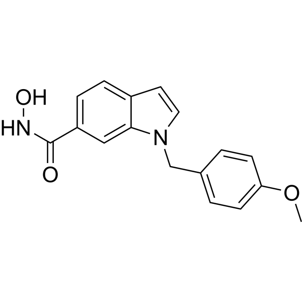
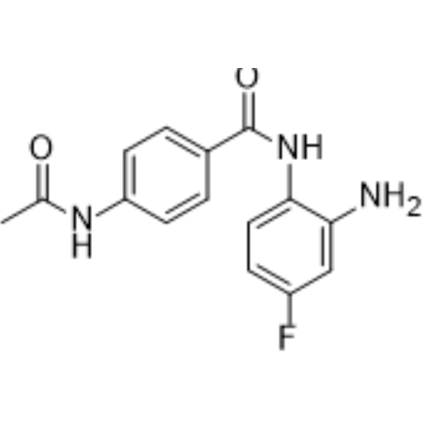
(Pink: HDAC3 and hHDAC8 ligand (HY-168287); Blue: VHL ligand (HY-112078); Black: linker (HY-W007700)).
For research use only. We do not sell to patients.
- Purity : 98.06%
- CAS No.: 2742732-92-7
- Formula: C45H66N8O5S
- Molecular Weight:831.12
-
Storage:Powder -20°C, 3 years , 4°C, 2 years ; In solvent -80°C, 6 months , -20°C, 1 month


To place orders, for customer services and technical support, please contact: MedChemExpress USA
Tel: 609-228-6898 E-mail: [email protected] [email protected]


-
 Biological Activity
-
Chemical Information
-
Solvent & Solubility
- Protocols
- Purity & Documentation
- References
-
 Help & FAQs


Publications Citing Use of MedChemExpress (MCE) YX968
MoreAll PROTACs Isoforms
More

Biological Activity
Description
|
HDAC3 1.7 nM (DC50) |
HDAC8 6.1 nM (DC50) |
In Vitro
YX968 (100 nM) achieves efficient loading into EVs via μDES electroporation, with the loaded EVs maintaining high loading stability across multiple storage conditions, though fresh EVs are optimal due to aggregation-related concentration loss[2].
YX968 forms strong ternary complexes with both purified HDAC3-VHL and HDAC8-VHL protein pairs in an in vitro cell-free system[3].
YX968 (30 min) inhibits purified human HDAC1, HDAC3, and HDAC8 enzymes with micromolar potency, and its inhibitory activity against HDAC3 and HDAC8 is enhanced in the presence of the VHL complex[4].
YX968 (0.1-100000 nM; 45 min) selectively forms stable ternary complexes with purified human HDAC3 and HDAC8 (but not HDAC1) and the VHL-elongin B-elongin C complex, with peak formation at 10 nM[4].
YX968 (16-250 nM; 8 h) is an HDAC3/HDAC8 dual degrader that achieves 91.7% HDAC8 and 94.3% HDAC3 degradation in MDA-MB-231 cells at 16 nM (8 h incubation), and >95% degradation of both isoforms at 250 nM (8 h incubation)[3].
YX968 (100 nM; 2 h) induces degradation of both HDAC3 and HDAC8 in MDA-MB-231 cells when treated with 100 nM for 2 h, as confirmed by TMT-based proteomic profiling[3].
YX968 induces strong antiproliferative activity in MDA-MB-231 cells via dual degradation of HDAC3 and HDAC8, which is more potent than targeting either isoform individually[3].
YX968 (0.063 nM-64 μM; 2-24 h) potently and selectively degrades HDAC3 and HDAC8 in MDA-MB-231 cells via the ubiquitin-proteasome system, with DC50 values of 1.7 nM for HDAC3 and 6.1 nM for HDAC8 after 8 h treatment, and the degradation is reversible upon drug washout[4].
YX968 (14 h) selectively degrades HDAC3 and HDAC8 in MCF7 cells, with histone acetylation induction only at concentrations far higher than those needed for target degradation[4].
YX968 (100 nM; 2 h) induces selective downregulation of HDAC8 and NCOR1 in MDA-MB-231 cells, with no significant off-target protein degradation detected across >7,000 quantified proteins[4].
YX968 (50 nM; 3 h) induces selective downregulation of HDAC3 and NCOR1 (along with three other low-fold-change proteins) in MM.1S cells, with no significant off-target protein degradation detected across ~6,400 quantified proteins[4].
YX968 (3-50 nM; 6-24 h)-mediated degradation of HDAC3 and HDAC8 in MDA-MB-231 cells does not cause significant transcriptomic perturbation, in stark contrast to the broad gene expression changes induced by pan-class I HDAC inhibition[4].
YX968 (125 nM-3 μM; continuous incubation until colony formation) potently suppresses clonogenic growth of multiple breast and lung cancer cell lines, with greater efficacy than HDAC3/HDAC8 inhibitors alone or in combination, and greater potency than HDAC3-selective PROTAC XZ9002[4].
MedChemExpress (MCE) has not independently confirmed the accuracy of these methods. They are for reference only.
-
Cell Line:MDA-MB-231 human triple negative breast cancer (TNBC) cells
-
Concentration:8 nM; 16 nM; 100 nM
-
Incubation Time:2 h; 4 h; 6 h; 8 h; 10 h
-
Result:Induced HDAC3 degradation in MDA-MB-231 cells at 8 nM, a lower concentration than required for free YX968.
Caused significant dose-dependent degradation of both HDAC3 and HDAC8 at 16 nM and 100 nM.
Started degradative effects as early as 2 h post-treatment, with maximum HDAC3 and HDAC8 degradation observed within 10 h.
-
Cell Line:MDA-MB-231 human triple negative breast cancer cells
-
Concentration:8-800 nM (degradation assay); 16-100 nM (EV loading concentration)
-
Incubation Time:8 h (fixed-time degradation assay); 2-10 h (time-course analysis)
-
Result:Induced significant HDAC3/8 degradation at 800 nM when used alone or loaded into EVs via incubation, lipofection, or cuvette-based electroporation.
Maintained HDAC3/8 degradation activity at 8 nM only when loaded into EVs via electroporation.
Increased HDAC3/8 degradation with increasing loading concentration from 16 nM to 100 nM, with the highest EV concentration yielding the greatest HDAC8 degradation.
Showed higher HDAC3/8 degradation efficacy when loaded into μDES-produced EVs than conventional electroporation-produced EVs.
Induced HDAC3/8 degradation as early as 2 hrs post-treatment via μDES-produced EVs, with maximum degradation observed within 10 hrs and no reversal of effects detected.
Exhibited stronger HDAC3 degradation when loaded into EVs via cuvette-based electroporation than via lipofection.
-
Cell Line:MDA-MB-231 human triple-negative breast cancer cells
-
Concentration:16 nM, 250 nM
-
Incubation Time:8 h
-
Result:Degraded 91.7% of HDAC8 and 94.3% of HDAC3 at 16 nM.
Achieved >95% degradation of both HDAC8 and HDAC3 at 250 nM.
-
Cell Line:MDA-MB-231 triple-negative breast cancer cells
-
Concentration:1-16 nM (14 h incubation); 1.7 nM, 6.1 nM (8 h incubation for DC50 determination); 16 nM (washout experiment); 0.063-64 μM (16 h incubation); 100 nM (2 h incubation); 30 nM (14 h incubation); 50 nM (24 h incubation)
-
Incubation Time:8 h (DC50 determination); 14 h; 16 h; 24 h; 2 h; 0-24 h (drug-free culture after 24 h treatment)
-
Result:Degraded HDAC3 with a DC50 of 1.7 nM and HDAC8 with a DC50 of 6.1 nM after 8 h treatment.
Caused ~75% reduction of HDAC3 at 1 nM; caused >90% reduction of HDAC3 at 16 nM.
Initiated degradation within 1 h, reaching maximum by 4 h.
Led to HDAC3 recovery to 72% and HDAC8 recovery to 84% of steady-state levels by 24 h after washout.
Blocked degradation by proteasome inhibitors (MG132 at 10 μM, bortezomib), neddylation inhibitor MLN4924, and VHL ligand VH032; MG132 at 3 μM only moderately reversed HDAC3 degradation.
Showed no effect on degradation with autophagy inhibitors (3-methyladenine, bafilomycin A1).
Observed no hook effect up to 1 μM, but a hook effect at 64 μM for both HDAC3 and HDAC8.
Did not degrade HDAC1, HDAC2, or HDAC6 at any tested concentration.
-
Cell Line:MCF7, T47D, BT549, MDA-MB-468, HCC1806, MDA-MB-231, A549, H1299 cancer cell lines
-
Concentration:125 nM; sub-μM concentrations; 3 μM (comparative XZ9002 testing)
-
Incubation Time:continuous incubation until colony formation (15-20 days total)
-
Result:Effectively inhibited growth of estrogen receptor-positive breast cancer cell lines (MCF7, T47D) and triple-negative breast cancer cell lines (BT549, MDA-MB-468, HCC1806) at 125 nM.
Showed effective growth suppression of MDA-MB-231 and A549 cells at sub-μM concentrations.
Showed no effect on H1299 cells (resistant).
Demonstrated YX968-NC was 2-4-fold less potent than YX968.
Was significantly more potent than HDAC3-selective inhibitor RGFP-966, HDAC8-selective inhibitor PCI 34051, or their combination, and more potent than first-generation HDAC3 PROTAC XZ9002.
-
Cell Line:MDA-MB-231 triple-negative breast cancer cells
-
Concentration:0.063-64 μM; 10 μM birinapant
-
Incubation Time:16 h
-
Result:Induced dose-dependent cleavage (activation) of effector caspase 3, with weak activation detectable at 63 nM.
Induced dose-dependent accumulation of p21 and H3K27ac, with weak induction detectable at 63 nM for H3K27ac and 250 nM for p21.
In Vivo
YX968 (100 nM; 1.4×10⁶ EVs/mL; i.p.; 3×/week; ~5 weeks)-loaded EVs delivered via µDES significantly enhance intratumoral HDAC8 degradation and reduce tumour growth in BT-549 TNBC xenograft mice[1].
YX968 (100 nM; 1.4×10⁶ EVs/mL; i.p.; single dose)-loaded EVs delivered via µDES induce targeted, time-dependent degradation of HDAC3 and HDAC8 in human MDA-MB-231 tumours, with no off-target effects in murine PY8119 tumours or spleen tissue in a multi-tumour TNBC mouse model[1].
μDES-produced YX968-loaded EVs (100 nM; intraperitoneal injection; 3 times per week; nearly 5 weeks) deliver significant in vivo HDAC3/HDAC8 degradation in MDA-MB-231 TNBC tumors, reduce tumor growth, and extend mouse median survival by 4.5 days compared to controls[2].
μDES-produced YX968-loaded EVs (100 nM; intraperitoneal injection; single dose) distribute to MDA-MB-231 TNBC tumors in a multi-tumor mouse model, inducing selective HDAC3/8 degradation in human tumors without off-target effects in the spleen or murine tumors[2].
High-dose μDES-produced YX968-loaded EVs (1000 nM; intraperitoneal injection; 2 doses on days 0 and 7) are well-tolerated in healthy C57BL/6J mice, with no observed body weight loss, hematological abnormalities, or tissue damage, unlike YX968 alone which causes kidney and liver toxicity[2].
MedChemExpress (MCE) has not independently confirmed the accuracy of these methods. They are for reference only.
-
Animal Model:NOD-SCID interleukin-2 receptor gamma null (NSG) mice[1]
-
Dosage:100 nM (starting concentration); 1.4×10¹⁰ EVs/mL
-
Administration:i.p.; 3×/week; ~5 weeks
-
Result:Reduced tumour size and growth rate significantly compared to free YX968, unloaded EVs, or PBS controls.
Extended mouse survival to 39 days, compared to 30 days for the free YX968 group (p < 0.0005).
Induced significant degradation of HDAC3 and HDAC8 in tumour lysates; free YX968 did not induce significant HDAC degradation in tumours.
Detected no HDAC degradation in spleen tissue.\nReduced tumour size significantly compared to free YX968 controls.
Induced significant degradation of HDAC8 in tumour lysates; free YX968 did not induce significant HDAC degradation in tumours.
Detected no HDAC degradation in spleen tissue.
-
Animal Model:NOD-SCID interleukin-2 receptor gamma null (NSG) mice[1]
-
Dosage:100 nM (starting concentration); 1.4×10¹⁰ EVs/mL
-
Administration:i.p.; single dose
-
Result:Induced significant degradation of HDAC3 and HDAC8 in MDA-MB-231 tumours at 15 and 24 hours post-injection.
Did not induce noticeable HDAC degradation in MDA-MB-231 tumours for free YX968 and unloaded EVs.
Detected no HDAC degradation in PY8119 tumours or spleen tissue across any treatment group or time point.
-
Animal Model:C57BL/6J mice (male and female, 7-9 weeks old)[1]
-
Dosage:1000 nM (starting concentration); 1.4×10¹¹ EVs/mL
-
Administration:i.p.; 2 doses (Day 0 and Day 7)
-
Result:Observed no decline in body weight in any treatment group.
Detected no significant changes in haematological parameters (white blood cells, lymphocytes, neutrophils, red blood cells, platelets).
Showed no tissue damage or toxicity in kidney, liver, and small intestine via histological analysis.
-
Animal Model:NOD-SCID interleukin-2 receptor gamma null (NSG) (age 7-9 weeks, subcutaneous implantation of 1 × 10⁶ MDA-MB-231 human breast cancer cells)[2]
-
Dosage:100 nM YX968 loaded into EVs at a concentration of 1.4 × 10¹⁰ EVs/mL
-
Administration:intraperitoneal injection; 3 times per week; nearly 5 weeks
-
Result:Reduced final tumor volume significantly compared to vehicle, unloaded EV, and YX968-only groups.
Induced considerable HDAC3/8 degradation in tumors.
Prolonged median survival time by 4.5 days compared to control groups.
-
Animal Model:NOD-SCID interleukin-2 receptor gamma null (NSG) (age 7-9 weeks, subcutaneous implantation of 1 × 10⁶ MDA-MB-231 human breast cancer cells and 5 × 10⁵ PY8119 murine breast cancer cells)[2]
-
Dosage:100 nM YX968 loaded into EVs at a concentration of 1.4 × 10¹⁰ EVs/mL
-
Administration:intraperitoneal injection; single dose
-
Result:Showed strong fluorescent signal in vivo for up to 48 hours post-injection.
Accumulated primarily in spleen, liver, and both tumors in ex vivo imaging.
Induced HDAC3/8 degradation in MDA-MB-231 tumors at 15 and 24 hours post-injection.
Caused no HDAC3/8 degradation in spleen or PY8119 tumors at any time point.
-
Animal Model:C57BL/6J (age 7-9 weeks, both male and female)[2]
-
Dosage:1000 nM YX968 loaded into EVs at a concentration of 1.4 × 10¹¹ EVs/mL
-
Administration:intraperitoneal injection; 2 doses on days 0 and 7
-
Result:Caused no decline in body weight in male or female mice.
Kept hematological parameters (white blood cells, lymphocytes, neutrophils, red blood cells, platelets) relatively unchanged compared to controls.
Showed no observable tissue damage to kidney, liver, or small intestine.
- Detection of Apoptosis
- TUNEL staining for apoptotic DNA fragmentation
- Annexin V plus membrane-impermeant dye apoptosis staining
- Protocol for Cell Counting and Cell Density Analysis
- Somatic Cell Culture
- Apoptosis Solutions
- CCK-8/WST-8 Cell Proliferation Assay
- Cell Counting-Based Growth Curve Assay
- MTT Cell Proliferation Assay
- Breast Cancer Modeling
- Cell Viability Determination by MTT Colorimetric Assay


Chemical Information
-
CAS No. 2742732-92-7
-
Appearance Solid
-
Molecular Weight 831.12
-
Formula C45H66N8O5S
-
Color White to off-white
-
SMILES
CCCNNC(C(C=C1)=CC=C1N(CC2)CCN2CCCCCCCC(N[C@@H](C(C)(C)C)C(N3[C@@H](C[C@H](C3)O)C(N[C@H](C4=CC=C(C5=C(N=CS5)C)C=C4)C)=O)=O)=O)=O
-
Shipping
Room temperature in continental US; may vary elsewhere.
-
Storage
Powder -20°C 3 years 4°C 2 years In solvent -80°C 6 months -20°C 1 month


Publications (1)
-
Journal Impact Factor
-
Most Recent
-
Sci Adv
2026 Apr 10;12(15):eaec0149. PMID: 41961925


Solvent & Solubility
In Vitro:
DMSO : 100 mg/mL (120.32 mM; Need ultrasonic; Hygroscopic DMSO has a significant impact on the solubility of product, please use newly opened DMSO)
Please refer to the solubility information to select the appropriate solvent. Once prepared, please aliquot and store the solution to prevent product inactivation from repeated freeze-thaw cycles.
Storage method and period of stock solution: -80°C, 6 months; -20°C, 1 month. When stored at -80°C, please use it within 6 months. When stored at -20°C, please use it within 1 month.
Please refer to the solubility information to select the appropriate solvent. Once prepared, please aliquot and store the solution to prevent product inactivation from repeated freeze-thaw cycles.
Storage method and period of stock solution: -80°C, 6 months; -20°C, 1 month. When stored at -80°C, please use it within 6 months. When stored at -20°C, please use it within 1 month.
Concentration (start) × Volume (start) = Concentration (final) × Volume (final)
In Vivo:
Select the appropriate dissolution method based on your experimental animal and administration route.
- For the following dissolution methods, please ensure to first prepare a clear stock solution using an In Vitro approach and then sequentially add co-solvents:
- To ensure reliable experimental results, the clarified stock solution can be appropriately stored based on storage conditions. As for the working solution for In Vivo experiments, it is recommended to prepare freshly and use it on the same day.
- The percentages shown for the solvents indicate their volumetric ratio in the final prepared solution. If precipitation or phase separation occurs during preparation, heat and/or sonication can be used to aid dissolution.
Add each solvent one by one: 10% DMSO 40% PEG300 5% Tween-80 45% Saline
Solubility: 5 mg/mL (6.02 mM); Clear solution; Need ultrasonic and warming and heat to 60°C
This protocol yields a clear solution of 5 mg/mL.
Taking 1 mL working solution as an example, add 100 μL DMSO stock solution (50.0 mg/mL) to 400 μL PEG300, and mix evenly; then add 50 μL Tween-80 and mix evenly; then add 450 μL Saline to adjust the volume to 1 mL.
Preparation of Saline: Dissolve 0.9 g sodium chloride in ddH₂O and dilute to 100 mL to obtain a clear Saline solution.
Add each solvent one by one: 10% DMSO 90% (20% SBE-β-CD in Saline)
Solubility: 5 mg/mL (6.02 mM); Clear solution; Need ultrasonic and warming and heat to 60°C
This protocol yields a clear solution of 5 mg/mL.
Taking 1 mL working solution as an example, add 100 μL DMSO stock solution (50.0 mg/mL) to 900 μL 20% SBE-β-CD in Saline, and mix evenly.
Preparation of 20% SBE-β-CD in Saline (4°C, storage for one week): 2 g SBE-β-CD powder is dissolved in 10 mL Saline, completely dissolve until clear.
In Vivo Dissolution Calculator
Please enter the basic information of animal experiments:
-
-
-
-
Recommended: Prepare an additional quantity of animals to account for potential losses during experiments.
Please enter your animal formula composition:
-
%DMSO +
Recommended: Keep the proportion of DMSO in working solution below 2% if your animal is weak.
-
%+
-
+%Tween-80 + +
-
%Saline +
The co-solvents required include: DMSO, . All of co-solvents are available by MedChemExpress (MCE). , Tween 80. All of co-solvents are available by MedChemExpress (MCE).
Working solution concentration: 0.22 mg/mL
Method for preparing stock solution: mg drug dissolved in μL DMSO. Stock solution concentration: mg/mL.
1. Take μL DMSO stock solution;
2. Add μL .
μL , mix evenly;
3. Then add μL Tween 80, mix evenly;
4. Then add μL
Please ensure that the stock solution in the first step is dissolved to a clear state, and add co-solvents in sequence. You can use ultrasonic heating (ultrasonic cleaner, recommended frequency 20-40 kHz), vortexing, etc. to assist dissolution.


Purity & Documentation
-
Data Sheet (294 KB)
-
SDS (254 KB)
- English - EN (254 KB)
- Français - FR (254 KB)
- Deutsch - DE (254 KB)
- Norwegian - NO (254 KB)
- Español - ES (254 KB)
- Swedish - SV (254 KB)
- Italian - IT (254 KB)
- Korean - KR (254 KB)
- Portuguese - PT (254 KB)
-
Handling Instructions (2659 KB)
References
[1]. Erwin N, et al. Proteolysis Targeting Chimera Loaded Extracellular Vesicles for Developing Triple Negative Breast Cancer Treatment. Journal of extracellular vesicles. 2025 Dec;14(12):e70211. [Content Brief]
[3]. Xiao Y, et al. Discovery of a Highly Potent and Selective HDAC8 Degrader: Advancing the Functional Understanding and Therapeutic Potential of HDAC8. Journal of medicinal chemistry. 2024 Aug 08;67(15):12784-12806. [Content Brief]
[4]. Xiao Y, et al. HDAC3 and HDAC8 PROTAC dual degrader reveals roles of histone acetylation in gene regulation. Cell chemical biology. 2023 Nov 16;30(11):1421-1435.e12. [Content Brief]


Complete Stock Solution Preparation Table
Please refer to the solubility information to select the appropriate solvent. Once prepared, please aliquot and store the solution to prevent product inactivation from repeated freeze-thaw cycles.
Storage method and period of stock solution: -80°C, 6 months; -20°C, 1 month. When stored at -80°C, please use it within 6 months. When stored at -20°C, please use it within 1 month.
| Optional Solvent | Concentration Solvent Mass | 1 mg | 5 mg | 10 mg | 25 mg |
|---|---|---|---|---|---|
| DMSO | 1 mM | 1.2032 mL | 6.0160 mL | 12.0320 mL | 30.0799 mL |
| 5 mM | 0.2406 mL | 1.2032 mL | 2.4064 mL | 6.0160 mL | |
| 10 mM | 0.1203 mL | 0.6016 mL | 1.2032 mL | 3.0080 mL | |
| 15 mM | 0.0802 mL | 0.4011 mL | 0.8021 mL | 2.0053 mL | |
| 20 mM | 0.0602 mL | 0.3008 mL | 0.6016 mL | 1.5040 mL | |
| 25 mM | 0.0481 mL | 0.2406 mL | 0.4813 mL | 1.2032 mL | |
| 30 mM | 0.0401 mL | 0.2005 mL | 0.4011 mL | 1.0027 mL | |
| 40 mM | 0.0301 mL | 0.1504 mL | 0.3008 mL | 0.7520 mL | |
| 50 mM | 0.0241 mL | 0.1203 mL | 0.2406 mL | 0.6016 mL | |
| 60 mM | 0.0201 mL | 0.1003 mL | 0.2005 mL | 0.5013 mL | |
| 80 mM | 0.0150 mL | 0.0752 mL | 0.1504 mL | 0.3760 mL | |
| 100 mM | 0.0120 mL | 0.0602 mL | 0.1203 mL | 0.3008 mL |


YX968 Related Classifications
HY-164233 Related Classifications
 Powered by Bioz