



Icariside I
Based on 3 publication(s) in Google Scholar
Icariside I (GH01) is an orally active metabolite of icalin. Icariside I improves estrogen deficiency-induced osteoporosis by simultaneously regulating osteoblast and osteoclast differentiation. Icariside I promotes ATP (HY-B2176) or Nigericin (HY-127019)-induced mtROS production and NLRP3 inflammasome activation and causes idiosyncratic hepatotoxicity. Icariside I does not alter the activation of NLRC4 and AIM2 inflammasomes. Icariside I inhibits breast cancer proliferation, apoptosis, invasion, and metastasis by targeting the IL-6/STAT3 pathway. Icariside I is a kynurenine-AhR pathway inhibitor that alleviates cancer by blocking tumor immune escape.
연구목적의 판매만을 진행합니다. 환자를 대상으로 한 판매는 하지 않습니다.
- Purity : 98.76%
- CAS No.: 56725-99-6
- 화학식: C27H30O11
- 분자량:530.52
-
보관:
4°C, sealed storage, away from moisture and light
* In solvent : -80°C, 6 months; -20°C, 1 month (sealed storage, away from moisture and light)

To place orders, for customer services and technical support, please contact: MedChemExpress USA
Tel: 609-228-6898 E-mail: [email protected] [email protected]


-
 Biological Activity
-
Chemical Information
-
용액&용해도
- Protocol
- 순도&문서
- References
-
 Help & FAQs


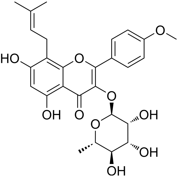
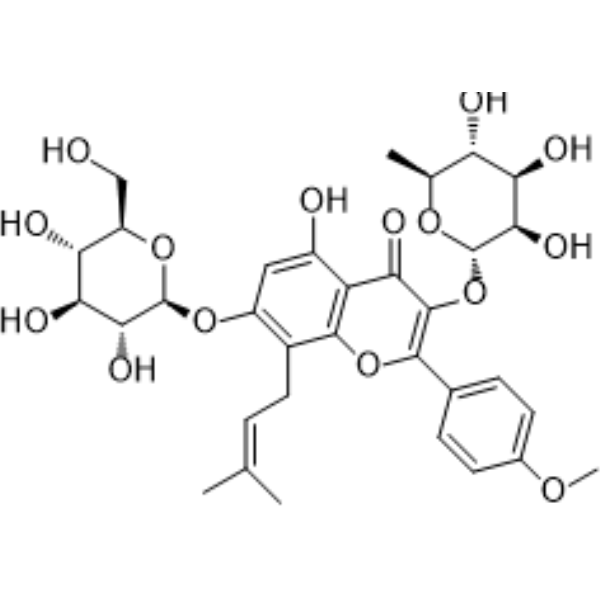

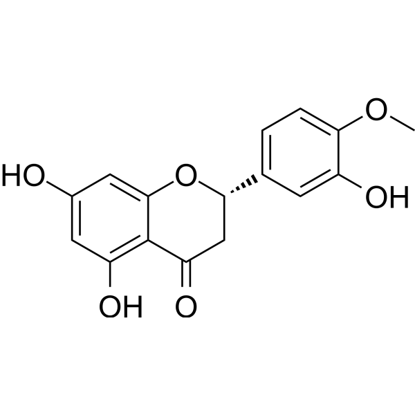
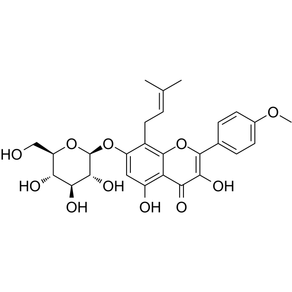
Publications Citing Use of MedChemExpress (MCE) Icariside I
MoreAll Caspase Isoforms
More

Biological Activity
제품 설명
IC50 & Target
[1]|
NLRP3 |
Cellular Effect
|
Cell Line

|
Type | Value | Description | References |
|---|---|---|---|---|
| NCI/ADR-RES | IC50 |
60.78 μM
Compound: 4
|
Cytotoxicity against human MCF7/ADR cells after 72 hrs by MTT assay
Cytotoxicity against human MCF7/ADR cells after 72 hrs by MTT assay
|
[PMID: 19523827] |
In Vitro
Icariside I (10-40 μM, 1 h) enhances NLRP3 inflammasome activation dependent on mitochondrial ROS production triggered by ATP (HY-B2176) and Nigericin (HY-127019), but not SiO2, Poly(I:C) (HY-107202) and cytosolic LPS (HY-D1056), has no effect on NLRC4 and AIM2 inflammasomes activation in BMDMs[1].
Icariside I (20 μM, 1 h) promotes ATP or Nigericin-induced ASC oligomerization but has no effect on K+ efflux in BMDMs[1].
Icariside I (0-100 μM, 12 h) does not impair the viability of 4T1 cells at a concentration lower than 60 μM[2].
Icariside I (0-40 μM, 1 h) inhibits IL-6/STAT3 signaling in 4T1-luc cells[2].
Icariside I (0-40 μM, 1-5 days) suppresses proliferation and migration, reduces the mRNA level of MMP2 and MMP9 in 4T1-luc cells[2].
Icariside I (0-40 μM, 1 h) can hinder the cell cycle and reduces Cyclin D1, CDK4, bcl-2, and increases bax mRNA expression to regulate proliferation and survival of 4T1-luc cells[2].
Icariside I (5-20 μM, 24 h) inhibits Kyn-AhR pathway as well as reduction of tumor cell viability in B16F10-cells[3].
Icariside I (0.1-100 nM) represses osteoclast differentiation and resorption by suppressing MAPK-p38-NFATc1 cascade in primary BMMs[4].
Icariside I (0.1-1000 nM, 3-14 days) promotes differentiation and formation of osteoblasts, upregulated downstream signal factors such as RUNX2 in primary BMMs[4].
MedChemExpress (MCE) has not independently confirmed the accuracy of these methods. They are for reference only.
-
Cell Line:BMDMs
-
Concentration:20 μM
-
Incubation Time:1 h
-
Result:Promoted caspase-1 activation and IL-1β production, was not affected the expression of NLRP3, ASC, pro-IL-1β, and pro-caspase-1 (p45) induced by ATP in LPS-primed.
Had no effect on caspase-1 cleavage, IL-1β secretion and LDH release triggered by SiO2 and poly.
Did not alter caspase-1 cleavage, IL-1β secretion, and LDH release in Pam3CSK4 (HY-P1180)-primed BMDMs transfected with LPS, did not affect the expression of pro-IL-1β, ASC, NLRP3 and pro-caspase-1.
Did not alter the release of caspase-1 cleavage, IL-1β secretion, LDH and ASC oligomerization release in response to Salmonella typhimurium infection and triggered by Poly (dA:dT) (HY-138646) transfection, was not affected the expression of NLRP3, ASC, pro-IL-1β and pro-caspase-1 (p45) in cell lysate.
Promoted ATP-induced ASC oligomerization and the production of caspase-1 and IL-1β induced by Nigericin but not SiO2, poly(I:C) and intracellular LPS in LPS-primed.
-
Cell Line:4T1-luc cells
-
Concentration:0 μM, 10 μM, 20 μM, 40 μM
-
Incubation Time:1 h
-
Result:Reduced the phosphorylation of STAT3 induced by IL-6, decreased p-STAT3 (Tyr705) exposed to IL-6 (50 ng/mL) after 12 h. Decreased the protein level of vimentin in the presence of IL-6.
-
Cell Line:4T1-luc cells
-
Concentration:0 μM, 10 μM, 20 μM, 40 μM
-
Incubation Time:1 days, 5 days
-
Result:Inhibited IL-6-induced wound healing, the development of colonies.
-
Cell Line:4T1-luc cells
-
Concentration:0 μM, 10 μM, 20 μM, 40 μM
-
Incubation Time:1 h
-
Result:Entered a viable G1 arrest state, decreased IL-6-induced the proliferative phase of the cell cycle (S+G2/M), prevented cells from entering the S phase.
-
Cell Line:BMMs
-
Concentration:100 nM
-
Incubation Time:0 min, 5 min, 15 min, 30 min
-
Result:Reduced the level of phosphorylated p38 (p-p38) protein, had no change in JNK, P-JNK, ERK 1/2, and P-ERK 1/2 protein levels.
In Vivo
Icariside I (25-50 mg/kg, p.o., once a days, 27 days) suppresses tumor growth and lung metastasis in the 4T1 breast cancer model through IL-6/STAT3 pathway[2].
Icariside I (5-20 mg/kg, p.o., 7 days) downregulates SLC7A8 and PAT4 transporters and AhR, thus inhibiting nuclear PD-1 in CTLs, through activation of JAK1-STAT1 signaling, thus inducing tumor cell apoptosis in tumor-bearing mice[3].
Icariside I (5-50 mg/kg, i.g., 6 days per week, 4 weeks) ameliorates estrogen deficiency-induced osteoporosis without significant hepatotoxicity in ovariectomy (OVX)-induced osteoporosis mouse model[4].
MedChemExpress (MCE) has not independently confirmed the accuracy of these methods. They are for reference only.
-
Animal Model:LPS-mediated susceptible C57BL/6 mice (Female 6-8-week-old) model of IDILI[1]
-
Dosage:25 mg/kg, 50 mg/kg, 100 mg/kg
-
Administration:i.p., once
-
Result:Induced the elevation of ALT and AST serum levels, increased the production of IL-1β and TNF-α production.
Did not alter liver tissue structure Icariside I and LPS alone treatment, led to a trend of hepatocyte focal necrosis and inflammatory cell infiltration in the liver tissue used in combination with LPS.
Facilitated the number of macrophages, leucocytes, macrophages and neutrophils used in combination with LPS.
-
Animal Model:4T1-luc cells (1 × 10⁵) xenografts BALB/c mice (Female, 6 weeks)[2]
-
Dosage:25 mg/kg, 50 mg/kg
-
Administration:p.o., once a days, 27 days
-
Result:Reduced tumor weight and tumor volume, reduced phosphorylated STAT3 levels and IL-6.
Decreased the expressions of vimentin, bcl-2, Cyclin D1, and CDK4, increased the expressions of pro-apoptotic proteins bax and cleaved caspase 3.
Reduced lung metastasis and damage, decreased mRNA expression of the invasive proteins MMP9 and vimentin.
-
Animal Model:Tumor-bearing mice (C57BL/6 female, 18-22 g)[3]
-
Dosage:5 mg/kg, 20 mg/kg
-
Administration:p.o., 7 days
-
Result:Reduced tumor volume and tumor weight without body weight changes, inhibited tumor cell proliferation and promoted cell shrinkage, nuclear condensation and necrosis of mouse tumor cells.
Restored the size and weight of immune organs (thymus and spleen), alleviated thymus atrophy and splenomegaly in tumor-bearing mice, and improved the total T cells, CD4+T cells, and CD8+T cells and their ratios in the peripheral blood of the tumor group, enhanced the proportion of TILs and CD8+ T cells and up-regulated the CD8+/CD4+ ratio in tumor tissues.
Upregulated mRNAs levels of CCL4 and CCL8 and CXCL9 and CXCL10, as well as IFN-γ and GZMB and CD69 and Klrk1.
Rescued the levels of intermediate metabolites including Trp, Kyn, kynurenic acid, xanthurenic acid, 3 H-KYN, 3-HAA, quinolinic acid and picolinic acid involved in Trp-Kyn pathway in plasma, reduced the levels of Kyn, kynurenic acid and xanthurenic acid and ratio of Kyn to Trp, downregulated mRNA levels of multiple genes including Ido1, Kat1, Kat3, Kmo, Kynu and Ahr involved in Kyn-AhR pathway.
Down-regulated the mRNA level of SLC7A8, PAT4 transporters of Kyn, kynurenic acid and xanthurenic acid, inhibited AhR, down-regulated the mRNA level of PD-1, p27.
-
Animal Model:OVX-induced osteoporosis mouse (C57BL/6 12 week-old female) model[4]
-
Dosage:5 mg/kg, 50 mg/kg
-
Administration:i.g., 6 days per week, 4 weeks
-
Result:Improved liver injury, ameliorates estrogen deficiency-induced osteoporosis without significant hepatotoxicity.
- Detection of Apoptosis
- TUNEL staining for apoptotic DNA fragmentation
- Annexin V plus membrane-impermeant dye apoptosis staining
- Research Protocol for Endocrine Diseases
- Apoptosis Solutions
- Cell invasion
- Hepatotoxicity Study
- Breast Cancer Modeling
- Orthotopic Cell-Line Xenograft
- Osteoclast differentiation from monocyte/macrophage precursors
- Pyroptosis Solutions
- Cell Viability Determination by MTT Colorimetric Assay


Chemical Information
-
CAS No. 56725-99-6
-
Appearance Solid
-
분자량 530.52
-
화학식 C27H30O11
-
Color Light yellow to yellow
-
SMILES
O=C1C(O)=C(C2=CC=C(OC)C=C2)OC3=C(C/C=C(C)\C)C(O[C@H]4[C@@H]([C@H]([C@@H]([C@@H](CO)O4)O)O)O)=CC(O)=C13
-
Synonyms
Icarisid I
-
Structure Classification
-
Initial Source
-
선적
Room temperature in continental US; may vary elsewhere.
-
보관
4°C, sealed storage, away from moisture and light
* In solvent : -80°C, 6 months; -20°C, 1 month (sealed storage, away from moisture and light)


Publications (3)
-
Journal Impact Factor
-
Most Recent
-
Phytomedicine
Novel strategies for identification and prevention of idiosyncratic liver injury caused by TCM compatibility: Exemplification by Epimedii Folium and Psoraleae Fructus. [Abstract]2026 Jan 16:153:157841. PMID: 41655541 -
Food Chem
Effects of sun drying combined with baking processes on the flavor quality of Chongqing Tuocha raw tea. [Abstract]2025 Dec 30:497:146992. PMID: 41285060 -
Food Chem
Flavonoid-mediated metabolic underpinning quality variation in red bud-sport pear mutants. [Abstract]2025 Oct 15:489:144992. PMID: 40466530


용액&용해도
In Vitro:
DMSO : 62.5 mg/mL (117.81 mM; Need ultrasonic; Hygroscopic DMSO has a significant impact on the solubility of product, please use newly opened DMSO)
Please refer to the solubility information to select the appropriate solvent. Once prepared, please aliquot and store the solution to prevent product inactivation from repeated freeze-thaw cycles.
Storage method and period of stock solution: -80°C, 6 months; -20°C, 1 month (sealed storage, away from moisture and light). When stored at -80°C, please use it within 6 months. When stored at -20°C, please use it within 1 month.
Please refer to the solubility information to select the appropriate solvent. Once prepared, please aliquot and store the solution to prevent product inactivation from repeated freeze-thaw cycles.
Storage method and period of stock solution: -80°C, 6 months; -20°C, 1 month (sealed storage, away from moisture and light). When stored at -80°C, please use it within 6 months. When stored at -20°C, please use it within 1 month.
Concentration (start) × Volume (start) = Concentration (final) × Volume (final)
In Vivo:
Select the appropriate dissolution method based on your experimental animal and administration route.
- For the following dissolution methods, please ensure to first prepare a clear stock solution using an In Vitro approach and then sequentially add co-solvents:
- To ensure reliable experimental results, the clarified stock solution can be appropriately stored based on storage conditions. As for the working solution for In Vivo experiments, it is recommended to prepare freshly and use it on the same day.
- The percentages shown for the solvents indicate their volumetric ratio in the final prepared solution. If precipitation or phase separation occurs during preparation, heat and/or sonication can be used to aid dissolution.
Add each solvent one by one: 10% DMSO 40% PEG300 5% Tween-80 45% Saline
Solubility: 2.08 mg/mL (3.92 mM); Suspended solution; Need ultrasonic
This protocol yields a suspended solution of 2.08 mg/mL. Suspended solution can be used for oral and intraperitoneal injection.
Taking 1 mL working solution as an example, add 100 μL DMSO stock solution (20.8 mg/mL) to 400 μL PEG300, and mix evenly; then add 50 μL Tween-80 and mix evenly; then add 450 μL Saline to adjust the volume to 1 mL.
Preparation of Saline: Dissolve 0.9 g sodium chloride in ddH₂O and dilute to 100 mL to obtain a clear Saline solution.
Add each solvent one by one: 10% DMSO 90% (20% SBE-β-CD in Saline)
Solubility: 2.08 mg/mL (3.92 mM); Suspended solution; Need ultrasonic
This protocol yields a suspended solution of 2.08 mg/mL. Suspended solution can be used for oral and intraperitoneal injection.
Taking 1 mL working solution as an example, add 100 μL DMSO stock solution (20.8 mg/mL) to 900 μL 20% SBE-β-CD in Saline, and mix evenly.
Preparation of 20% SBE-β-CD in Saline (4°C, storage for one week): 2 g SBE-β-CD powder is dissolved in 10 mL Saline, completely dissolve until clear.
In Vivo Dissolution Calculator
Please enter the basic information of animal experiments:
-
-
-
-
Recommended: Prepare an additional quantity of animals to account for potential losses during experiments.
Please enter your animal formula composition:
-
%DMSO +
Recommended: Keep the proportion of DMSO in working solution below 2% if your animal is weak.
-
%+
-
+%Tween-80 + +
-
%Saline +
The co-solvents required include: DMSO, . All of co-solvents are available by MedChemExpress (MCE). , Tween 80. All of co-solvents are available by MedChemExpress (MCE).
Working solution concentration: 0.22 mg/mL
Method for preparing stock solution: mg drug dissolved in μL DMSO. Stock solution concentration: mg/mL. * In solvent : -80°C, 6 months; -20°C, 1 month (sealed storage, away from moisture and light)
1. Take μL DMSO stock solution;
2. Add μL .
μL , mix evenly;
3. Then add μL Tween 80, mix evenly;
4. Then add μL
Please ensure that the stock solution in the first step is dissolved to a clear state, and add co-solvents in sequence. You can use ultrasonic heating (ultrasonic cleaner, recommended frequency 20-40 kHz), vortexing, etc. to assist dissolution.


순도&문서
-
Data Sheet (290 KB)
-
SDS (396 KB)
- English - EN (396 KB)
- Français - FR (396 KB)
- Deutsch - DE (396 KB)
- Norwegian - NO (396 KB)
- Español - ES (396 KB)
- Swedish - SV (396 KB)
- Italian - IT (396 KB)
- Korean - KR (396 KB)
- Portuguese - PT (396 KB)
-
Handling Instructions (2659 KB)
References
[1]. Gao Y, et al. Icariside I specifically facilitates ATP or nigericin-induced NLRP3 inflammasome activation and causes idiosyncratic hepatotoxicity. Cell Commun Signal. 2021 Feb 11;19(1):13. [Content Brief]
[2]. Hou M, et al. Icariside I reduces breast cancer proliferation, apoptosis, invasion, and metastasis probably through inhibiting IL-6/STAT3 signaling pathway. J Pharm Pharmacol. 2024 May 3;76(5):499-513. [Content Brief]
[3]. Chen G, et al. Icariside I - A novel inhibitor of the kynurenine-AhR pathway with potential for cancer therapy by blocking tumor immune escape. Biomed Pharmacother. 2022 Sep;153:113387. [Content Brief]
[4]. Chen C, et al. A Novel Prenylflavonoid Icariside I Ameliorates Estrogen Deficiency-Induced Osteoporosis via Simultaneous Regulation of Osteoblast and Osteoclast Differentiation. ACS Pharmacol Transl Sci. 2023 Jan 13;6(2):270-280. [Content Brief]


Complete Stock Solution Preparation Table
Please refer to the solubility information to select the appropriate solvent. Once prepared, please aliquot and store the solution to prevent product inactivation from repeated freeze-thaw cycles.
Storage method and period of stock solution: -80°C, 6 months; -20°C, 1 month (sealed storage, away from moisture and light). When stored at -80°C, please use it within 6 months. When stored at -20°C, please use it within 1 month.
| Optional Solvent | Concentration Solvent Mass | 1 mg | 5 mg | 10 mg | 25 mg |
|---|---|---|---|---|---|
| DMSO | 1 mM | 1.8849 mL | 9.4247 mL | 18.8494 mL | 47.1236 mL |
| 5 mM | 0.3770 mL | 1.8849 mL | 3.7699 mL | 9.4247 mL | |
| 10 mM | 0.1885 mL | 0.9425 mL | 1.8849 mL | 4.7124 mL | |
| 15 mM | 0.1257 mL | 0.6283 mL | 1.2566 mL | 3.1416 mL | |
| 20 mM | 0.0942 mL | 0.4712 mL | 0.9425 mL | 2.3562 mL | |
| 25 mM | 0.0754 mL | 0.3770 mL | 0.7540 mL | 1.8849 mL | |
| 30 mM | 0.0628 mL | 0.3142 mL | 0.6283 mL | 1.5708 mL | |
| 40 mM | 0.0471 mL | 0.2356 mL | 0.4712 mL | 1.1781 mL | |
| 50 mM | 0.0377 mL | 0.1885 mL | 0.3770 mL | 0.9425 mL | |
| 60 mM | 0.0314 mL | 0.1571 mL | 0.3142 mL | 0.7854 mL | |
| 80 mM | 0.0236 mL | 0.1178 mL | 0.2356 mL | 0.5890 mL | |
| 100 mM | 0.0188 mL | 0.0942 mL | 0.1885 mL | 0.4712 mL |


Icariside I Related Classifications
HY-N1939 Related Classifications
 Powered by Bioz