



Plantainoside D
Based on 2 publication(s) in Google Scholar
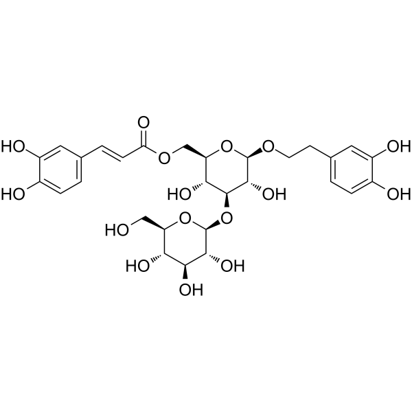
Plantainoside D, a phenylethanoid glycosides, is a IKK-β inhibitor with diverse biological activities. Plantainoside D shows inhibitory activity of angiotensin-converting enzyme (ACE) with an IC50 of 2.17 mM. Plantainoside D significantly reduces the release of glutamate from nerve terminals in the cerebral cortex of rats by inhibiting the voltage-dependent calcium channel (VDCCs) and protein kinase C (PKC) signaling cascade. Plantainoside D significantly alleviates cell apoptosis by inhibiting the generation of ROS and the activation of NF-κB. Plantainoside D significantly improves acute lung injury (ALI) induced by sepsis by regulating the Sirt3/NLRP3 signaling pathway. Plantainoside D can be used for the study of neuroprotection, antioxidant, anti-inflammation, antihypertension.
Nur für Forschungszwecke. Wir verkaufen nicht an Patienten.
- Reinheit : 99.15%
- CAS. Nr.: 147331-98-4
- Formel: C29H36O16
- Molecular Weight:640.59
-
Speicherung:
4°C, protect from light
* In solvent : -80°C, 6 months; -20°C, 1 month (protect from light)

To place orders, for customer services and technical support, please contact: MedChemExpress USA
Tel: 609-228-6898 E-mail: [email protected] [email protected]


-
 Biologische Aktivität
-
Chemical Information
-
Lösungsmittel & Löslichkeit
- Protokoll
- Reinheit & Dokumentation
- Verweise
-
 Help & FAQs


-
HY-B0627
MetforminAntihyperglycemic Agent -
HY-N0029
Forsythoside BTNF Inhibitor -
HY-N0031
PlantamajosideNF-κB Inhibitor -
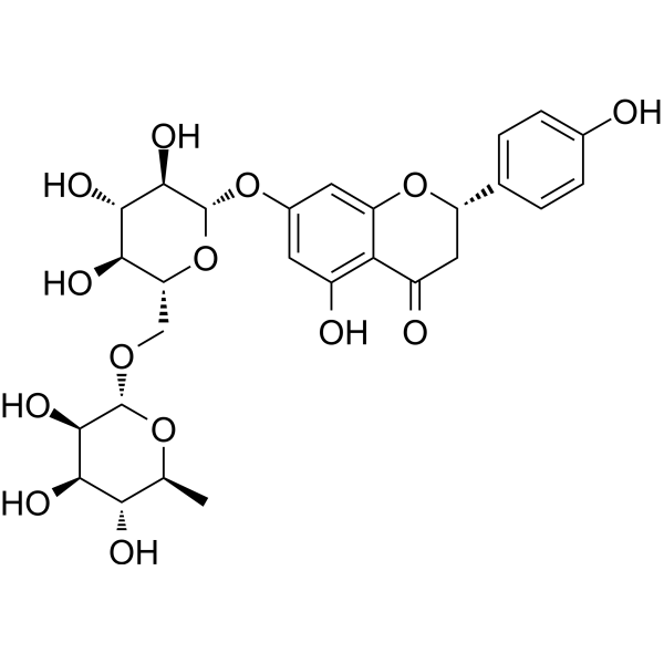
HY-N0804
NarirutinShikimate Kinase Inhibitor -
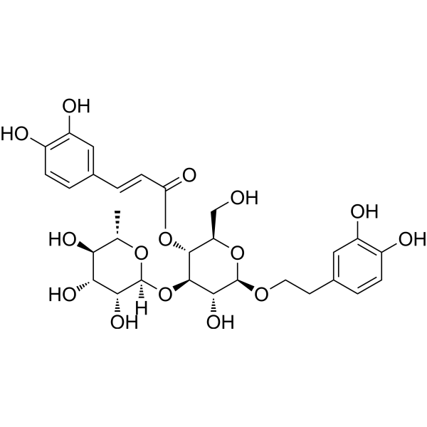
HY-N0021
VerbascosideBacterial Inhibitor -
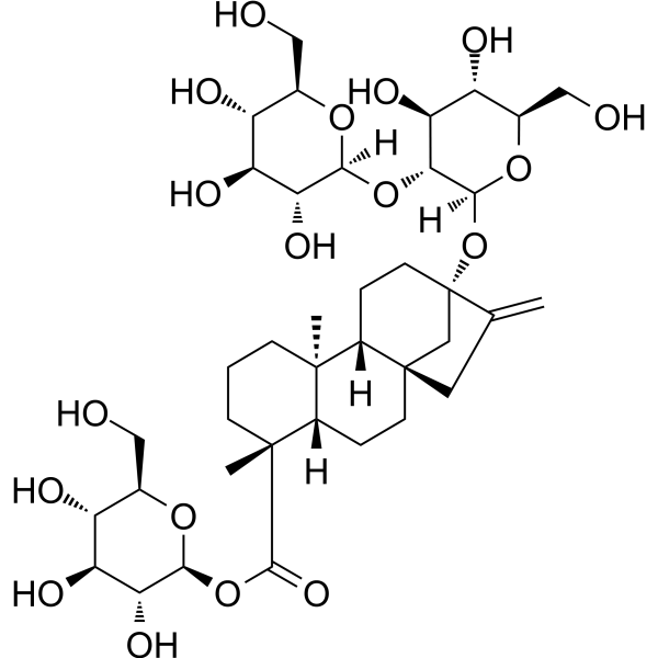
HY-N0669
Steviosidesweetener -
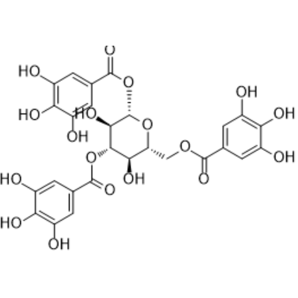
HY-N6006
1,3,6-Tri-O-galloyl-beta-D-glucosePhenolic Compound -
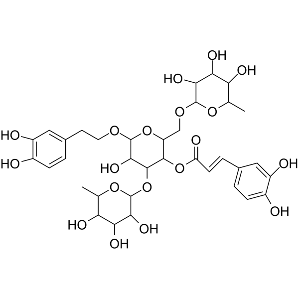
HY-N0033
PoliumosideAldose Reductase Inhibitor -
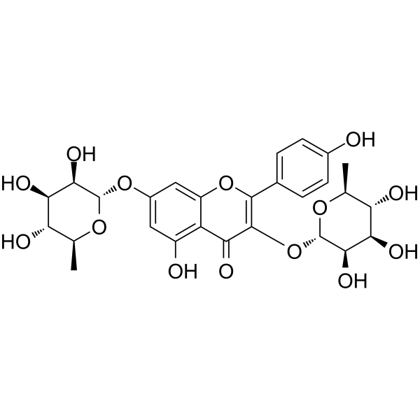
HY-N0628
KaempferitrinInsulin Receptor Activator -
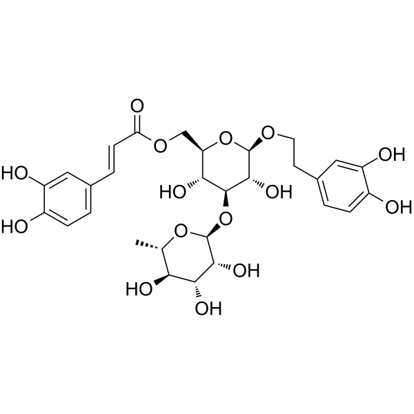
HY-N0022
IsoacteosideNatural Product -
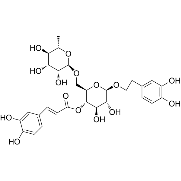
HY-N0028
Forsythiaside AAnti-Inflammatory Agent -
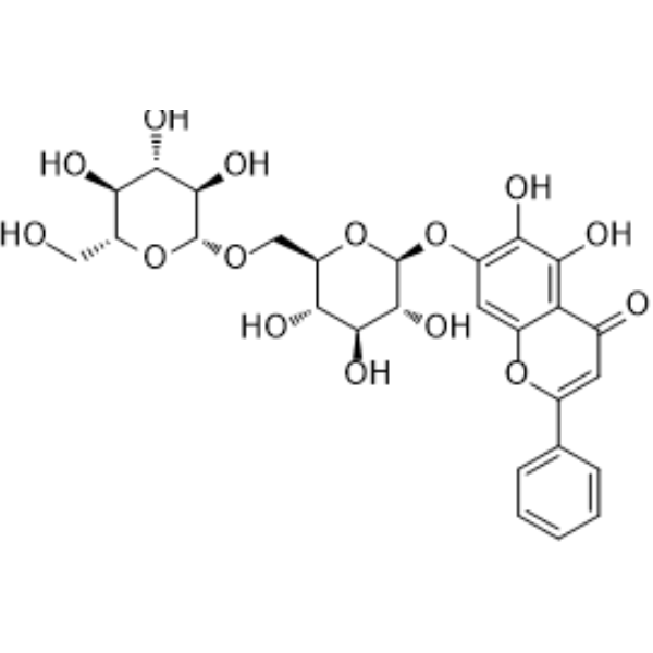
HY-N1435
Oroxin BAntitumor Agent
Publications Citing Use of MedChemExpress (MCE) Plantainoside D
More Customer Validation & Images

-
IP
-
In Vivo Efficacy Study
-
Histological Imaging/Staining
-
IF
-
ELISA
Alle Calcium Channel Isoform-spezifische Produkte anzeigen
More

Biologische Aktivität
Beschreibung
|
IKK-β |
Calcium Channel |
NF-κB |
NLRP3 |
ACE 2.17 mM (IC50) |
PKC |
In Vitro
Plantainoside D (0-50 μM, 10 min) significantly inhibits the release of glutamate induced by 4-aminopyridine (4-AP) (HY-B0604) via vesicle exocytosis pathway with an IC50 of 32 μM in rat synaptosome[1].
Plantainoside D (30 μM, 10 min) reduces the Ca2+ influx induced by 4-AP through N-type calcium channels, and does not alter the membrane potential in rat synaptosome[1].
Plantainoside D (30 μM, 20 min) acts through PKC-α/SNAP-25 phosphorylation as its key downstream mechanism in rat synaptosome[1].
Plantainoside D (5 μM, 24 h) significantly improves sepsis induced ALI by regulation of Sirt3/NLRP3 pathway in MLE-12 cells[2].
Plantainoside D (1-20 μg/mL, 1-4 h) significantly reduces the apoptosis induced by Doxorubicin (ADR) (HY-15142A) by inhibiting the generation of ROS and the activation of NF-κB[3].
Plantainoside D exhibits weak inhibitory effects of CYP1A2 (IC50 = 12.83 μM), CYP2D6 (IC50 = 8.39 μM) and CYP3A4 (IC50 = 14.66 μM)[4].
MedChemExpress (MCE) has not independently confirmed the accuracy of these methods. They are for reference only. Further protocols information, click here.
-
Cell Line:Rat cerebral cortical synaptosomes
-
Concentration:30 μM
-
Incubation Time:20 min
-
Result:Reduced the phosphorylation of PKC-α (from 239.2% to 54.1%) and SNAP-25 (from 228.9% to 89.7%) induced by 4-AP.
-
Cell Line:MLE-12 cells induced by LPS (HY-D1056)
-
Concentration:5 μM
-
Incubation Time:24 h
-
Result:Increased cell vitality, reduced ROS and MDA.
-
Cell Line:H9c2 cell with ADR
-
Concentration:1, 5, 10, 20 μg/mL
-
Incubation Time:1 h
-
Result:Reduced the cell mortality rate with 20 μg/mL pretreatment.
Reduced apoptotic morphology (nuclear enrichment, fragmentation).
Increased Bcl-2/Bax ratio and inhibited caspase-3.
-
Cell Line:H9c2 cell with ADR
-
Concentration:2 μM
-
Incubation Time:15 min, 30 min, 1h, 2h, 4h
-
Result:Abrogated the activation of NF-κB.
The glycoside reduced the degradation of IkBα in ADR-treated cells.
In Vivo
MedChemExpress (MCE) has not independently confirmed the accuracy of these methods. They are for reference only.
-
Animal Model:LPS-induced ALI model established in ICR WT male mice (6 weeks, 16-18 g)[2]
-
Dosage:50 mg/kg
-
Administration:Intraperitoneal injection (i.p.), once daily for 7 days
-
Result:Significantly decreased the levels of w/d, ROS and MPO.
Significantly decreased MDA and increased SOD and GSH-px in serum and lung tissues.
Significantly restored oxidative stress and neutrophil infiltration.
Significantly decreased the levels of cytokines TNF-α, IL-1β, IL-6, NLRP3, ASC and caspase-1.


Chemical Information
-
CAS. Nr. 147331-98-4
-
Appearance Solid
-
Molecular Weight 640.59
-
Formel C29H36O16
-
Color White to light yellow
-
SMILES
O[C@H]([C@H]([C@@H]([C@@H](COC(/C=C/C1=CC=C(O)C(O)=C1)=O)O2)O)O[C@H]3[C@@H]([C@H]([C@@H]([C@@H](CO)O3)O)O)O)[C@@H]2OCCC4=CC=C(O)C(O)=C4
-
Structure Classification
-
Initial Source
-
Versand
Room temperature in continental US; may vary elsewhere.
-
Speicherung
4°C, protect from light
* In solvent : -80°C, 6 months; -20°C, 1 month (protect from light)


Publications (2)
-
Journal Impact Factor
-
Most Recent
-
Redox Biol
Extracellular vesicle-packaged GBP2 from macrophages aggravates sepsis-induced acute lung injury by promoting ferroptosis in pulmonary vascular endothelial cells. [Abstract]2025 May:82:103614. PMID: 40156957
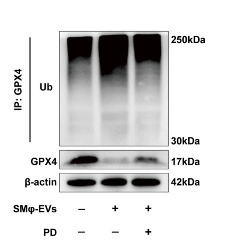
Plantainoside D purchased from MedChemExpress. Usage Cited in: Redox Biol. 2025 May:82:103614. [Abstract]
Representative images of immunoblotting of GPX4 protein and its ubiquitination levels in HPMECs co-cultured with SMφ-EVs, with or without Plantainoside D (PD) (10 μM) treatment.
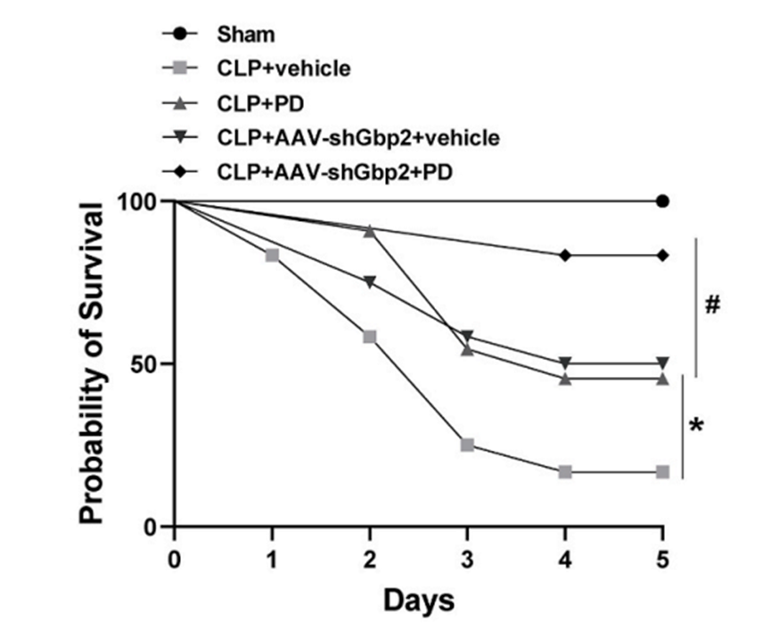
Plantainoside D purchased from MedChemExpress. Usage Cited in: Redox Biol. 2025 May:82:103614. [Abstract]
Survival rate of mice treated with or without CLP following administration of AAV-shGbp2 or Plantainoside D PD (30 mg/kg, i.v.) treatment.
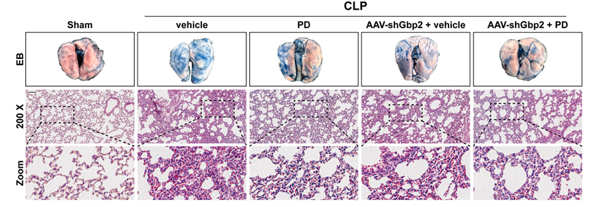
Plantainoside D purchased from MedChemExpress. Usage Cited in: Redox Biol. 2025 May:82:103614. [Abstract]
Representative images of Evans blue staining and HE staining of mouse lungs treated with Plantainoside D PD (30 mg/kg, i.v.).
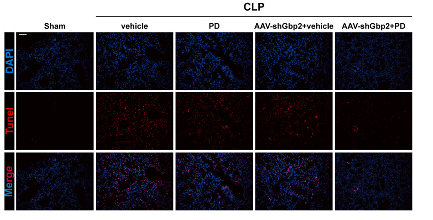
Plantainoside D purchased from MedChemExpress. Usage Cited in: Redox Biol. 2025 May:82:103614. [Abstract]
Representative images and quantification of Tunel staining of mouse lungs treated with Plantainoside D PD (30 mg/kg, i.v.).
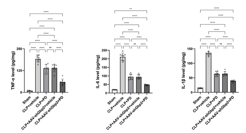
Plantainoside D purchased from MedChemExpress. Usage Cited in: Redox Biol. 2025 May:82:103614. [Abstract]
Quantification of IL-6, TNF-α and IL-1β in mouse lungs treated with Plantainoside D PD (30 mg/kg, i.v.).
-
Biochem Pharmacol
KCMF1 regulates HRI ubiquitination to inhibit the integrated stress response in ovarian cancer. [Abstract]2026 Mar:245:117634. PMID: 41391693


Lösungsmittel & Löslichkeit
In Vitro:
DMSO : 250 mg/mL (390.27 mM; Need ultrasonic; Hygroscopic DMSO has a significant impact on the solubility of product, please use newly opened DMSO)
Please refer to the solubility information to select the appropriate solvent. Once prepared, please aliquot and store the solution to prevent product inactivation from repeated freeze-thaw cycles.
Storage method and period of stock solution: -80°C, 6 months; -20°C, 1 month (protect from light). When stored at -80°C, please use it within 6 months. When stored at -20°C, please use it within 1 month.
Please refer to the solubility information to select the appropriate solvent. Once prepared, please aliquot and store the solution to prevent product inactivation from repeated freeze-thaw cycles.
Storage method and period of stock solution: -80°C, 6 months; -20°C, 1 month (protect from light). When stored at -80°C, please use it within 6 months. When stored at -20°C, please use it within 1 month.
Konzentration (Stammlösung) × Volumen (Stammlösung) = Konzentration (Ziellösung) × Volumen (Ziellösung)
In Vivo:
Select the appropriate dissolution method based on your experimental animal and administration route.
- For the following dissolution methods, please ensure to first prepare a clear stock solution using an In Vitro approach and then sequentially add co-solvents:
- To ensure reliable experimental results, the clarified stock solution can be appropriately stored based on storage conditions. As for the working solution for In Vivo experiments, it is recommended to prepare freshly and use it on the same day.
- The percentages shown for the solvents indicate their volumetric ratio in the final prepared solution. If precipitation or phase separation occurs during preparation, heat and/or sonication can be used to aid dissolution.
Add each solvent one by one: 10% DMSO 90% (20% SBE-β-CD in Saline)
Solubility: ≥ 2.08 mg/mL (3.25 mM); Clear solution
This protocol yields a clear solution of ≥ 2.08 mg/mL (saturation unknown).
Taking 1 mL working solution as an example, add 100 μL DMSO stock solution (20.8 mg/mL) to 900 μL 20% SBE-β-CD in Saline, and mix evenly.
Preparation of 20% SBE-β-CD in Saline (4°C, storage for one week): 2 g SBE-β-CD powder is dissolved in 10 mL Saline, completely dissolve until clear.
In Vivo Dissolution Calculator
Please enter the basic information of animal experiments:
-
-
-
-
Recommended: Prepare an additional quantity of animals to account for potential losses during experiments.
Please enter your animal formula composition:
-
%DMSO +
Recommended: Keep the proportion of DMSO in working solution below 2% if your animal is weak.
-
%+
-
+%Tween-80 + +
-
%Saline +
The co-solvents required include: DMSO, . All of co-solvents are available by MedChemExpress (MCE). , Tween 80. All of co-solvents are available by MedChemExpress (MCE).
Working solution concentration: 0.22 mg/mL
Method for preparing stock solution: mg drug dissolved in μL DMSO. Stock solution concentration: mg/mL. * In solvent : -80°C, 6 months; -20°C, 1 month (protect from light)
1. Take μL DMSO stock solution;
2. Add μL .
μL , mix evenly;
3. Then add μL Tween 80, mix evenly;
4. Then add μL
Please ensure that the stock solution in the first step is dissolved to a clear state, and add co-solvents in sequence. You can use ultrasonic heating (ultrasonic cleaner, recommended frequency 20-40 kHz), vortexing, etc. to assist dissolution.

Protokoll
-
Apoptosis
Apoptosis, also called programmed cell death, is generally characterized by distinct morphological characteristics.
-
TUNEL staining for apoptotic DNA fragmentation
TUNEL staining detects DNA strand breaks by using terminal deoxynucleotidyl transferase to add labeled nucleotides to exposed 3′-OH DNA termini, generating either microscopic staining in fixed cells or tissue sections, or fluorescence/cytometric signal in cell suspensions. TUNEL positivity reflects DNA fragmentation but should not be interpreted alone as definitive apoptosis, because TUNEL can also label necrotic, autolytic, mechanically damaged, or DNA-repair-associated DNA breaks.
-
Cell Cytotoxicity Assay
Cytotoxicity assays are usually based on the assessment of cell membrane damage, which can also be indirectly detected by measuring cell viability. Detection methods include MTT assay, CKK-8 assay, LDH assay and ATP assay, etc.
-
LPS-Induced Endotoxemia/Systemic Inflammation
Lipopolysaccharide (LPS)-induced endotoxemia is a widely used in vivo model of acute systemic inflammation in which LPS, a Gram-negative bacterial endotoxin, activates innate immune signaling primarily through TLR4, leading to rapid and transient induction of pro-inflammatory cytokines such as TNF-α, IL-6, and IL-1β in circulation and tissues. This cytokine surge is commonly used as a measurable readout of systemic inflammatory activation and immune dysregulation, and is typically assessed within hours after intraperitoneal LPS administration in mouse models of endotoxemia. The model captures key features of systemic inflammatory response syndrome, including cytokine release, immune cell activation, and downstream tissue responses, and has been used to evaluate anti-inflammatory interventions such as cytokine modulation, lipid mediators, and immune cell-targeting therapies.
-
Annexin V plus membrane-impermeant dye apoptosis staining
Annexin V-based apoptosis assays rely on the detection of phosphatidylserine (PS) externalization from the inner leaflet of the plasma membrane to the outer leaflet, an early biochemical hallmark of apoptosis. Fluorescently labeled Annexin V binds PS in a calcium-dependent manner, enabling identification of early apoptotic cells by flow cytometry or fluorescence microscopy. When combined with a membrane-impermeant DNA-binding dye (e. g. , propidium iodide), this approach allows discrimination between viable (Annexin V−/dye−), early apoptotic (Annexin V+/dye−), and late apoptotic or necrotic (Annexin V+/dye+) cell populations by assessing membrane integrity and PS exposure.
-
ROS/oxidative-stress fluorescent staining
ROS/oxidative-stress fluorescent staining uses cell-permeant fluorogenic probes that become fluorescent after oxidation inside cells or tissues; commonly used examples include DCFH-DA/DCFDA for broad cellular oxidant detection, DHE for superoxide-related signal detection, MitoSOX for mitochondrial superoxide-related signal detection, and CellROX probes for oxidative-stress-associated fluorescence readouts. The assay detects probe oxidation rather than a single ROS species unless the probe and analysis method have been chemically validated for that species. DCFH-DA enters cells, is deacetylated by intracellular esterases to DCFH, and produces fluorescent DCF after oxidation, so the readout is used as an operational measure of total cellular oxidative stress rather than a species-specific ROS measurement. DHE and MitoSOX can report superoxide-related oxidation, but red fluorescence alone can include non-specific ethidium-like oxidation products; HPLC or optimized spectral approaches are
-
Apoptosis Solutions
Apoptosis is a regulated, generally non-lytic cell-death pathway that removes unwanted, damaged, infected, or abnormal cells through coordinated morphological changes, caspase activation, DNA fragmentation, and membrane remodeling. The intrinsic apoptosis pathway is controlled mainly by mitochondrial outer membrane permeabilization, BCL-2 family proteins, cytochrome c release, apoptosome formation, caspase-9 activation, and downstream executioner caspase-3/7 activation. The extrinsic apoptosis pathway is initiated by death receptors such as Fas, TNFR, and TRAIL receptors, which recruit adaptor proteins and activate caspase-8 before engaging executioner caspases or mitochondrial amplification through BID cleavage. Apoptosis is linked to many phenotypes, including cancer cell killing, tissue homeostasis, immune regulation, neurodegeneration, infection response, and treatment-induced cytotoxicity; unresolved questions include how apoptosis interacts with necroptosis, pyroptosis, ferroptos
-
Research Protocol for Inflammation-related Diseases
The NLRP3 inflammasome is a cytosolic innate immune signaling platform that integrates priming signals and danger-signal activation to promote caspase-1 activation, maturation of IL-1β and IL-18, and gasdermin D-mediated pyroptotic cell death. The core experimental logic is to determine whether inflammatory disease phenotypes are driven by increased NLRP3 expression, ASC-containing inflammasome assembly, caspase-1 cleavage, GSDMD cleavage, and extracellular release of IL-1β/IL-18 rather than by nonspecific cell injury alone. The pathway is strongly linked to inflammation-related disease phenotypes because monosodium urate crystals activate NALP3/NLRP3 inflammasome signaling in gout-like crystal inflammation, cholesterol crystals activate NLRP3 inflammasomes in atherogenesis models, and DSS-induced intestinal inflammation has been reported to involve NLRP3 inflammasome activity. However, experimental colitis studies also show context-dependent protective effects of NLRP3 inflammasome co
-
Inhalation Toxicity Study
Inhalation toxicity studies expose rodents to a controlled aerosol, vapor, gas, or smoke atmosphere and assess respiratory and systemic toxicity using exposure-atmosphere characterization, clinical observations, body and organ weights, bronchoalveolar lavage fluid, histopathology, blood chemistry, hematology, and, when included, molecular endpoints such as transcriptomics, proteomics, lipidomics, or tissue burden analysis. The primary biological readouts are airway irritation, pulmonary inflammation, cytotoxicity, altered surfactant or lipid homeostasis, impaired particle clearance, and tissue remodeling, reflected by BALF cell differentials, BALF protein, LDH, phosphatase activities, cytokines, lung weight, microscopic respiratory-tract lesions, and retained lung burden.


Reinheit & Dokumentation
-
Data Sheet (283 KB)
-
SDS (252 KB)
- English - EN (252 KB)
- Français - FR (252 KB)
- Deutsch - DE (252 KB)
- Norwegian - NO (252 KB)
- Español - ES (252 KB)
- Swedish - SV (252 KB)
- Italian - IT (252 KB)
- Korean - KR (252 KB)
- Portuguese - PT (252 KB)
-
Handling Instructions (2659 KB)
Verweise
[1]. Chiu KM, et al. Plantainoside D Reduces Depolarization-Evoked Glutamate Release from Rat Cerebral Cortical Synaptosomes. Molecules. 2023 Jan 30;28(3):1313. [Content Brief]
[2]. Wang J, et al. Sirt3 regulates NLRP3 and participates in the effects of plantainoside D on acute lung injury sepsis. Aging (Albany NY). 2023 Apr 5;15(14):6710-6720. [Content Brief]
[3]. Kim DS, et al. Plantainoside D protects adriamycin-induced apoptosis in H9c2 cardiac muscle cells via the inhibition of ROS generation and NF-kappaB activation. Life Sci. 2007 Jan 2;80(4):314-23. [Content Brief]
[4]. Zhou J, et al. Human liver microsomes study on the inhibitory effect of plantainoside D on the activity of cytochrome P450 activity. BMC Complement Med Ther. 2022 Jul 23;22(1):197. [Content Brief]
[5]. Geng F, Yang L, Chou G, Wang Z. Bioguided isolation of angiotensin-converting enzyme inhibitors from the seeds of Plantago asiatica L. Phytother Res. 2010;24(7):1088-1094. [Content Brief]
[6]. Zhou P, Hua F, Wang X, Huang JL. Therapeutic potential of IKK-β inhibitors from natural phenolics for inflammation in cardiovascular diseases. Inflammopharmacology. 2020;28(1):19-37. [Content Brief]


Complete Stock Solution Preparation Table
Please refer to the solubility information to select the appropriate solvent. Once prepared, please aliquot and store the solution to prevent product inactivation from repeated freeze-thaw cycles.
Storage method and period of stock solution: -80°C, 6 months; -20°C, 1 month (protect from light). When stored at -80°C, please use it within 6 months. When stored at -20°C, please use it within 1 month.
| Optional Solvent | Concentration Solvent Mass | 1 mg | 5 mg | 10 mg | 25 mg |
|---|---|---|---|---|---|
| DMSO | 1 mM | 1.5611 mL | 7.8053 mL | 15.6106 mL | 39.0265 mL |
| 5 mM | 0.3122 mL | 1.5611 mL | 3.1221 mL | 7.8053 mL | |
| 10 mM | 0.1561 mL | 0.7805 mL | 1.5611 mL | 3.9027 mL | |
| 15 mM | 0.1041 mL | 0.5204 mL | 1.0407 mL | 2.6018 mL | |
| 20 mM | 0.0781 mL | 0.3903 mL | 0.7805 mL | 1.9513 mL | |
| 25 mM | 0.0624 mL | 0.3122 mL | 0.6244 mL | 1.5611 mL | |
| 30 mM | 0.0520 mL | 0.2602 mL | 0.5204 mL | 1.3009 mL | |
| 40 mM | 0.0390 mL | 0.1951 mL | 0.3903 mL | 0.9757 mL | |
| 50 mM | 0.0312 mL | 0.1561 mL | 0.3122 mL | 0.7805 mL | |
| 60 mM | 0.0260 mL | 0.1301 mL | 0.2602 mL | 0.6504 mL | |
| 80 mM | 0.0195 mL | 0.0976 mL | 0.1951 mL | 0.4878 mL | |
| 100 mM | 0.0156 mL | 0.0781 mL | 0.1561 mL | 0.3903 mL |


Plantainoside D Related Classifications
HY-N5063 Related Classifications
 Powered by Bioz