



MELK-8a
Based on 3 publication(s) in Google Scholar
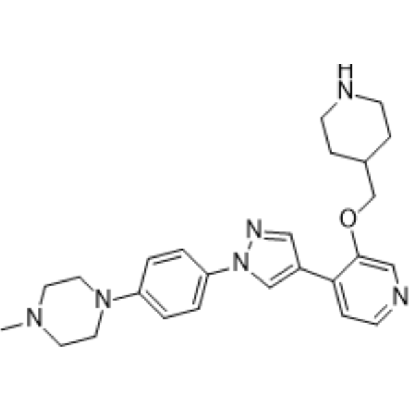
MELK-8a (NVS-MELK8a) is an ATP-competitive, selective MELK inhibitor with an IC50 value of 0.002 μM. MELK-8a inhibits NF-κB signaling. MELK-8a induces G2/M checkpoint activation, delayed mitotic entry, G2 phase prolongation, and delayed activation of Aurora A/B and CDK1. MELK-8a inhibits melanoma. MELK-8a can be used for research on triple-negative breast cancer.
For research use only. We do not sell to patients.
- Purity : 98.32%
- CAS No.: 1922153-17-0
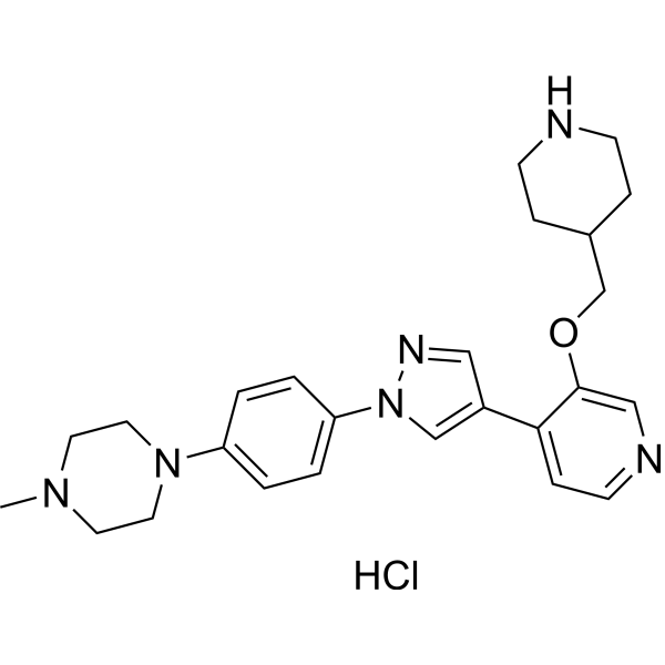
- Formula: C25H32N6O
- Molecular Weight:432.56
-
Storage:Powder -20°C, 3 years , 4°C, 2 years ; In solvent -80°C, 6 months , -20°C, 1 month

To place orders, for customer services and technical support, please contact: MedChemExpress USA
Tel: 609-228-6898 E-mail: [email protected] [email protected]


-
 Biological Activity
-
Chemical Information
-
Solvent & Solubility
- Protocols
- Purity & Documentation
- References
-
 Help & FAQs



Publications Citing Use of MedChemExpress (MCE) MELK-8a
More Customer Validation & Images

-
WB

-
WB

-
WB

-
WB

-
WB

All Aurora Kinase Isoforms
More

Biological Activity
Description
IC50 & Target
[1]|
CDK1 |
Aurora A |
Aurora B |
Cellular Effect
|
Cell Line

|
Type | Value | Description | References |
|---|---|---|---|---|
| MDA-MB-468 | IC50 |
0.11 μM
|
Antiproliferative activity against human MDA-MB-468 cells assessed as viability reduction after 7-day incubation by ATP-based luminescence assay.
Antiproliferative activity against human MDA-MB-468 cells assessed as viability reduction after 7-day incubation by ATP-based luminescence assay.
|
27187609 |
| MCF7 | IC50 |
3.68 μM
|
Antiproliferative activity against human MCF-7 cells assessed as viability reduction after 7-day incubation by ATP-based luminescence assay.
Antiproliferative activity against human MCF-7 cells assessed as viability reduction after 7-day incubation by ATP-based luminescence assay.
|
27187609 |
| MDA-MB-231 | IC50 |
1.7 μM
|
Reduction of viability of human MDA-MB-231 cells after 72 hrs exposure assessed by resazurin reduction fluorometrically.
Reduction of viability of human MDA-MB-231 cells after 72 hrs exposure assessed by resazurin reduction fluorometrically.
|
31896573 |
| MDA-MB-468 | IC50 |
2.3 μM
|
Reduction of viability of human MDA-MB-468 cells after 72 hrs exposure assessed by resazurin reduction fluorometrically.
Reduction of viability of human MDA-MB-468 cells after 72 hrs exposure assessed by resazurin reduction fluorometrically.
|
31896573 |
| HeLa S3 | IC50 |
1.9 μM
|
Reduction of viability of human HeLaS3 cells after 72 hrs exposure assessed by resazurin reduction fluorometrically.
Reduction of viability of human HeLaS3 cells after 72 hrs exposure assessed by resazurin reduction fluorometrically.
|
31896573 |
In Vitro
MELK-8a is highly selective for MELK in MDA-MB-468 cells, with MELK being the only kinase significantly displaced at 1 µM, nearly complete loss of MELK binding at 3 µM, and broad off-target effects occurring only at 10 µM[2].
MELK-8a (1 nM-30 µM; 72 h) reduces the viability of MDA-MB-231 (IC50 = 1.7 µM), MDA-MB-468 (IC50 = 2.3 µM), and HeLaS3 (IC50 = 1.9 µM) cells after 72 h[2].
MELK-8a (0.1-10 µM; 8-14 days) reduces long-term colony formation of MDA-MB-231 and MDA-MB-468 cells, with an IC50 between 1 and 3 µM[2].
NVS-MELK8a reduces the viability of TNBC cells at concentrations at which it is highly selective for MELK[3].
MELK-8a selectively inhibits the proliferation of MELK-dependent TNBC MDA-MB-468 cells, with minimal effect on MELK-independent cells[4].
MELK-8a attenuates NF-kB signaling by disrupting MELK-mediated SQSTM1/p62 phosphorylation and inhibits melanoma cell proliferation[4].
MELK-8a (0.5-3 µM; 1-24 h) induces a dose-dependent decrease in p-H3 (Ser-10) in HeLaS3 cells without PARP cleavage or changes in cyclin[2].
MELK-8a delays the activation of Aurora A/B and CDK1 and mitotic entry by approximately 2.5-3 h in synchronized HeLaS3 cells[2].
MELK-8a delays mitotic entry, but synchronized HeLaS3 cells complete M phase and re-enter the cell cycle with only about a 2 h delay[2].
MELK-8a dose-dependently prolongs G2 phase in U2OS cells (median 342 and 687 min versus 261 min for the control), whereas M phase prolongation is minimal[2].
MELK-8a (60 s) binds to MELK with KD = 2 nM and slow dissociation (t1/2 = 60 s)[1].
MELK-8a (1 μM) is highly selective across 456 kinases, inhibiting only seven off-target kinases by >85% at 1 μM, with ≥90-fold selectivity for MELK over the tested off-target kinases[1].
MELK-8a (up to 4 h) exhibits good Caco-2 permeability, moderate to high mouse microsomal extraction rates, and mouse plasma stability for up to 4 h[1].
MELK-8a binds MELK in a cell-free KINOMEscan assay with Kd = 14 mM, shows no detectable STK11 binding (Kd > 30 µM), and exhibits weak binding to MAP2K4 (Kd = 17 µM)[2].
MELK-8a (1.2-10 µM) inhibits MELK in cell-free assays with IC50 = 17 nM, inhibits Aurora B only with IC50 = 24 µM, and does not inhibit Aurora A below 10 µM[2].
MELK-8a inhibits MELK catalytic activity with an IC50 of approximately 2 nM[4].
MedChemExpress (MCE) has not independently confirmed the accuracy of these methods. They are for reference only.
-
Cell Line:MDA-MB-231, MDA-MB-468, and HeLaS3
-
Concentration:1 nM to 30 µM
-
Incubation Time:72 h
-
Result:Viability decreased with IC50 = 1.7 µM in MDA-MB-231 cells, IC50 = 2.3 µM in MDA-MB-468 cells, and IC50 = 1.9 µM in HeLaS3 cells.
-
Cell Line:MDA-MB-468
-
Concentration:0.5, 1, and 3 µM
-
Incubation Time:1, 6, and 24 h
-
Result:No PARP cleavage was observed at any concentration or time point.
No effect on cyclin E1, cyclin A2, or cyclin B1 abundance was observed.
A dose-dependent decrease in p-H3 (Ser-10) was observed as early as 1 h and maintained through 24 h.
-
Cell Line:HeLaS3
-
Concentration:0.5, 1, and 3 µM
-
Incubation Time:1, 2, 4, 8, 12, and 24 h
-
Result:The same dose-dependent decrease in p-H3 (Ser-10) was caused in HeLaS3 cells as in MDA-MB-468 cells, with no effect on PARP cleavage or abundance of the measured cyclins.


Chemical Information
-
CAS No. 1922153-17-0
-
Appearance Solid
-
Molecular Weight 432.56
-
Formula C25H32N6O
-
Color Off-white to light yellow
-
SMILES
CN(CC1)CCN1C2=CC=C(N3C=C(C4=CC=NC=C4OCC5CCNCC5)C=N3)C=C2
-
Synonyms
NVS-MELK8a
-
Shipping
Room temperature in continental US; may vary elsewhere.
-
Storage
Powder -20°C 3 years 4°C 2 years In solvent -80°C 6 months -20°C 1 month


Publications (3)
-
Journal Impact Factor
-
Most Recent
-
Cell Rep
2017 Dec 5;21(10):2829-2841. PMID: 29212029
MELK-8a purchased from MedChemExpress. Usage Cited in: Cell Rep. 2017 Dec 5;21(10):2829-2841. [Abstract]
A375 cells are treated with MELK-8a for 48 h and analyzed for indicated proteins by immunoblot analysis. ACTINB is used for as a loading control.
-
J Biol Chem
Mass spectrometry-based selectivity profiling identifies a highly selective inhibitor of the kinase MELK that delays mitotic entry in cancer cells. [Abstract]2020 Feb 21;295(8):2359-2374. PMID: 31896573
MELK-8a purchased from MedChemExpress. Usage Cited in: J Biol Chem. 2020 Feb 21;295(8):2359-2374. [Abstract]
HeLaS3 cells treated with 8a complete mitosis and begin the subsequent cell cycle following delayed mitotic entry. HeLaS3 cells are synchronized at G1/S with double thymidine block and released into DMSO or 3 μM 8a. Cells are collected starting at 6h post-release, with subsequent collections completed every 2h over a 20h timecourse. Cells are lysed and immunoblotted with the indicated antibodies. The split in the image indicates that the samples from DMSO and 3 μM 8a conditions are blotted sepa
MELK-8a purchased from MedChemExpress. Usage Cited in: J Biol Chem. 2020 Feb 21;295(8):2359-2374. [Abstract]
Delayed mitotic entry caused by 8a is associated with delayed activation of Aurora A, Aurora B, and CDK1. HeLaS3 cells are synchronized at G1/S with double thymidine block and released into DMSO or 3 μM 8a. Cells are collected starting at 6h post-release, with subsequent collections completed every 0.5 or 1h, over a 12h timecourse. Cells are lysed and immunoblotted with the indicated antibodies. The split in the image indicates that the samples from DMSO and 3 μM 8a conditions are blotted sepa.
MELK-8a purchased from MedChemExpress. Usage Cited in: J Biol Chem. 2020 Feb 21;295(8):2359-2374. [Abstract]
MELK inhibition with 3 μM 8a causes delayed mitotic entry. HeLaS3 cells are synchronized at G1/S with double thymidine block and released into DMSO, 1 μM, or 3 μM 8a. Cells are collected every 2h over a 12h timecourse. Cells are lysed and immunoblotted with the indicated antibodies. The splits in the image indicate that the samples from DMSO, 1 μM, and 3 μM 8a conditions are immunoblotted separately due to space constraints. Asynchronous HeLaS3 lysate is loaded in the lanes labeled “A” in order
MELK-8a purchased from MedChemExpress. Usage Cited in: J Biol Chem. 2020 Feb 21;295(8):2359-2374. [Abstract]
Asynchronous MDA-MB-468 cells are treated with DMSO or 0.5, 1, or 3 μM 8a for 1, 6, or 24h. Cells are harvested and immunoblotted with the indicated antibodies.
-


Solvent & Solubility
In Vitro:
DMSO : 10 mg/mL (23.12 mM; ultrasonic and warming and heat to 60°C; Hygroscopic DMSO has a significant impact on the solubility of product, please use newly opened DMSO)
Please refer to the solubility information to select the appropriate solvent. Once prepared, please aliquot and store the solution to prevent product inactivation from repeated freeze-thaw cycles.
Storage method and period of stock solution: -80°C, 6 months; -20°C, 1 month. When stored at -80°C, please use it within 6 months. When stored at -20°C, please use it within 1 month.
Please refer to the solubility information to select the appropriate solvent. Once prepared, please aliquot and store the solution to prevent product inactivation from repeated freeze-thaw cycles.
Storage method and period of stock solution: -80°C, 6 months; -20°C, 1 month. When stored at -80°C, please use it within 6 months. When stored at -20°C, please use it within 1 month.
Concentration (start) × Volume (start) = Concentration (final) × Volume (final)
In Vivo Dissolution Calculator
Please enter the basic information of animal experiments:
-
-
-
-
Recommended: Prepare an additional quantity of animals to account for potential losses during experiments.
Working solution concentration: 0.22 mg/mL


Purity & Documentation
-
Data Sheet (284 KB)
-
SDS (251 KB)
- English - EN (251 KB)
- Français - FR (251 KB)
- Deutsch - DE (251 KB)
- Norwegian - NO (251 KB)
- Español - ES (251 KB)
- Swedish - SV (251 KB)
- Italian - IT (251 KB)
- Korean - KR (251 KB)
- Portuguese - PT (251 KB)
-
Handling Instructions (2659 KB)
References
[1]. Touré BB, et al. Toward the Validation of Maternal Embryonic Leucine Zipper Kinase: Discovery, Optimization of Highly Potent and Selective Inhibitors, and Preliminary Biology Insight. Journal of medicinal chemistry. 2016 May 26;59(10):4711-23. [Content Brief]
[2]. McDonald IM, et al. Mass spectrometry-based selectivity profiling identifies a highly selective inhibitor of the kinase MELK that delays mitotic entry in cancer cells. The Journal of biological chemistry. 2020 Feb 21;295(8):2359-2374. [Content Brief]


Complete Stock Solution Preparation Table
Please refer to the solubility information to select the appropriate solvent. Once prepared, please aliquot and store the solution to prevent product inactivation from repeated freeze-thaw cycles.
Storage method and period of stock solution: -80°C, 6 months; -20°C, 1 month. When stored at -80°C, please use it within 6 months. When stored at -20°C, please use it within 1 month.
| Optional Solvent | Concentration Solvent Mass | 1 mg | 5 mg | 10 mg | 25 mg |
|---|---|---|---|---|---|
| DMSO | 1 mM | 2.3118 mL | 11.5591 mL | 23.1182 mL | 57.7955 mL |
| 5 mM | 0.4624 mL | 2.3118 mL | 4.6236 mL | 11.5591 mL | |
| 10 mM | 0.2312 mL | 1.1559 mL | 2.3118 mL | 5.7795 mL | |
| 15 mM | 0.1541 mL | 0.7706 mL | 1.5412 mL | 3.8530 mL | |
| 20 mM | 0.1156 mL | 0.5780 mL | 1.1559 mL | 2.8898 mL |


MELK-8a Related Classifications
HY-100368 Related Classifications
 Powered by Bioz
Keywords
- MELK-8a
- 1922153-17-0
- NVS-MELK8a
- MELK
- Mitosis
- NF-κB
- Aurora Kinase
- CDK
- Maternal embryonic leucine zipper kinase
- Nuclear factor-κB
- Nuclear factor-kappaB
- Cyclin dependent kinase
- Aurora A
- Aurora B
- CDK1
- G2/M checkpoint
- NF-κB signaling
- breast cancer
- melanoma
- serine/threonine kinase
- triple-negative breast cancer
- Inhibitor
- inhibitor
- inhibit