



Sinefungin
Based on 4 publication(s) in Google Scholar
Sinefungin is a potent inhibitor of virion mRNA(guanine-7-)-methyltransferase, mRNA(nucleoside-2'-)-methyltransferase, and viral multiplication. Sinefungin, a SET7/9 inhibitor, ameliorates renal fibrosis by inhibiting H3K4 methylation.
For research use only. We do not sell to patients.
- Purity : 98.00%
- CAS No.: 58944-73-3
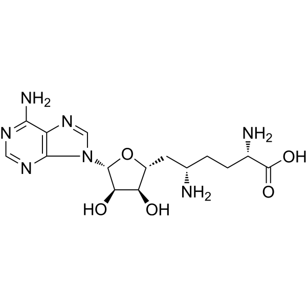
- Formula: C15H23N7O5
- Molecular Weight:381.39
-
Storage:Powder -20°C, 3 years , 4°C, 2 years ; In solvent -80°C, 6 months , -20°C, 1 month

To place orders, for customer services and technical support, please contact: MedChemExpress USA
Tel: 609-228-6898 E-mail: [email protected] [email protected]


-
 Biological Activity
-
Chemical Information
-
Solvent & Solubility
- Protocols
- Purity & Documentation
- References
-
 Help & FAQs


-
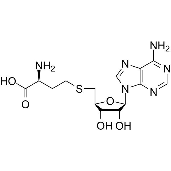
HY-19528
SAHMETTL3-14 Inhibitor -
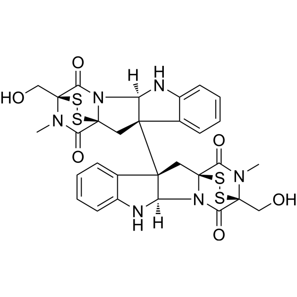
HY-N2019
ChaetocinSU(VAR)3-9 Inhibitor -

HY-15371
ForskolinAdenylate Cyclase Activator -
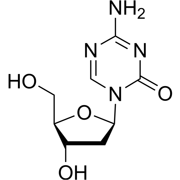
HY-A0004
DecitabineDNMT Inhibitor -
HY-17031
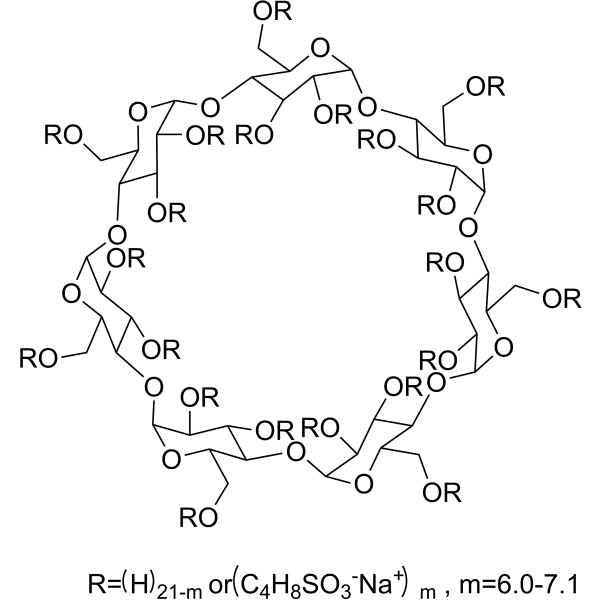
SBE-β-CDExcipient -
HY-134836
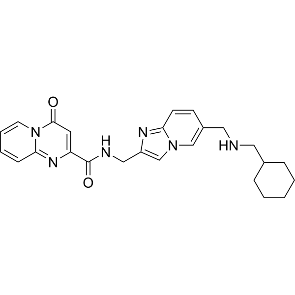
STM2457METTL3 Inhibitor -

HY-B0015
PaclitaxelAntitumor Agent -
HY-90006
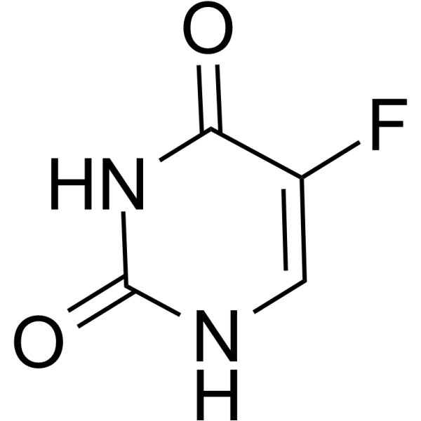
5-FluorouracilThymidylate Synthase Inhibitor -
HY-10586
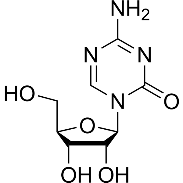
5-AzacytidineDNMT Inhibitor -

HY-13259
MG-132Proteasome Inhibitor -

HY-10071
Y-27632ROCK Inhibitor -
HY-16658
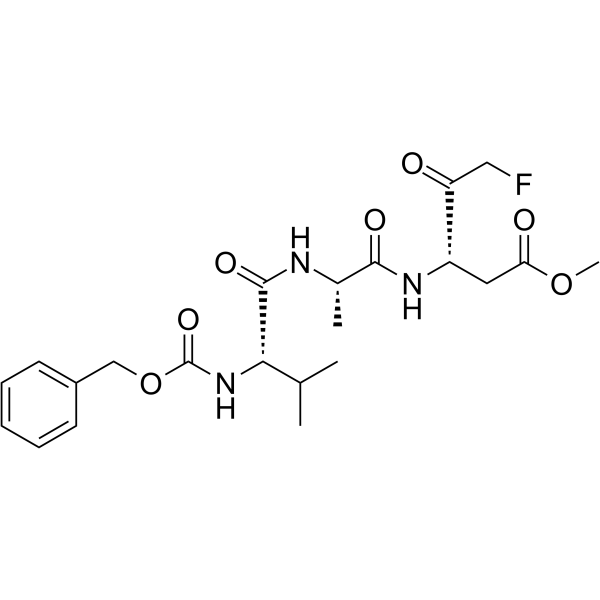
Z-VAD(OMe)-FMKPan-caspase Inhibitor
Publications Citing Use of MedChemExpress (MCE) Sinefungin
More Customer Validation & Images

-
RT-PCR
-
WB
-
Cell Proliferation/Viability Assay
All Histone Methyltransferase Isoforms
MoreAll Antibiotic Isoforms
MoreAll Parasite Isoforms
More

Biological Activity
Description
Cellular Effect
|
Cell Line

|
Type | Value | Description | References |
|---|---|---|---|---|
| A549 | CC50 |
72.93 μM
Compound: Sinefungin
|
Cytotoxicity against human A549 cells assessed as reduction in cell viability measured after 48 hrs by neutral red uptake assay
Cytotoxicity against human A549 cells assessed as reduction in cell viability measured after 48 hrs by neutral red uptake assay
|
[PMID: 34257831] |
| HEK293 | CC50 |
>500 μM
Compound: SFG
|
Cytotoxicity against human HEK293 cells assessed as cell viability at 100 uM measured after 24 hrs by MTT assay
Cytotoxicity against human HEK293 cells assessed as cell viability at 100 uM measured after 24 hrs by MTT assay
|
[PMID: 35849534] |
| HepG2 | CC50 |
>100 μM
Compound: Sinefungin
|
Cytotoxicity against human HepG2 cells assessed as reduction in cell viability measured after 48 hrs by neutral red uptake assay
Cytotoxicity against human HepG2 cells assessed as reduction in cell viability measured after 48 hrs by neutral red uptake assay
|
[PMID: 34257831] |
| Huh-7 | GI50 |
24.8 μM
Compound: Sinefungin
|
Growth inhibition of human Huh-7 cells
Growth inhibition of human Huh-7 cells
|
[PMID: 38805939] |
| Sf9 | IC50 |
8.6 μM
Compound: 1
|
Inhibition of full-length N-terminal FLAG-tagged PRMT5 (unknown origin) (1 to 637 residues) expressed in baculovirus infected Sf9 insect cells using histone H4 as peptide after 5 hrs in presence of [H3]AdoMet by scintillation proximity assay
Inhibition of full-length N-terminal FLAG-tagged PRMT5 (unknown origin) (1 to 637 residues) expressed in baculovirus infected Sf9 insect cells using histone H4 as peptide after 5 hrs in presence of [H3]AdoMet by scintillation proximity assay
|
[PMID: 30366617] |
In Vitro
Sinefungin (0.5 or 1.0 μg/mL, 60 minutes) ameliorates the TGF-β1-induced increase of α-SMA and inhibits the upregulation of histone H3K4 monomethylation in renal epithelial cells and renal fibroblast cells[2].
MedChemExpress (MCE) has not independently confirmed the accuracy of these methods. They are for reference only.
-
Cell Line:Renal epithelial cells.
-
Concentration:0.5 or 1.0 μg/mL.
-
Incubation Time:Pretreatment 60 minutes before TGF-β1 (10 ng/mL).
-
Result:Significantly reduced TGF-β1-inducedα-SMA protein expression and inhibited H3K4me1 in a dose-dependent manner in both NRK-52E and NRK-49F cells.
In Vivo
MedChemExpress (MCE) has not independently confirmed the accuracy of these methods. They are for reference only.
-
Animal Model:Male C57BL/6J mice (8 weeks of age)[2].
-
Dosage:10 mg/kg
-
Administration:Administered intraperitoneally per day immediately after UUO (prepared as a suspension in distilled water and 0.9% NaCl solution).
-
Result:Inhibited α-SMA protein expression.
Ameliorated those (α-SMA, FSP-1, collagen 1, collagen 3) both at 3 and 7 days after UUO.


Chemical Information
-
CAS No. 58944-73-3
-
Appearance Solid
-
Molecular Weight 381.39
-
Formula C15H23N7O5
-
Color White to yellow
-
SMILES
O[C@H]([C@@H]1O)[C@@H](O[C@@H]1C[C@@H](N)CC[C@H](N)C(O)=O)N2C(N=CN=C3N)=C3N=C2
-
Synonyms
Adenosyl-Ornithine; A-9145; Antibiotic 32232RP
-
Shipping
Room temperature in continental US; may vary elsewhere.
-
Storage
Powder -20°C 3 years 4°C 2 years In solvent -80°C 6 months -20°C 1 month


Publications (4)
-
Journal Impact Factor
-
Most Recent
-
Genomics Proteomics Bioinformatics
2025 May 10;23(1):qzaf008. PMID: 39957240
Sinefungin purchased from MedChemExpress. Usage Cited in: Genomics Proteomics Bioinformatics. 2025 May 10;23(1):qzaf008. [Abstract]
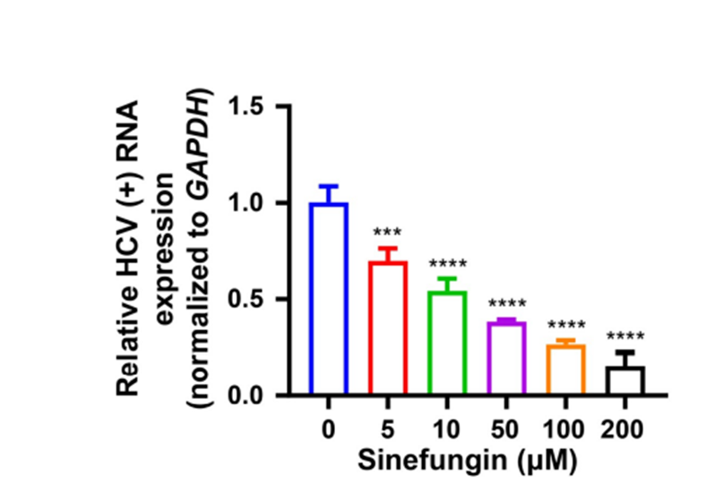
Quantification of HCV positive-strand (+) RNA via RT-qPCR in Huh7.5.1 cells treated with Sinefungin (5, 10, 50, 100, 200 μM) at 6 h post infection, and then harvested at 72 h.
Sinefungin purchased from MedChemExpress. Usage Cited in: Genomics Proteomics Bioinformatics. 2025 May 10;23(1):qzaf008. [Abstract]
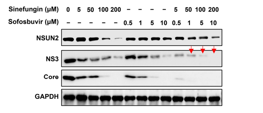
WB assays of the expression of NSUN2 as well as HCV NS3 and Core proteins in Huh7.5.1 cells treated with Sofosbuvir or in combination Sinefungin (5, 50, 100, 200 μM) at 6 h post infection, and then harvested at 72 h post infection.
-
Emerg Microbes Infect
SARS-CoV-2 and HCoV-OC43 regulate host m6A modification via activation of the mTORC1 signaling pathway to facilitate viral replication. [Abstract]2025 Dec;14(1):2447620. PMID: 39745173 -
Curr Res Microb Sci
Development of a luminescence-based method for measuring West Nile Virus MTase activity and its application to screen for antivirals. [Abstract]2024 Oct 2:7:100282. PMID: 39445035
Sinefungin purchased from MedChemExpress. Usage Cited in: Curr Res Microb Sci. 2024 Oct 2:7:100282. [Abstract]
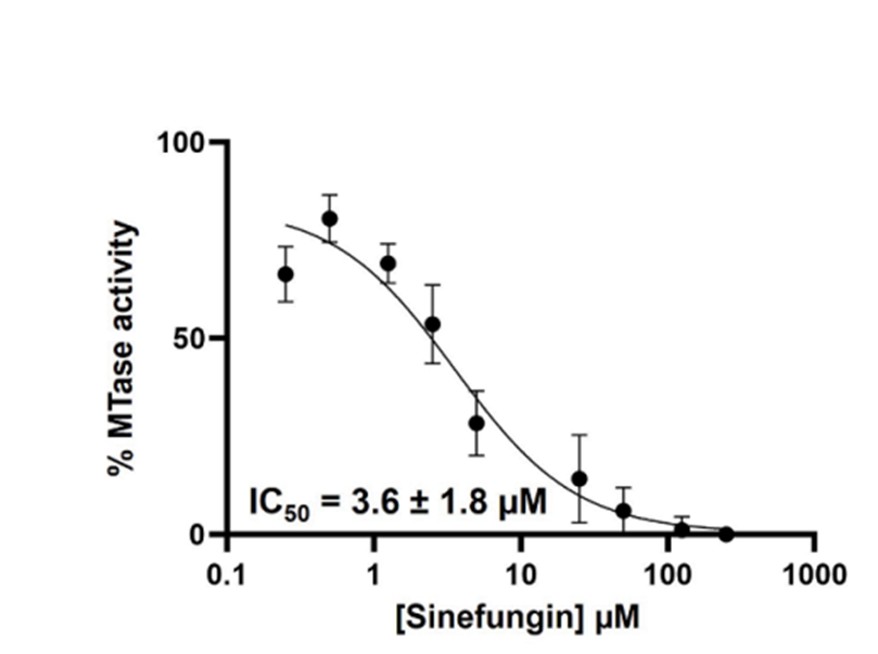
MTase activity dose-response inhibition curves and IC50 values of the WNV NS5 WT exerted by Sinefungin (1, 10, 100, 1000 μM).
-


Solvent & Solubility
In Vitro:
DMSO : 100 mg/mL (262.20 mM; Need ultrasonic; Hygroscopic DMSO has a significant impact on the solubility of product, please use newly opened DMSO)
H2O : 100 mg/mL (262.20 mM; Need ultrasonic)
Please refer to the solubility information to select the appropriate solvent. Once prepared, please aliquot and store the solution to prevent product inactivation from repeated freeze-thaw cycles.
Storage method and period of stock solution: -80°C, 6 months; -20°C, 1 month. When stored at -80°C, please use it within 6 months. When stored at -20°C, please use it within 1 month.
* Note: If you choose water as the stock solution, please dilute it to the working solution, then filter and sterilize it with a 0.22 μm filter before use.
Please refer to the solubility information to select the appropriate solvent. Once prepared, please aliquot and store the solution to prevent product inactivation from repeated freeze-thaw cycles.
Storage method and period of stock solution: -80°C, 6 months; -20°C, 1 month. When stored at -80°C, please use it within 6 months. When stored at -20°C, please use it within 1 month.
* Note: If you choose water as the stock solution, please dilute it to the working solution, then filter and sterilize it with a 0.22 μm filter before use.
Concentration (start) × Volume (start) = Concentration (final) × Volume (final)


Purity & Documentation
-
Data Sheet (275 KB)
-
SDS (392 KB)
- English - EN (392 KB)
- Français - FR (392 KB)
- Deutsch - DE (392 KB)
- Norwegian - NO (392 KB)
- Español - ES (392 KB)
- Swedish - SV (392 KB)
- Italian - IT (392 KB)
- Korean - KR (392 KB)
- Portuguese - PT (392 KB)
-
Handling Instructions (2659 KB)
References
[1]. Pugh CS, et al. Sinefungin, a potent inhibitor of virion mRNA(guanine-7-)-methyltransferase, mRNA(nucleoside-2'-)-methyltransferase, and viral multiplication. J Biol Chem. 1978 Jun 25;253(12):4075-7. [Content Brief]
[2]. Sasaki K, et al. Inhibition of SET Domain-Containing Lysine Methyltransferase 7/9 Ameliorates Renal Fibrosis. J Am Soc Nephrol. 2016 Jan;27(1):203-15. [Content Brief]


Complete Stock Solution Preparation Table
Please refer to the solubility information to select the appropriate solvent. Once prepared, please aliquot and store the solution to prevent product inactivation from repeated freeze-thaw cycles.
Storage method and period of stock solution: -80°C, 6 months; -20°C, 1 month. When stored at -80°C, please use it within 6 months. When stored at -20°C, please use it within 1 month.
| Optional Solvent | Concentration Solvent Mass | 1 mg | 5 mg | 10 mg | 25 mg |
|---|---|---|---|---|---|
| DMSO / H2O | 1 mM | 2.6220 mL | 13.1099 mL | 26.2199 mL | 65.5497 mL |
| 5 mM | 0.5244 mL | 2.6220 mL | 5.2440 mL | 13.1099 mL | |
| 10 mM | 0.2622 mL | 1.3110 mL | 2.6220 mL | 6.5550 mL | |
| 15 mM | 0.1748 mL | 0.8740 mL | 1.7480 mL | 4.3700 mL | |
| 20 mM | 0.1311 mL | 0.6555 mL | 1.3110 mL | 3.2775 mL | |
| 25 mM | 0.1049 mL | 0.5244 mL | 1.0488 mL | 2.6220 mL | |
| 30 mM | 0.0874 mL | 0.4370 mL | 0.8740 mL | 2.1850 mL | |
| 40 mM | 0.0655 mL | 0.3277 mL | 0.6555 mL | 1.6387 mL | |
| 50 mM | 0.0524 mL | 0.2622 mL | 0.5244 mL | 1.3110 mL | |
| 60 mM | 0.0437 mL | 0.2185 mL | 0.4370 mL | 1.0925 mL | |
| 80 mM | 0.0328 mL | 0.1639 mL | 0.3277 mL | 0.8194 mL | |
| 100 mM | 0.0262 mL | 0.1311 mL | 0.2622 mL | 0.6555 mL |
* Note: If you choose water as the stock solution, please dilute it to the working solution, then filter and sterilize it with a 0.22 μm filter before use.


Sinefungin Related Classifications
HY-101938 Related Classifications
 Powered by Bioz