



Herbimycin A

Based on 1 Customer Validation
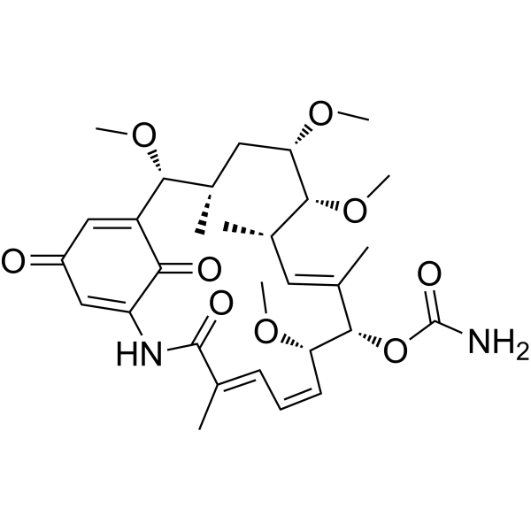
Herbimycin A is an antibiotic and protein tyrosine kinase inhibitor. Herbimycin A directly inhibits the autophosphorylation of p210 BCR-ABL with an IC50 of approximately 5 μM, and reduces Src kinase activity. Herbimycin A also induces the degradation of receptor tyrosine kinases such as insulin-like growth factor 1 receptor (IGF-1R), insulin receptor (IR) and epidermal growth factor receptor (EGFR) via the ubiquitin-20S proteasome pathway. Herbimycin A directly modifies NF-κB p50, with the main target site involving Cys62, thereby blocking the DNA binding of p50 and NF-κB-driven gene expression. Herbimycin A can be used in studies related to tyrosine kinase signaling, chronic myeloid leukemia, NF-κB signaling, osteoclast function, apoptosis and cellular stress.
For research use only. We do not sell to patients.
- Purity : 99%
- CAS No.: 70563-58-5
- Formula: C30H42N2O9
- Molecular Weight:574.66
-
Storage:Powder -20°C, 3 years ; In solvent -80°C, 6 months , -20°C, 1 month

To place orders, for customer services and technical support, please contact: MedChemExpress USA
Tel: 609-228-6898 E-mail: [email protected] [email protected]


-
 Biological Activity
-
Chemical Information
-
Solvent & Solubility
- Protocols
- Purity & Documentation
- References
-
 Help & FAQs


-
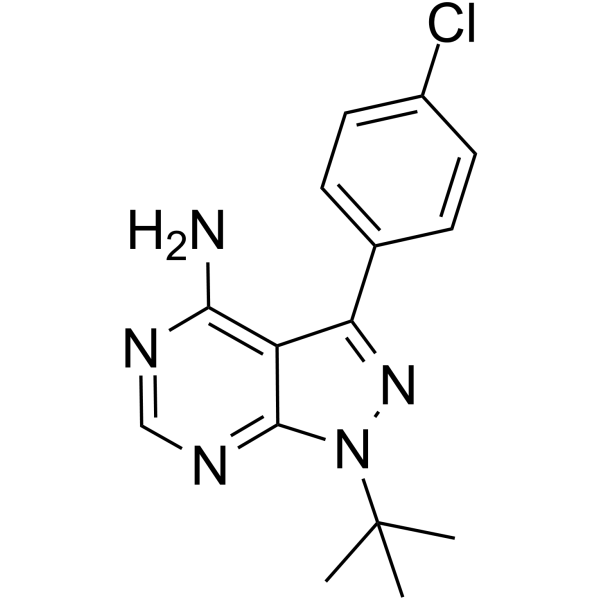
HY-13805
PP2Src Inhibitor -
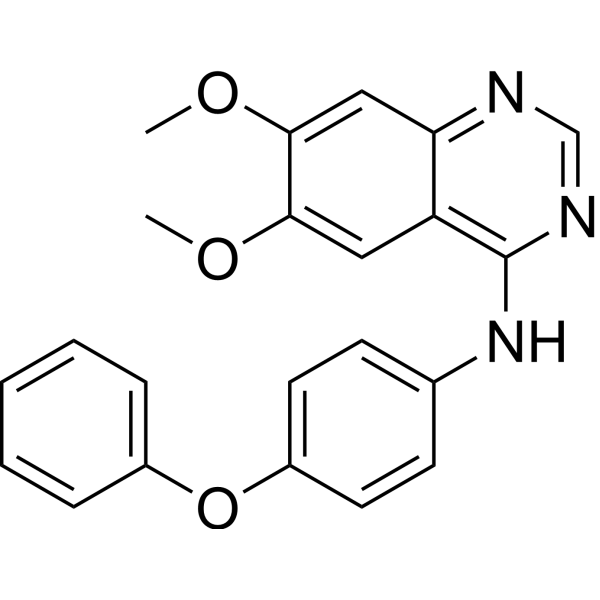
HY-101053
Src Inhibitor 1Src Inhibitor -
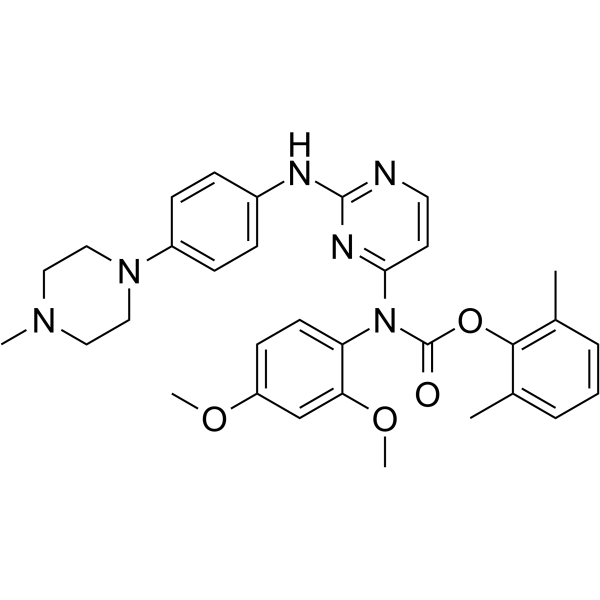
HY-12299
WH-4-023Src Inhibitor -
HY-13804
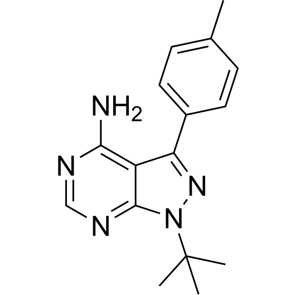
PP1Src Inhibitor -
HY-59047
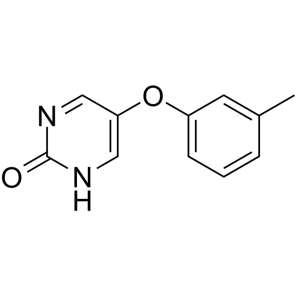
TolimidoneSrc Activator -
HY-15764A
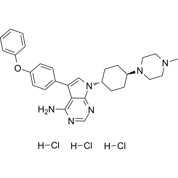
A 419259 trihydrochlorideSrc Inhibitor -
HY-108484
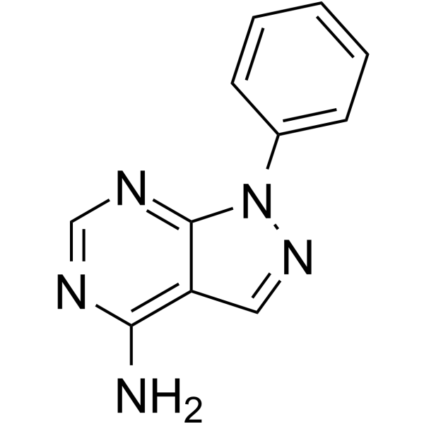
PP 3EGFR Inhibitor -
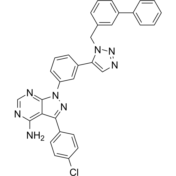
HY-108488
KB SRC 4c-Src Inhibitor -
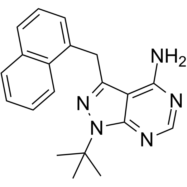
HY-13942
1-NM-PP1Src Inhibitor -
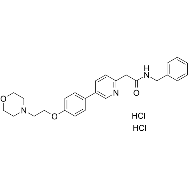
HY-10340A
Tirbanibulin dihydrochloride -
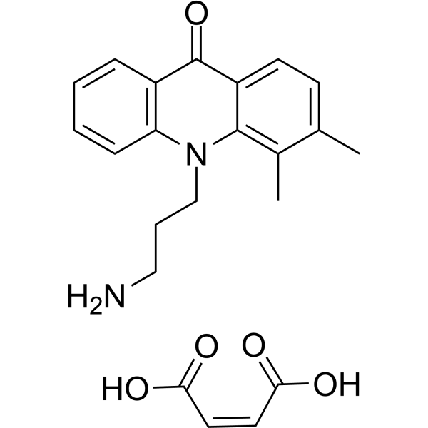
HY-108489
ER-27319 maleateSYK Iinhibitor -

HY-13805R
PP2 (Standard)Src Inhibitor


Biological Activity
Description
IC50 & Target
[6]|
p210 BCR-ABL 5 μM (IC50) |
Cellular Effect
|
Cell Line

|
Type | Value | Description | References |
|---|---|---|---|---|
| K562 | IC50 |
95 nM
|
Growth inhibition activity against human leukemia K562 cells assessed by cell number counting after 4 days of incubation.
Growth inhibition activity against human leukemia K562 cells assessed by cell number counting after 4 days of incubation.
|
2910452 |
| HL-60 | IC50 |
1030 nM
|
Growth inhibition activity against human leukemia HL-60 cells assessed by cell number counting after 4 days of incubation.
Growth inhibition activity against human leukemia HL-60 cells assessed by cell number counting after 4 days of incubation.
|
2910452 |
| THP-1 | IC50 |
1130 nM
|
Growth inhibition activity against human leukemia THP-1 cells assessed by cell number counting after 4 days of incubation.
Growth inhibition activity against human leukemia THP-1 cells assessed by cell number counting after 4 days of incubation.
|
2910452 |
| U-937 | IC50 |
1750 nM
|
Growth inhibition activity against human leukemia U937 cells assessed by cell number counting after 4 days of incubation.
Growth inhibition activity against human leukemia U937 cells assessed by cell number counting after 4 days of incubation.
|
2910452 |
In Vitro
Herbimycin A (2 μM; 1 h pre-incubation) inhibits IL-1α- or PMA-induced IL-2 production in EL4.NOB-1 cells, reducing IL-1α-induced IL-2 levels from 0.9 ng/mL to <0.1 ng/mL, and decreasing PMA-induced IL-2 levels from 50 ng/mL to 3.25 ng/mL[3].
Herbimycin A (0.1-10 μg/mL; 24 h) reduces IGF-IR levels in MCF-7 cells and IR and EGFR levels in MDA-MB-468 cells in a concentration-dependent manner; in MCF-7 cells, Herbimycin A (5 μg/mL) shortens the apparent half-life of IGF-IR from >24 h to approximately 6-7 h, and its degradation can be blocked by 20S proteasome inhibitors[1].
Herbimycin A (up to 1 μg/mL; 16-24 h) reduces foci of Rous sarcoma virus-transformed chicken embryo fibroblasts in a concentration-dependent manner, with complete disappearance of transformed foci at 1 μg/mL; the foci can reappear after the removal of Herbimycin A. It reverses transformation driven by tyrosine kinase oncogenes such as src, yes, fps, ros, abl and erbB, but exerts no similar effect on transformation driven by ras and myc[5].
Herbimycin A (50 nM; 1-3 h) rapidly reduces the tyrosine phosphorylation levels of p210 BCR-ABL and cellular proteins in K562 cells, with approximately 55% reduction in p210 BCR-ABL phosphorylation at 1 h, and tyrosine phosphorylation signals become almost undetectable after 3 h; Herbimycin A also induces erythroid differentiation of K562 cells in a concentration-dependent manner[2].
Herbimycin A (4 d) inhibits the growth of K562 cells with an IC50 of 95 nM; its erythroid differentiation-inducing effect reaches the maximum at 1 × 10⁻⁷ M[2].
Herbimycin A (1-100 ng/mL) inhibits osteoclastic bone resorption in mouse bone marrow cultures, isolated rat osteoclasts and fetal mouse long bone organ cultures in a concentration-dependent manner; treatment with 100 ng/mL for 24 h also significantly reduces PTH-stimulated pp60c-Src tyrosine kinase activity in mouse bone marrow cells[4].
Herbimycin A (1 μg/mL; 6 h) increases the expression level of the 70-kDa HSP70 to approximately 30 times that of untreated cells, and the induced HSP70 is mainly distributed in the cytoplasm[8].
Herbimycin A inhibits p210 BCR-ABL autophosphorylation in a concentration-dependent manner in cell-free immune complex kinase assays, with an IC50 of approximately 5 μM; the inhibition persists after washing away free Herbimycin A prior to the kinase reaction, supporting its direct action on p210 BCR-ABL[6].
MedChemExpress (MCE) has not independently confirmed the accuracy of these methods. They are for reference only.
-
Cell Line:murine EL4.NOB-1 thymoma cells
-
Concentration:0.02-2 μM
-
Incubation Time:1 h (preincubation prior to 24 h IL-1α stimulation)
-
Result:Caused concentration-dependent inhibition of IL-1α-induced IL-2 production.
Reduced IL-2 levels from 0.9 ng/mL to less than 0.1 ng/mL at 2 μM.
-
Cell Line:Recombinant human p50
-
Concentration:10-100 μM
-
Incubation Time:1 h (preincubation prior to 24 h PMA stimulation)
-
Result:Inhibited p50 binding to the NF-κB consensus DNA sequence, whereas the C62S p50 mutant was insensitive.
-
Cell Line:K562
-
Concentration:50 nM
-
Incubation Time:1-3 h
-
Result:Reduced p210 BCR-ABL phosphorylation by approximately 55% at 1 h, and tyrosine phosphorylation was barely detectable after 3 h.
-
Cell Line:NRK-52E
-
Concentration:500 ng/ mL
-
Incubation Time:24 h
-
Result:Reduced okadaic acid-induced apoptosis by approximately 50% and inhibited p38 activation and ATF-2 phosphorylation.
In Vivo
Herbimycin A (100 μg/mouse/injection; i.p.; 2 times/day; 8 d, i.e., 200 μg/mouse/day) begins to reduce blood Ca2+ levels on day 4 in hypercalcemic nude mice bearing human squamous cell carcinoma MH-85, with a significant reduction observed on day 8, but does not affect MH-85 tumor growth[4].
MedChemExpress (MCE) has not independently confirmed the accuracy of these methods. They are for reference only.
-
Animal Model:ICR Swiss white mice (male, 5-6 weeks old, hypercalcemia induced by subcutaneous recombinant human interleukin-1α injections)[4]
-
Dosage:10 μg/mouse per injection
-
Administration:s.c.; four times a day
-
Result:Markedly prevented the development of recombinant human interleukin-1α-induced hypercalcemia.
Maintained ionized calcium level near baseline by day 4, compared to ≈1.6 mmol/L in interleukin-1α-only treated mice.
-
Animal Model:BALB/c nu/nu nude mice (hypercalcemia induced by subcutaneous MH-85 human squamous cancer inoculation)[4]
-
Dosage:100 μg/mouse per injection; 200 μg/mouse total daily dose
-
Administration:i.p.; twice a day; 8 days
-
Result:Lowered blood ionized calcium levels by day 4, with a significant decrease to ≈1.55 mmol/L observed by day 8, compared to ≈1.95 mmol/L in DMSO-treated control mice.
Did not affect MH-85 tumor growth.
-
Animal Model:Sprague-Dawley (male, 250-300 g)[10]
-
Dosage:2 mL/kg of 1 mg/6 mL solution
-
Administration:i.p.; single dose
-
Result:Induced liver hsp70 expression to peak levels (35 arbitrary % volume) at 12 h post-administration, with expression significantly higher than at 6, 18, and 24 h.
Induced 20 arbitrary % volume of hsp70 in liver at 12 h post-administration, which was higher than levels in vehicle- and saline-treated rats.
Reduced peak core temperature to 41.16 °C during heat stress, lower than the peak temperatures of vehicle- and saline-treated rats.
Decreased the percentage of apoptotic hepatocytes significantly compared to vehicle- and saline-treated rats.
Increased levels of procaspase-3 (32 kDa) and decreased levels of active caspase-3 (20 kDa) in liver tissue compared to vehicle- and saline-treated rats.
- Protocol for Electrophoretic Mobility Shift Assay (EMSA)
- RT-PCR
- RNA extraction experimental
- Annexin V plus membrane-impermeant dye apoptosis staining
- Research Protocol for Inflammation-related Diseases
- Real Time qPCR (Q-PCR)
- Kinase activity and phosphorylation assays
- Detection of Apoptosis
- TUNEL staining for apoptotic DNA fragmentation
- Research Protocol for Endocrine Diseases
- Apoptosis Solutions
- Protocol for Kinase activity and phosphorylation assays
- Osteoclast differentiation from monocyte/macrophage precursors


Chemical Information
-
CAS No. 70563-58-5
-
Appearance Solid
-
Molecular Weight 574.66
-
Formula C30H42N2O9
-
Color Light yellow to yellow
-
SMILES
CO[C@H]([C@H](C[C@@H]([C@@H]([C@H](/C=C([C@@H]1OC(N)=O)\C)C)OC)OC)C)C(C(C(NC(/C(C)=C/C=C\[C@@H]1OC)=O)=C2)=O)=CC2=O
-
Structure Classification
-
Initial Source
Streptomyces spp.
-
Shipping
Room temperature in continental US; may vary elsewhere.
-
Storage
Powder -20°C 3 years In solvent -80°C 6 months -20°C 1 month


Solvent & Solubility
In Vitro:
DMSO : 1 mg/mL (1.74 mM; Need ultrasonic and warming; Hygroscopic DMSO has a significant impact on the solubility of product, please use newly opened DMSO)
Please refer to the solubility information to select the appropriate solvent. Once prepared, please aliquot and store the solution to prevent product inactivation from repeated freeze-thaw cycles.
Storage method and period of stock solution: -80°C, 6 months; -20°C, 1 month. When stored at -80°C, please use it within 6 months. When stored at -20°C, please use it within 1 month.
Please refer to the solubility information to select the appropriate solvent. Once prepared, please aliquot and store the solution to prevent product inactivation from repeated freeze-thaw cycles.
Storage method and period of stock solution: -80°C, 6 months; -20°C, 1 month. When stored at -80°C, please use it within 6 months. When stored at -20°C, please use it within 1 month.
Concentration (start) × Volume (start) = Concentration (final) × Volume (final)


Purity & Documentation
-
Data Sheet (308 KB)
-
SDS (251 KB)
- English - EN (251 KB)
- Français - FR (251 KB)
- Deutsch - DE (251 KB)
- Norwegian - NO (251 KB)
- Español - ES (251 KB)
- Swedish - SV (251 KB)
- Italian - IT (251 KB)
- Korean - KR (251 KB)
- Portuguese - PT (251 KB)
-
Handling Instructions (2659 KB)
References


Complete Stock Solution Preparation Table
Please refer to the solubility information to select the appropriate solvent. Once prepared, please aliquot and store the solution to prevent product inactivation from repeated freeze-thaw cycles.
Storage method and period of stock solution: -80°C, 6 months; -20°C, 1 month. When stored at -80°C, please use it within 6 months. When stored at -20°C, please use it within 1 month.
| Optional Solvent | Concentration Solvent Mass | 1 mg | 5 mg | 10 mg | 25 mg |
|---|---|---|---|---|---|
| DMSO | 1 mM | 1.7402 mL | 8.7008 mL | 17.4016 mL | 43.5040 mL |


Herbimycin A Related Classifications
HY-108486 Related Classifications
 Powered by Bioz