



TAPI-1
Based on 16 publication(s) in Google Scholar
TAPI-1 is a broad-spectrum MMP inhibitor and NF-κB p65 inhibitor that targets ADAM17/TACE, ADAM10 and other proteins. TAPI-1 reduces the proteolytic cleavage of membrane-bound TNF-α, decreases TNF-α levels, inhibits NF-κB pathway activation, and downregulates profibrotic markers. TAPI-1 reduces the proportion of proinflammatory immune cells, alleviates cardiac and airway fibrosis, and improves cardiac function after myocardial infarction. Meanwhile, TAPI-1 inhibits the viability, migration and invasion of esophageal squamous cell carcinoma cells, enhances the chemosensitivity of Cisplatin (HY-17394), induces apoptosis, and shows low toxicity to normal esophageal epithelial cells. TAPI-1 can be widely used in studies related to myocardial infarction-induced heart failure, severe traumatic tracheal stenosis, esophageal squamous cell carcinoma and other conditions.
For research use only. We do not sell to patients.
- Purity : 99.36%
- CAS No.: 163847-77-6
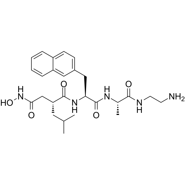
- Formula: C26H37N5O5
- Molecular Weight:499.60
-
Storage:Powder -20°C, 3 years , 4°C, 2 years ; In solvent -80°C, 2 years , -20°C, 1 year

To place orders, for customer services and technical support, please contact: MedChemExpress USA
Tel: 609-228-6898 E-mail: [email protected] [email protected]


-
 Biological Activity
-
Chemical Information
-
Solvent & Solubility
- Protocols
- Purity & Documentation
- References
-
 Help & FAQs


-
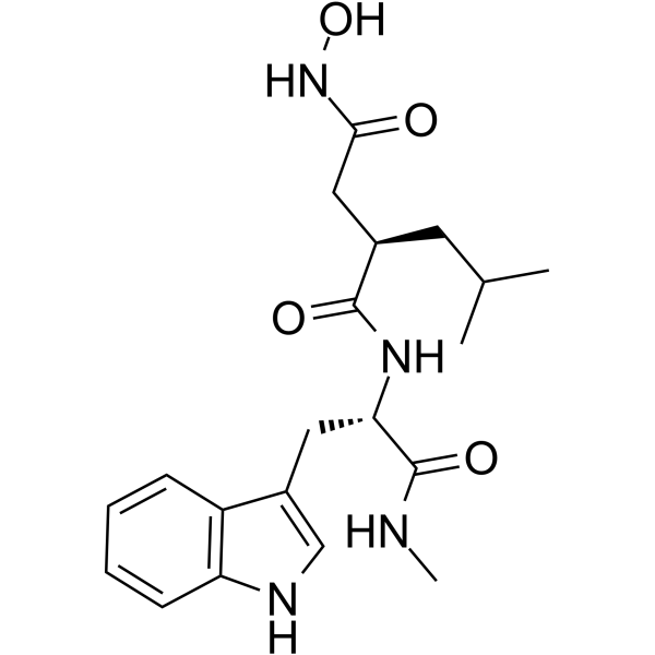
HY-15768
IlomastatMMP Inhibitor -
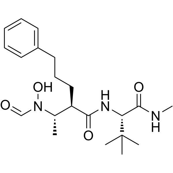
HY-19956
GI254023XADAM10/MMP9 Inhibitor -
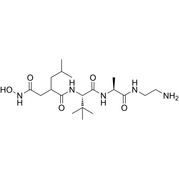
HY-100211
TAPI-2MMP/TACE/ADAM Inhibitor -
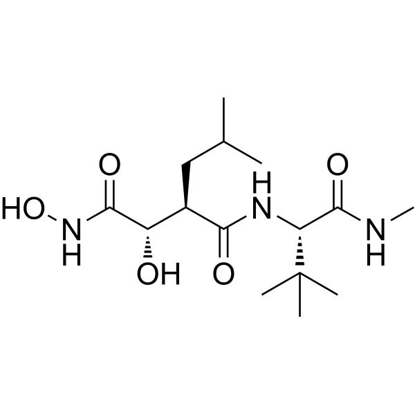
HY-12169
MarimastatMMP Inhibitor -
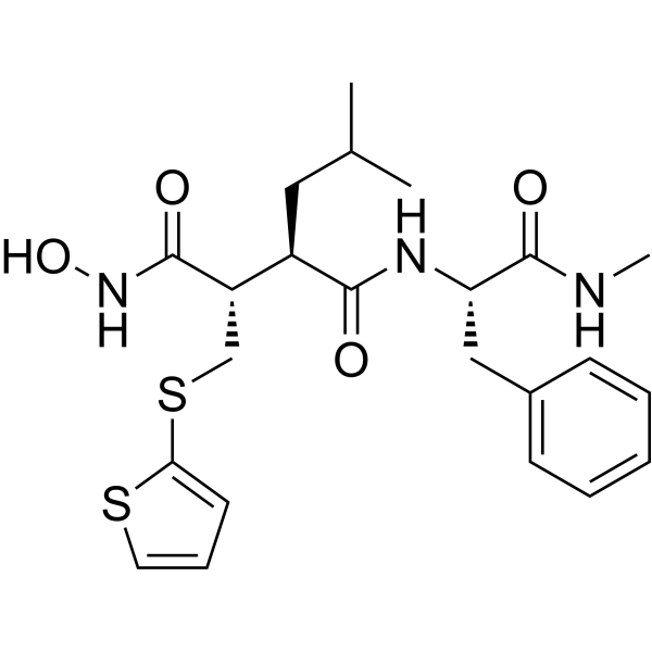
HY-13564
BatimastatMMP Inhibitor -
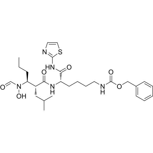
HY-115670
GW280264XADAM10/17 Inhibitor -
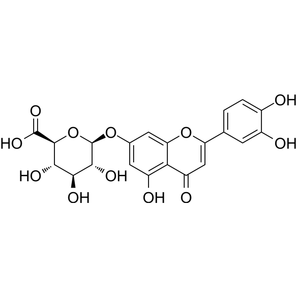
HY-N1463
Luteolin 7-O-glucuronideMMP Inhibitor -

HY-18739
Phorbol 12-myristate 13-acetatePKC Activator -
HY-16658B
Z-VAD-FMKPan-Caspase Inhibitor -

HY-10182
LaduviglusibGSK-3 Inhibitor -
HY-13027
DAPTγ-secretase Inhibitor -

HY-108841
RaleukinIL-1R Antagonist
Publications Citing Use of MedChemExpress (MCE) TAPI-1
More- Cell. 2026 Jun 25;189(13):3883-3902.e23. [Abstract]
- Exp Hematol Oncol. 2025 Jul 24;14(1):100. [Abstract]
- MedComm. 2023 Jul 8;4(4):e320. [Abstract]
- Adv Healthc Mater. 2025 Jul 16:e2501440. [Abstract]
- Neural Regen Res. 2026 Feb 1;21(2):730-741. [Abstract]
- Cell Syst. 2025 Mar 19;16(3):101203. [Abstract]
- Int Immunopharmacol. 2025 May 16:155:114571. [Abstract]
- J Cell Mol Med. 2026 Mar;30(5):e71076. [Abstract]
- J Cell Mol Med. 2025 Mar;29(5):e70466. [Abstract]
- Immun Inflamm Dis. 2025 Jul;13(7):publicationMeta. [Abstract]
- Dig Dis Sci. 2024 Jan;69(1):81-94. [Abstract]
- Int J Endocrinol. 2017:2017:9501792. [Abstract]
- J Neurovirol. 2023 Jun;29(3):283-296. [Abstract]
- Princeton University. 2025.
- bioRxiv. 2024 May 31:2024.05.30.596676. [Abstract]
- Research Square Preprint. 2022 Jan.
 Customer Validation & Images

-
Bio/Physico-chemical Assay
-
IF
-
WB
-
WB
-
WB


Biological Activity
Description
IC50 & Target
|
ADAM17 |
ADAM10 |
In Vitro
TAPI-1 (1.25-20 μM; 24-72 h) inhibits viability of TE-1 and Eca109 ESCC cells in a dose- and time-dependent manner without affecting viability of normal Het-1A esophageal epithelial cells[3].
TAPI-1 (10 μM; 24 h) upregulates TRAIL mRNA in TE-1 ESCC cells but does not alter apoptosis-related gene expression in Eca109 ESCC cells; TAPI-1 (5 μM; 12 h) downregulates multiple Cisplatin (HY-17394) resistance-related genes in both TE-1 and Eca109 ESCC cells[3].
TAPI-1 (10 μM; 72 h) does not induce apoptosis in TE-1 or Eca109 ESCC cells, but TAPI-1 (5 μM; 48 h) enhances cisplatin-induced apoptosis in both TE-1 and Eca109 ESCC cells[3].
TAPI-1 (5 μM; 48 h) enhances the sensitivity of TE-1 and Eca109 ESCC cells to cisplatin, as shown by significantly reduced cisplatin IC50 values[3].
TAPI-1 (10 μM; 12 h) suppresses NF-κB signaling in TE-1 and Eca109 ESCC cells by reducing NF-κB p65 phosphorylation and nuclear translocation[3].
MedChemExpress (MCE) has not independently confirmed the accuracy of these methods. They are for reference only.
-
Cell Line:human esophageal squamous cell carcinoma (ESCC) TE-1, Eca109 cells; human normal esophageal epithelial Het-1A cells
-
Concentration:1.25-20 μM (24 h viability assay); 10 μM (time-course viability assay)
-
Incubation Time:24 h (dose-response); 24, 48, 72 h (10 μM time-course)
-
Result:Inhibited viability of TE-1 and Eca109 cells in a dose-dependent manner, with significant decreases observed at 10 and 20 μM after 24 h.
Reduced viability of TE-1 and Eca109 cells significantly at 24 and 48 h at 10 μM, with no further change at 72 h.
Caused no significant viability changes in Het-1A cells across any tested concentration or time point.
-
Cell Line:ESCC TE-1, Eca109 cells
-
Concentration:10 μM (apoptosis-related gene assay); 5 μM (cisplatin resistance-related gene assay)
-
Incubation Time:24 h (apoptosis-related gene assay); 12 h (cisplatin resistance-related gene assay)
-
Result:Upregulated pro-apoptosis TRAIL mRNA significantly in TE-1 cells at 10 μM for 24 h, with no changes in BIM or BCL2 mRNA levels.
Caused no significant changes in BIM, TRAIL, or BCL2 mRNA levels in Eca109 cells at 10 μM for 24 h.
Downregulated 29 cisplatin resistance-related genes in TE-1 cells at 5 μM for 12 h.
Downregulated 21 cisplatin resistance-related genes in Eca109 cells at 5 μM for 12 h, with 20 genes downregulated in both cell lines.
In Vivo
TAPI-1 (0.8 mg/mL; local tracheal spray; once weekly; 3 weeks), either alone or combined with a silicone stent, significantly alleviates severe traumatic tracheal stenosis in Beagle dogs by inhibiting the ADAM17/TGF-β1 pathway, with TAPI-1 alone achieving the lowest degree of tracheal stenosis[2].
MedChemExpress (MCE) has not independently confirmed the accuracy of these methods. They are for reference only.
-
Animal Model:Beagle (male, 12 months old, 10-12 kg, traumatic tracheal stenosis model established via rigid bronchoscope-guided argon plasma coagulation + balloon compression)[2]
-
Dosage:0.8 mg/mL (1 mL volume)
-
Administration:local tracheal spray; once weekly; 3 weeks (initiated first week post-modeling); silicone stent implantation (third week post-modeling, for combined group)
-
Result:Significantly reduced tracheal stenosis degree.
Significantly downregulated tracheal mRNA and protein levels of ADAM17, TGF-β1, and fibronectin 1, with levels lower than those in the mitomycin treatment group.
Alleviated pathological tracheal changes including reduced airway epithelial hyperplasia, submucosal thickening, fibrous tissue proliferation, and lymphocytic infiltration.
When combined with silicone stent, significantly reduced tracheal stenosis degree, with no significant difference compared to TAPI-1 alone group.
When combined with silicone stent, significantly downregulated tracheal mRNA and protein levels of ADAM17, TGF-β1, and fibronectin 1, but levels were significantly higher than those in the TAPI-1 alone group.
When combined with silicone stent, alleviated pathological tracheal changes including reduced airway epithelial hyperplasia, submucosal thickening, fibrous tissue proliferation, and lymphocytic infiltration.
- Collagen: Sirius Red Staining
- Mitochondrial membrane-potential fluorescent assay
- Annexin V plus membrane-impermeant dye apoptosis staining
- Cell migration
- Connective Tissue: Masson's Trichrome/Collagen Trichrome Staining
- Research Protocol for Cardiovascular Diseases
- Research Protocol for Inflammation-related Diseases
- Cell Viability Determination by MTT Colorimetric Assay
- Fibrosis/Collagen Morphometry
- Detection of Apoptosis
- TUNEL staining for apoptotic DNA fragmentation
- Mitochondrial membrane-potential and mitochondrial mass staining
- Apoptosis Solutions
- Cell invasion


Chemical Information
-
CAS No. 163847-77-6
-
Appearance Solid
-
Molecular Weight 499.60
-
Formula C26H37N5O5
-
Color White to light yellow
-
SMILES
O=C(N[C@@H](CC1=CC=C2C=CC=CC2=C1)C(N[C@@H](C)C(NCCN)=O)=O)[C@H](CC(C)C)CC(NO)=O
-
Shipping
Room temperature in continental US; may vary elsewhere.
-
Storage
Powder -20°C 3 years 4°C 2 years In solvent -80°C 2 years -20°C 1 year


Publications (16)
-
Journal Impact Factor
-
Most Recent
-
Cell
Cell-autonomous control of CAR signaling and receptor shedding via ADAM17-mediated proteolysis. [Abstract]2026 Jun 25;189(13):3883-3902.e23. PMID: 42143019 -
Exp Hematol Oncol
2025 Jul 24;14(1):100. PMID: 40708008 -
MedComm
Identification of circulating T-cell immunoglobulin and mucin domain 4 as a potential biomarker for coronary heart disease. [Abstract]2023 Jul 8;4(4):e320. PMID: 37426678
TAPI-1 purchased from MedChemExpress. Usage Cited in: MedComm. 2023 Jul 8;4(4):e320. [Abstract]
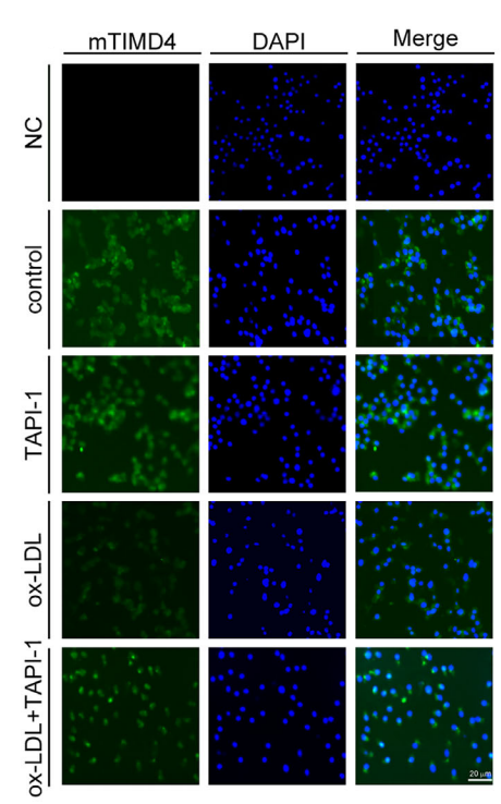
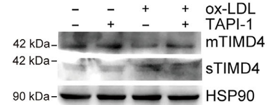
With TAPI-1 (1 μM) treatment, mTIMD4 protein expression increased whiles sTIMD4 level decreased noticeably.
TAPI-1 purchased from MedChemExpress. Usage Cited in: MedComm. 2023 Jul 8;4(4):e320. [Abstract]
With TAPI-1 (1 μM) treatment, mTIMD4 protein expression increased whiles sTIMD4 level decreased noticeably.
TAPI-1 purchased from MedChemExpress. Usage Cited in: MedComm. 2023 Jul 8;4(4):e320. [Abstract]
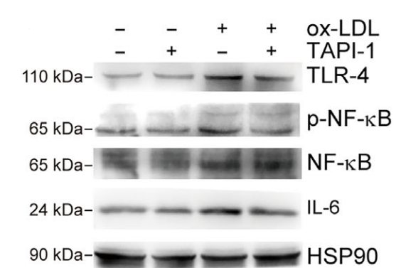
TAPI-1 (1 μM) abolished the protein expression of phosphorylated nuclear factor kappa B (p-NF-κB), anti-Toll-like receptor 4 (TLR-4) and IL‐6 that were upregulated by ox-LDL.
TAPI-1 purchased from MedChemExpress. Usage Cited in: MedComm. 2023 Jul 8;4(4):e320. [Abstract]
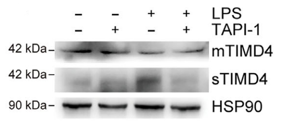
RAW264.7 cells were treated with LPS (1 μg/mL) and TAPI-1 (1 μM) for 24 h, and the protein expression of mTIMD4, sTIMD4 by Western blotting.
-
Adv Healthc Mater
Triple-Tailored Analgesic Hydrogel System Targeting ADAM17 in Orofacial Inflammatory Pain. [Abstract]2025 Jul 16:e2501440. PMID: 40671384 -
Neural Regen Res
Fat mass and obesity-mediated m6A modification modulates neuroinflammatory responses after traumatic brain injury. [Abstract]2026 Feb 1;21(2):730-741. PMID: 39248160 -
Cell Syst
Large-scale control over collective cell migration using light-activated epidermal growth factor receptors. [Abstract]2025 Mar 19;16(3):101203. PMID: 40037348
TAPI-1 purchased from MedChemExpress. Usage Cited in: Cell Syst. 2025 Mar 19;16(3):101203. [Abstract]
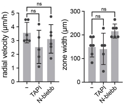
Quantification of peak radial velocity (left) and migration zone width (right) at 4 h post illumination for illuminated tissues treated with each compound. N=6,5,6 tissues for control, TAPI-1 (10 μM), and N-blebbistatin, respectively.
-
Int Immunopharmacol
Paeoniflorin alleviated STZ-induced diabetic retinopathy via regulation of the PDI/ADAM17/MerTK pathway. [Abstract]2025 May 16:155:114571. PMID: 40209310 -
J Cell Mol Med
Alpha-Fetoprotein Stimulates Cleavage of Membranal MICA/B on Liver Cancer Cell Lead to Escape Immune Surveillance of Natural Killer Cells. [Abstract]2026 Mar;30(5):e71076. PMID: 41749070 -
J Cell Mol Med
2025 Mar;29(5):e70466. PMID: 40077919 -
Immun Inflamm Dis
Amphiregulin Promotes Proliferation and Migration of the Damaged Endothelial Cells in Kawasaki Disease Cell Models. [Abstract]2025 Jul;13(7):publicationMeta. PMID: 40660822 -
Dig Dis Sci
TAPI-1 Exhibits Anti-tumor Efficacy in Human Esophageal Squamous Cell Carcinoma Cells via Suppression of NF-κB Signaling Pathway. [Abstract]2024 Jan;69(1):81-94. PMID: 38007701 -
Int J Endocrinol
Hydrogen Sulfide Inhibits High Glucose-Induced sFlt-1 Production via Decreasing ADAM17 Expression in 3T3-L1 Adipocytes. [Abstract]2017:2017:9501792. PMID: 28740508 -
J Neurovirol
Disruption of the ADAM17/NF-κB feedback loop in astrocytes ameliorates HIV-1 Tat-induced inflammatory response and neuronal death. [Abstract]2023 Jun;29(3):283-296. PMID: 37185939 -
-
bioRxiv
Large-scale control over collective cell migration using light-controlled epidermal growth factor receptors. [Abstract]2024 May 31:2024.05.30.596676. PMID: 38853934 -


Solvent & Solubility
In Vitro:
DMSO : 100 mg/mL (200.16 mM; Need ultrasonic; Hygroscopic DMSO has a significant impact on the solubility of product, please use newly opened DMSO)
Please refer to the solubility information to select the appropriate solvent. Once prepared, please aliquot and store the solution to prevent product inactivation from repeated freeze-thaw cycles.
Storage method and period of stock solution: -80°C, 2 years; -20°C, 1 year. When stored at -80°C, please use it within 2 years. When stored at -20°C, please use it within 1 year.
Please refer to the solubility information to select the appropriate solvent. Once prepared, please aliquot and store the solution to prevent product inactivation from repeated freeze-thaw cycles.
Storage method and period of stock solution: -80°C, 2 years; -20°C, 1 year. When stored at -80°C, please use it within 2 years. When stored at -20°C, please use it within 1 year.
Concentration (start) × Volume (start) = Concentration (final) × Volume (final)
In Vivo:
Select the appropriate dissolution method based on your experimental animal and administration route.
- For the following dissolution methods, please ensure to first prepare a clear stock solution using an In Vitro approach and then sequentially add co-solvents:
- To ensure reliable experimental results, the clarified stock solution can be appropriately stored based on storage conditions. As for the working solution for In Vivo experiments, it is recommended to prepare freshly and use it on the same day.
- The percentages shown for the solvents indicate their volumetric ratio in the final prepared solution. If precipitation or phase separation occurs during preparation, heat and/or sonication can be used to aid dissolution.
Add each solvent one by one: 10% DMSO 40% PEG300 5% Tween-80 45% Saline
Solubility: ≥ 2.08 mg/mL (4.16 mM); Clear solution
This protocol yields a clear solution of ≥ 2.08 mg/mL (saturation unknown).
Taking 1 mL working solution as an example, add 100 μL DMSO stock solution (20.8 mg/mL) to 400 μL PEG300, and mix evenly; then add 50 μL Tween-80 and mix evenly; then add 450 μL Saline to adjust the volume to 1 mL.
Preparation of Saline: Dissolve 0.9 g sodium chloride in ddH₂O and dilute to 100 mL to obtain a clear Saline solution.
Add each solvent one by one: 10% DMSO 90% (20% SBE-β-CD in Saline)
Solubility: ≥ 2.08 mg/mL (4.16 mM); Clear solution
This protocol yields a clear solution of ≥ 2.08 mg/mL (saturation unknown).
Taking 1 mL working solution as an example, add 100 μL DMSO stock solution (20.8 mg/mL) to 900 μL 20% SBE-β-CD in Saline, and mix evenly.
Preparation of 20% SBE-β-CD in Saline (4°C, storage for one week): 2 g SBE-β-CD powder is dissolved in 10 mL Saline, completely dissolve until clear.
In Vivo Dissolution Calculator
Please enter the basic information of animal experiments:
-
-
-
-
Recommended: Prepare an additional quantity of animals to account for potential losses during experiments.
Please enter your animal formula composition:
-
%DMSO +
Recommended: Keep the proportion of DMSO in working solution below 2% if your animal is weak.
-
%+
-
+%Tween-80 + +
-
%Saline +
The co-solvents required include: DMSO, . All of co-solvents are available by MedChemExpress (MCE). , Tween 80. All of co-solvents are available by MedChemExpress (MCE).
Working solution concentration: 0.22 mg/mL
Method for preparing stock solution: mg drug dissolved in μL DMSO. Stock solution concentration: mg/mL.
1. Take μL DMSO stock solution;
2. Add μL .
μL , mix evenly;
3. Then add μL Tween 80, mix evenly;
4. Then add μL
Please ensure that the stock solution in the first step is dissolved to a clear state, and add co-solvents in sequence. You can use ultrasonic heating (ultrasonic cleaner, recommended frequency 20-40 kHz), vortexing, etc. to assist dissolution.


Purity & Documentation
-
Data Sheet (282 KB)
-
SDS (479 KB)
- English - EN (479 KB)
- Français - FR (479 KB)
- Deutsch - DE (479 KB)
- Norwegian - NO (479 KB)
- Español - ES (479 KB)
- Swedish - SV (479 KB)
- Italian - IT (479 KB)
- Korean - KR (479 KB)
- Portuguese - PT (479 KB)
-
Handling Instructions (2659 KB)
References
[1]. Chen Q, et al. Targeted delivery of TAPI-1 via biomimetic nanoparticles ameliorates post-infarct left ventricle function and remodelling. Cardiovasc Res. 2025;121(5):760-774. [Content Brief]
[2]. Yang Z, et al. TAPI-1 Combined With Silicone Stents Alleviated Severe Traumatic Tracheal Stenosis via the ADAM17/TGF-β1 Pathway. Can Respir J. 2026;2026:9485331. Published 2026 Jan 11. [Content Brief]
[3]. Gao L, et al. TAPI-1 Exhibits Anti-tumor Efficacy in Human Esophageal Squamous Cell Carcinoma Cells via Suppression of NF-κB Signaling Pathway. Dig Dis Sci. 2024;69(1):81-94. [Content Brief]


Complete Stock Solution Preparation Table
Please refer to the solubility information to select the appropriate solvent. Once prepared, please aliquot and store the solution to prevent product inactivation from repeated freeze-thaw cycles.
Storage method and period of stock solution: -80°C, 2 years; -20°C, 1 year. When stored at -80°C, please use it within 2 years. When stored at -20°C, please use it within 1 year.
| Optional Solvent | Concentration Solvent Mass | 1 mg | 5 mg | 10 mg | 25 mg |
|---|---|---|---|---|---|
| DMSO | 1 mM | 2.0016 mL | 10.0080 mL | 20.0160 mL | 50.0400 mL |
| 5 mM | 0.4003 mL | 2.0016 mL | 4.0032 mL | 10.0080 mL | |
| 10 mM | 0.2002 mL | 1.0008 mL | 2.0016 mL | 5.0040 mL | |
| 15 mM | 0.1334 mL | 0.6672 mL | 1.3344 mL | 3.3360 mL | |
| 20 mM | 0.1001 mL | 0.5004 mL | 1.0008 mL | 2.5020 mL | |
| 25 mM | 0.0801 mL | 0.4003 mL | 0.8006 mL | 2.0016 mL | |
| 30 mM | 0.0667 mL | 0.3336 mL | 0.6672 mL | 1.6680 mL | |
| 40 mM | 0.0500 mL | 0.2502 mL | 0.5004 mL | 1.2510 mL | |
| 50 mM | 0.0400 mL | 0.2002 mL | 0.4003 mL | 1.0008 mL | |
| 60 mM | 0.0334 mL | 0.1668 mL | 0.3336 mL | 0.8340 mL | |
| 80 mM | 0.0250 mL | 0.1251 mL | 0.2502 mL | 0.6255 mL | |
| 100 mM | 0.0200 mL | 0.1001 mL | 0.2002 mL | 0.5004 mL |


TAPI-1 Related Classifications
HY-16657 Related Classifications
 Powered by Bioz