



Novobiocin sodium
Based on 14 publication(s) in Google Scholar
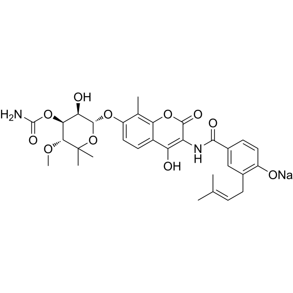
Novobiocin (Albamycin; Cathomycin) sodium is an orally active antibiotic and Hsp-90 antagonist that also inhibits the Polθ helicase/ATPase domain with IC50 values of 24 μM and 15-17 μM, respectively. Novobiocin sodium binds to Hsp90, GyrB, and POLθ, inhibiting their ATPase activity and ssDNA binding. Novobiocin sodium inhibits molecular chaperone function and DNA repair, and induces apoptosis and genomic instability. Novobiocin sodium exhibits bactericidal and antiparasitic activities. Novobiocin sodium inhibits tumor growth in vivo. Novobiocin sodium is used for research on equine piroplasmosis, ovarian cancer, breast cancer, anthrax, and melioidosis.
For research use only. We do not sell to patients.
- Purity : 99.07%
- CAS No.: 1476-53-5
- Formula: C31H35N2NaO11
- Molecular Weight:634.61
-
Storage:
4°C, sealed storage, away from moisture
* In solvent : -80°C, 1 year; -20°C, 6 months (sealed storage, away from moisture)

To place orders, for customer services and technical support, please contact: MedChemExpress USA
Tel: 609-228-6898 E-mail: [email protected] [email protected]


-
 Biological Activity
-
Chemical Information
-
Solvent & Solubility
- Protocols
- Purity & Documentation
- References
-
 Help & FAQs


-
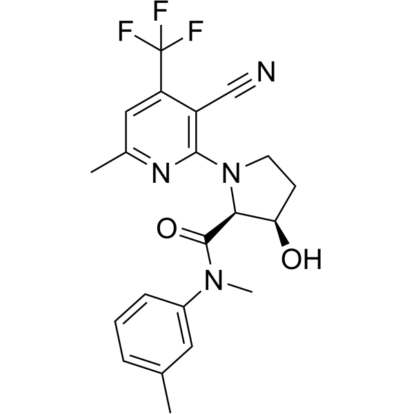
HY-141520
ART558Polθ Inhibitor -
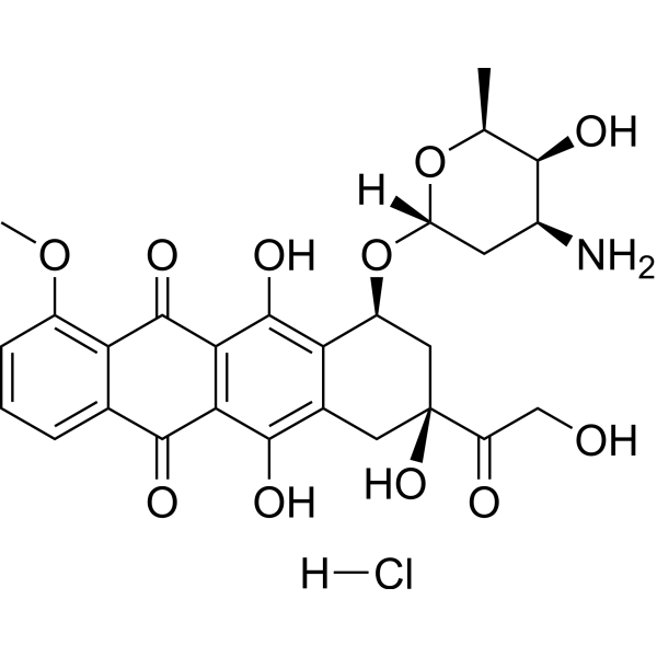
HY-15142
Doxorubicin hydrochlorideTopoisomerase Inhibitor -
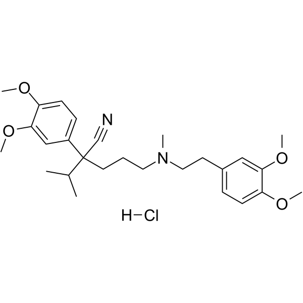
HY-A0064
Verapamil hydrochlorideCalcium Channel Blocker -
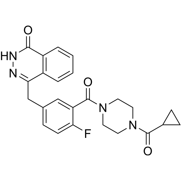
HY-10162
OlaparibPARP Inhibitor -
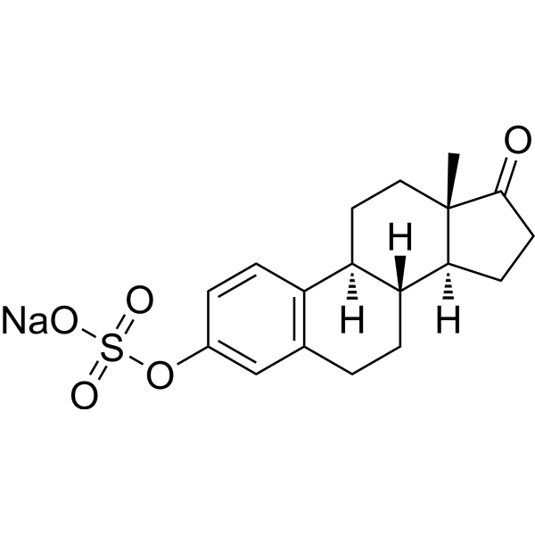
HY-113293B
Estrone sulfate sodiumEndogenous Estrogen -
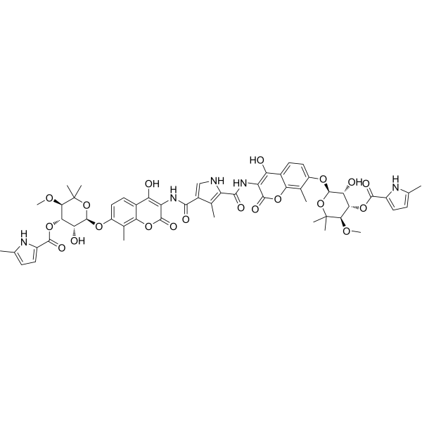
HY-N7452
Coumermycin A1JAK2 Signal Activator -
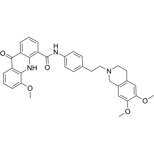
HY-50879
ElacridarP-gp And BCRP Dual-Inhibitor -
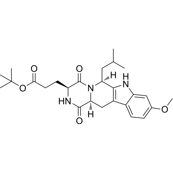
HY-10010
Ko 143BCRP/ABCG2 Inhibitor -

HY-10219
RapamycinmTOR Inhibitor -
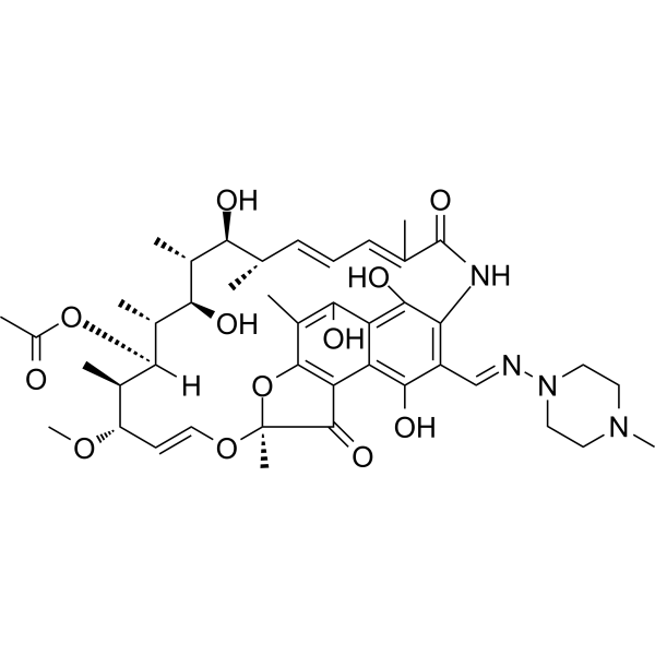
HY-B0272
RifampicinAntivirus Agent -
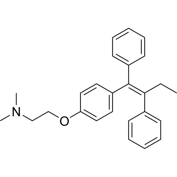
HY-13757A
TamoxifenEstrogen Receptor Modulator -
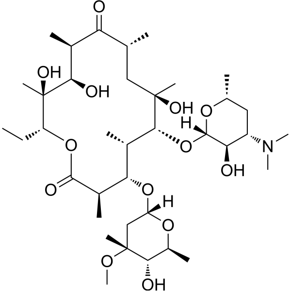
HY-B0220
ErythromycinAntibiotic
Publications Citing Use of MedChemExpress (MCE) Novobiocin sodium
More- Nat Methods. 2023 Sep;20(9):1388-1399. [Abstract]
- Blood. 2018 Jul 19;132(3):307-320. [Abstract]
- Adv Sci (Weinh). 2022 Dec;9(34):e2203088. [Abstract]
- Cell Rep Med. 2024 Jun 18;5(6):101592. [Abstract]
- Mol Ther. 2026 Jul 20:S1525-0016(26)00602-7.
- Mol Cancer Ther. 2026 Jul 15:10.1158/1535-7163.MCT-26-0182.
- Int J Mol Sci. 2019 Mar 5;20(5):1125. [Abstract]
- PLoS Pathog. 2026 Jan 26;22(1):e1013903. [Abstract]
- Mol Pharm. 2022 Nov 7;19(11):4320-4332. [Abstract]
- Cancer Res Commun. 2024 Apr 9;4(4):1024-1040. [Abstract]
- J Med Virol. 2025 Oct;97(10):e70655. [Abstract]
- Surgery. 2024 Nov;176(5):1380-1387. [Abstract]
- Vet Microbiol. 2026 May:316:110992. [Abstract]
- bioRxiv. 2025 Sep 15.
 Customer Validation & Images

-
Cell Imaging/Staining
-
Cell Proliferation/Viability Assay
-
Bio/Physico-chemical Assay
-
Cell Proliferation/Viability Assay
-
WB
All Antibiotic Isoforms
MoreAll DNA/RNA Synthesis Isoforms
MoreAll Parasite Isoforms
More

Biological Activity
Description
IC50 & Target
[1]|
β-lactam |
HSP90 |
In Vitro
Novobiocin (1-200 μM; 96 h) sodium inhibits the growth of Theileria equi and Babesia caballi in a dose-dependent manner, with IC50 values of 165 μM and 84.85 μM, respectively, and causes parasite death at high doses[1].
Novobiocin (1-2000 μM; 24 h) sodium shows low cytotoxicity against horse PBMCs, with a CC50 of 11.63 mM and an SSI of 70.47, indicating its safety for mammalian cells[1].
Novobiocin (1-2000 μM; 90 min) sodium shows extremely low hemolytic activity against horse erythrocytes, with a CC50 of 261.97 mM and an SSI of 1587, indicating its safety for host erythrocytes[1].
Novobiocin sodium inhibits MMEJ repair but not HR repair in U2OS cells[3].
Novobiocin sodium increases IR-induced RAD51 and γH2AX foci formation in U2OS cells[3].
Novobiocin sodium induces chromosomal aberrations and radial chromosomes in BRCA1-/- RPE1 cells[3].
Novobiocin (120 nM-1 mM; 1 h pre-incubation, 2 h reaction) sodium is a non-ATP-competitive inhibitor of Polθ-ATPase activity[4].
Novobiocin (4 mM; 30 min) sodium binds to the allosteric site of the Polθ-ATPase domain, affecting ATP hydrolysis and stabilizing the core of this domain[4].
Novobiocin sodium blocks the binding of Polθ to ssDNA in vitro[4].
Novobiocin (500 μM; 30 min) sodium stabilizes the Polθ-ATPase domain[4].
Novobiocin (500 μM; 1 h) sodium promotes tetramerization of the Polθ-ATPase domain[4].
Novobiocin (50-200 μM; 24 h) sodium blocks the binding of intracellular Polθ to ssDNA in a dose-dependent manner[4].
Novobiocin (200 μM; 2 h) sodium treatment leads to the accumulation of ssDNA gaps in RPE1 TP53-/- BRCA1-/- cells[4].
Novobiocin (1 day) sodium exhibits MIC values ranging from 0.25 to 16 μg mL-1 against N. gonorrhoeae DSM9188 and its mutant strains derived from single-colony isolation, with its primary target being the ParE subunit of DNA topoisomerase IV[2].
Novobiocin sodium exhibits low in vitro effective concentrations against both Bacillus anthracis and Burkholderia pseudomallei[5].
Novobiocin sodium specifically inhibits the ATPase activity of purified POLθ in vitro, with an IC50 of 24 μM[3].
Novobiocin (12-14 days) sodium selectively reduces the viability of BRCA1-/- and BRCA2-/- RPE1 cells compared with isogenic WT cells[3].
Novobiocin sodium shows increased resistance in POLQ-deficient U2OS cells[3].
Novobiocin sodium acts synergistically with Rucaparib (HY-10617A) to reduce the survival rate of BRCA1-/- RPE1 cells and decrease the IC50 of Rucaparib[3].
Novobiocin sodium synergizes with Olaparib in HR-deficient TOV21G + EV cells, reducing the IC50 of Olaparib by more than 40-fold[3].
Novobiocin sodium is cytotoxic to multiple PARPi-resistant clones derived from RPE1-BRCA1-/- cells[3].
Novobiocin (25-100 μM) sodium induces dose-dependent apoptosis in BRCA1-/- RPE1 cells but does not induce apoptosis in WT RPE1 cells[3].
MedChemExpress (MCE) has not independently confirmed the accuracy of these methods. They are for reference only. Further protocols information, click here.
-
Cell Line:Theileria equi and Babesia caballi parasitized horse red blood cells (RBCs)
-
Concentration:1, 5, 10, 20, 50, 100, 200 μM (T. equi); 10, 20, 50, 100, 200 μM (B. caballi)
-
Incubation Time:96 h
-
Result:Inhibited the in vitro growth of Theileria equi with an IC50 of 165 μM.
Inhibited the in vitro growth of Babesia caballi with an IC50 of 84.85 μM.
Showed significant growth inhibition of T. equi at 100 μM and 200 μM at 24 h and 48 h.
Showed significant growth inhibition of T. equi at ≥20 μM at 72 h and 96 h.
Showed significant growth inhibition of B. caballi at 100 and 200 μM at 24, 48, 72, and 96 h.
Caused parasite death at 100 μM and 200 μM after 96 h.
Failed to inhibit growth at 1-50 μM (T. equi) and 10-50 μM (B. caballi) in viability studies.
Induced dot-shaped/distorted nuclear material at 100 and 200 μM after 96 h.
Showed no recrudescence at 100 and 200 μM in viability testing.
-
Cell Line:Equine peripheral blood mononuclear cells (PBMCs)
-
Concentration:1, 5, 10, 25, 50, 100, 1000, 2000 μM
-
Incubation Time:24 h
-
Result:Showed less than 10% cytotoxicity at 1000 μM.
Exhibited a CC50 of 11.63 mM.
Demonstrated a specific selective index (SSI) of 70.47.
-
Cell Line:U2OS POLQ knockout cells expressing Myc-tagged Polθ
-
Concentration:50 μM, 100 μM, 200 μM
-
Incubation Time:24 h
-
Result:Reduced Polθ recruitment to ssDNA in cells in a dose-dependent manner.
Blocked 96% of Polθ from being recruited to damage sites at the highest dose of 200 μM.
In Vivo
Novobiocin (100 mg/kg; i.p.; twice daily; 35 days) sodium strongly inhibits the growth of Brca1-deficient GEMM-derived tumors and extends overall survival by 3-fold compared with the vehicle group[3].
Novobiocin (100 mg/kg; i.p.; twice daily; 4 weeks) sodium specifically inhibits the growth of FANCF-deficient HR-defective tumors in vivo [3].
Novobiocin (75 mg/kg; i.p.; twice daily; 4 weeks) sodium alone induces tumor regression and, in combination with Olaparib (HY-10162), achieves tumor regression in the HR-deficient DF83 PDX model[3].
Novobiocin (75 mg/kg; i.p.; twice daily; 4 weeks) sodium significantly inhibits tumor growth in the PARPi-resistant DF59 PDX model[3].
Novobiocin (75 mg/kg; i.p.; twice daily; 4 weeks) sodium does not inhibit tumor growth in the DF149 PDX model with normal HR function, which is associated with low POLθ expression[3].
Novobiocin (1.25-2.5 mg; i.p.; 1-3 times) sodium confers high protection against lethal intraperitoneal B. pseudomallei infection in BALB/c mice and promotes sterile immunity in most surviving mice[5].
Novobiocin (2.5 mg; i.p.; 2-5 doses) sodium significantly prolongs time to death but does not significantly improve overall survival in the highly lethal inhalational anthrax model in A/J mice[5].
Novobiocin (2.5 mg; i.p.; 4 doses; 7 days) sodium provides 70% protection against lethal aerosolized B. pseudomallei infection in BALB/c mice, and bacterial burden is typically undetectable in the tissues of surviving mice[5].
Novobiocin (2.5 mg; i.p.; 3 doses) sodium significantly synergizes with live attenuated vaccine, improving survival and prolonging time to death in a lethal aerosol B. pseudomallei model[5].
MedChemExpress (MCE) has not independently confirmed the accuracy of these methods. They are for reference only.
-
Animal Model:Mice (strain not specified)[1]
-
Dosage:5, 10, 20, 50, and 100 mg/kg
-
Administration:i.p.; single dose
-
Result:Induced a significant rise in serum glutamic oxaloacetic transaminase (SGOT) and serum glutamic pyruvic transaminase (SGPT) in mice at 100 mg/kg at 24 h, which decreased to non-significant levels thereafter.
Showed non-significant differences in kidney biochemical markers (creatinine, blood urea nitrogen) and other parameters (bilirubin and total protein) at different time intervals.
Exhibited minimal diffuse cytoplasmic rarefaction and vacuolation in hepatocytes at 100 mg/kg.
Showed no adverse histopathological changes in any organ at other doses.
Identified 50 mg/kg as the No Observed Adverse Effect Level (NOAEL).
-
Animal Model:FVB/129P2 (female, at least 6 weeks old, K14-Cre-Brca1^f/f;Trp53^f/f GEMM tumor transplanted into mammary fat pad)[3]
-
Dosage:100 mg/kg
-
Administration:i.p.; twice a day; 35 days
-
Result:Significantly suppressed tumor growth after 7 days of treatment and beyond.
Prolonged overall survival by 3-fold compared to vehicle, with median survival times of 29 and 10 days for treated and vehicle groups, respectively.
-
Animal Model:NU(NCr)-Foxn1^nu athymic nude (female)[3]
-
Dosage:100 mg/kg
-
Administration:i.p.; twice a day; 4 weeks
-
Result:Specifically impaired the growth of FANCF-deficient tumors, with no effect on FANCF-complemented tumors.
Strongly induced RAD51 foci in the treated tumors.
-
Animal Model:NSG (female, 8 weeks old)[3]
-
Dosage:75 mg/kg
-
Administration:i.p.; twice daily; 4 weeks
-
Result:Led to tumor regression as a single agent, while tumors in vehicle-treated mice showed exponential growth.
Achieved complete tumor regression in combination with olaparib, with few tumor cells detectable via BLI on day 28.\nSubstantially reduced tumor growth as monotherapy.
In combination with olaparib, further inhibited tumor growth with regression in the first two weeks.
Exhibited higher levels of phospho-RPA32 compared to vehicle-treated samples.
Exhibited a strong increase in RAD51 staining when treated alone or in combination with olaparib.\nDid not inhibit tumor growth in the HR-proficient DF149 PDX model.
-
Animal Model:BALB/c mice[5]
-
Dosage:1.25 mg; 2.5 mg
-
Administration:i.p.; 1, 2, or 3 doses
-
Result:Administered 1.25 mg or 2.5 mg in 1, 2, or 3 doses, achieving 70-90% protection except for a single 1.25 mg dose.
Administered 1.25 mg in 3 doses, achieving 90% protection.
Extended time-to-mortality in all treatment groups compared to PBS control.
Resulted in no detectable bacterial burden in spleens of most survivors.
-
Animal Model:A/J mice[5]
-
Dosage:2.5 mg
-
Administration:i.p.; 2, 3, 4, or 5 doses
-
Result:Administered 2.5 mg in 2 doses, resulting in a time-to-mortality of 5.8 days.
Administered 2.5 mg in 3 doses, resulting in a time-to-mortality of 6.7 days.
Administered 2.5 mg in 5 doses, resulting in a time-to-mortality of 6.9 days.
Administered 2.5 mg in 4 doses over 7 days, resulting in a time-to-mortality of 8.3 days.
Did not significantly improve overall survival rates by day 21.
-
Animal Model:BALB/c mice[5]
-
Dosage:2.5 mg
-
Administration:i.p.; 2, 3, 4, or 5 doses
-
Result:Administered 2.5 mg in 4 doses over 7 days, protecting 70% of mice.
Administered 2.5 mg in 5 doses over 3 days, protecting 60% of mice.
Administered 2.5 mg in 2 doses, protecting 22% of mice.
Administered 2.5 mg in 3 doses, protecting 20% of mice.
Resulted in no detectable bacteria in lung or spleen tissues of any survivors, except for one mouse in the 3-dose group with a bacterial burden of approximately 1.2 × 10^8 CFU/g in the spleen.
-
Animal Model:BALB/c mice (vaccinated)[5]
-
Dosage:2.5 mg
-
Administration:i.p.; 3 doses
-
Result:Achieved 80% survival at day 21.
Resulted in a geometric mean time-to-mortality of 35.1 days.
Yielded a synergy score of 3.15.


Chemical Information
-
CAS No. 1476-53-5
-
Appearance Solid
-
Molecular Weight 634.61
-
Formula C31H35N2NaO11
-
Color White to off-white
-
SMILES
CC1=C(OC2=O)C(C(O)=C2NC(C3=CC(C/C=C(C)/C)=C(O[Na])C=C3)=O)=CC=C1O[C@H](OC(C)(C)[C@H](OC)[C@H]4OC(N)=O)[C@@H]4O
-
Synonyms
Albamycin sodium; Cathomycin sodium
-
Initial Source
Streptomyces niveus
-
Shipping
Room temperature in continental US; may vary elsewhere.
-
Storage
4°C, sealed storage, away from moisture
* In solvent : -80°C, 1 year; -20°C, 6 months (sealed storage, away from moisture)


Publications (14)
-
Journal Impact Factor
-
Most Recent
-
Nat Methods
2023 Sep;20(9):1388-1399. PMID: 37474806
Novobiocin sodium purchased from MedChemExpress. Usage Cited in: Nat Methods. 2023 Sep;20(9):1388-1399. [Abstract]
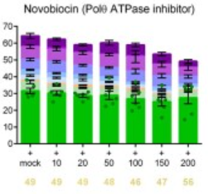
Editing efficiencies of the VCAN target using Cas9D10A double nicking in H9 hESCs carrying the K3753R mutation and additional small molecules to inhibit the MMEJ repair proteins Polϴ, PARP or Ligase I/III: Novobiocin.
-
Blood
Targeting HSP90 dimerization via the C terminus is effective in imatinib-resistant CML and lacks the heat shock response. [Abstract]2018 Jul 19;132(3):307-320. PMID: 29724897
Novobiocin sodium purchased from MedChemExpress. Usage Cited in: Blood. 2018 Jul 19;132(3):307-320. [Abstract]
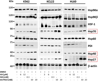
K562, KCL22 and HL60 are treated with the indicated (cytotoxic) concentration of Amidopyrine (AX), Novobiocin (NB) and AUY922 for 48h and later protein lysates are subjected to immunoblot analysis.
-
Adv Sci (Weinh)
SRSF5-Mediated Alternative Splicing of M Gene is Essential for Influenza A Virus Replication: A Host-Directed Target Against Influenza Virus. [Abstract]2022 Dec;9(34):e2203088. PMID: 36257906 -
Cell Rep Med
A CD36-dependent non-canonical lipid metabolism program promotes immune escape and resistance to hypomethylating agent therapy in AML. [Abstract]2024 Jun 18;5(6):101592. PMID: 38843841 -
-
-
Int J Mol Sci
Cisplatin Synergistically Enhances Antitumor Potency of Conditionally Replicating Adenovirus via p53 Dependent or Independent Pathways in Human Lung Carcinoma. [Abstract]2019 Mar 5;20(5):1125. PMID: 30841620
Novobiocin sodium purchased from MedChemExpress. Usage Cited in: Int J Mol Sci. 2019 Mar 5;20(5):1125. [Abstract]
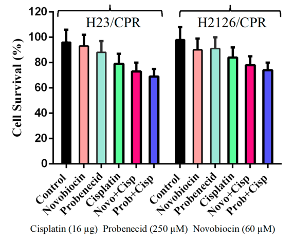
Cell viability assay to analyze the impact of Probenecid and Novobiocin treatments on resistance.
-
PLoS Pathog
Anti-orthopoxvirus drugs inhibit lumpy skin disease virus replication by targeting viral DNA polymerase. [Abstract]2026 Jan 26;22(1):e1013903. PMID: 41587213 -
Mol Pharm
2022 Nov 7;19(11):4320-4332. PMID: 36269563 -
Cancer Res Commun
Inhibition of NEK2 Promotes Chemosensitivity and Reduces KSHV-positive Primary Effusion Lymphoma Burden. [Abstract]2024 Apr 9;4(4):1024-1040. PMID: 38592451 -
J Med Virol
Drug Repurposing: In Vitro Evaluation of Simeprevir as a Novel Antiviral Drug Against Severe Fever With Thrombocytopenia Syndrome Virus. [Abstract]2025 Oct;97(10):e70655. PMID: 41117261
Novobiocin sodium purchased from MedChemExpress. Usage Cited in: J Med Virol. 2025 Oct;97(10):e70655. [Abstract]
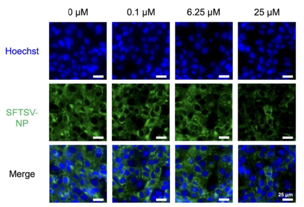
Novobiocin also reduced NP expression at 25 μM, with mean fluorescence intensities significantly lower than those of the untreated control.
Novobiocin sodium purchased from MedChemExpress. Usage Cited in: J Med Virol. 2025 Oct;97(10):e70655. [Abstract]
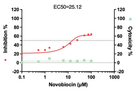
Cytoxicity and inhibition of Novobiocin, EC50 = 25.12 μM.
-
Surgery
Overexpression of human DNA polymerase theta is a biomarker of aggressive and DNA repair-deficient papillary thyroid cancers. [Abstract]2024 Nov;176(5):1380-1387. PMID: 38897886 -
Vet Microbiol
The Chinese medicine monomer Schisandrin C inhibits PRRSV infection by regulating the OGT-PI3K/AKT/mTOR signaling pathway. [Abstract]2026 May:316:110992. PMID: 41865607 -


Solvent & Solubility
In Vitro:
DMSO : ≥ 100 mg/mL (157.58 mM; Hygroscopic DMSO has a significant impact on the solubility of product, please use newly opened DMSO)
H2O : 50 mg/mL (78.79 mM; Need ultrasonic)
* "≥" means soluble, but saturation unknown.
Please refer to the solubility information to select the appropriate solvent. Once prepared, please aliquot and store the solution to prevent product inactivation from repeated freeze-thaw cycles.
Storage method and period of stock solution: -80°C, 1 year; -20°C, 6 months (sealed storage, away from moisture). When stored at -80°C, please use it within 1 year. When stored at -20°C, please use it within 6 months.
* Note: If you choose water as the stock solution, please dilute it to the working solution, then filter and sterilize it with a 0.22 μm filter before use.
Please refer to the solubility information to select the appropriate solvent. Once prepared, please aliquot and store the solution to prevent product inactivation from repeated freeze-thaw cycles.
Storage method and period of stock solution: -80°C, 1 year; -20°C, 6 months (sealed storage, away from moisture). When stored at -80°C, please use it within 1 year. When stored at -20°C, please use it within 6 months.
* Note: If you choose water as the stock solution, please dilute it to the working solution, then filter and sterilize it with a 0.22 μm filter before use.
Concentration (start) × Volume (start) = Concentration (final) × Volume (final)
In Vivo:
For the following dissolution methods, please prepare the working solution directly:
It is recommended to prepare fresh solutions and use them promptly within a short period of time.
The percentages shown for the solvents indicate their volumetric ratio in the final prepared solution. If precipitation or phase separation occurs during preparation, heat and/or sonication can be used to aid dissolution.
Add each solvent one by one: PBS
Solubility: 100 mg/mL (157.58 mM); Clear solution; Need ultrasonic and warming and heat to 60°C

Protocols
-
Apoptosis
Apoptosis, also called programmed cell death, is generally characterized by distinct morphological characteristics.
-
TUNEL staining for apoptotic DNA fragmentation
TUNEL staining detects DNA strand breaks by using terminal deoxynucleotidyl transferase to add labeled nucleotides to exposed 3′-OH DNA termini, generating either microscopic staining in fixed cells or tissue sections, or fluorescence/cytometric signal in cell suspensions. TUNEL positivity reflects DNA fragmentation but should not be interpreted alone as definitive apoptosis, because TUNEL can also label necrotic, autolytic, mechanically damaged, or DNA-repair-associated DNA breaks.
-
Cell Cytotoxicity Assay
Cytotoxicity assays are usually based on the assessment of cell membrane damage, which can also be indirectly detected by measuring cell viability. Detection methods include MTT assay, CKK-8 assay, LDH assay and ATP assay, etc.
-
Annexin V plus membrane-impermeant dye apoptosis staining
Annexin V-based apoptosis assays rely on the detection of phosphatidylserine (PS) externalization from the inner leaflet of the plasma membrane to the outer leaflet, an early biochemical hallmark of apoptosis. Fluorescently labeled Annexin V binds PS in a calcium-dependent manner, enabling identification of early apoptotic cells by flow cytometry or fluorescence microscopy. When combined with a membrane-impermeant DNA-binding dye (e. g. , propidium iodide), this approach allows discrimination between viable (Annexin V−/dye−), early apoptotic (Annexin V+/dye−), and late apoptotic or necrotic (Annexin V+/dye+) cell populations by assessing membrane integrity and PS exposure.
-
Patient-Derived Xenograft (PDX)
Patient-derived xenograft (PDX) models are generated by engrafting primary human tumor tissue directly into immunodeficient mice, allowing in vivo propagation of patient tumor biology without initial in vitro adaptation. These models are used to preserve key histopathological and molecular characteristics of the original tumor and enable assessment of tumor growth dynamics and therapeutic response in a living organism. The biological readout is tumor engraftment and subsequent growth in the murine host, which reflects the ability of human tumor cells to survive, vascularize, and expand in an immunocompromised microenvironment.
-
Apoptosis Solutions
Apoptosis is a regulated, generally non-lytic cell-death pathway that removes unwanted, damaged, infected, or abnormal cells through coordinated morphological changes, caspase activation, DNA fragmentation, and membrane remodeling. The intrinsic apoptosis pathway is controlled mainly by mitochondrial outer membrane permeabilization, BCL-2 family proteins, cytochrome c release, apoptosome formation, caspase-9 activation, and downstream executioner caspase-3/7 activation. The extrinsic apoptosis pathway is initiated by death receptors such as Fas, TNFR, and TRAIL receptors, which recruit adaptor proteins and activate caspase-8 before engaging executioner caspases or mitochondrial amplification through BID cleavage. Apoptosis is linked to many phenotypes, including cancer cell killing, tissue homeostasis, immune regulation, neurodegeneration, infection response, and treatment-induced cytotoxicity; unresolved questions include how apoptosis interacts with necroptosis, pyroptosis, ferroptos
-
Patient-Derived Orthotopic Xenograft (PDOX)
Patient-derived orthotopic xenograft (PDOX) modeling implants fresh patient tumor tissue or patient-derived tumor cells into the anatomically corresponding organ or tissue site of immunodeficient mice, usually by surgical orthotopic implantation, to preserve patient tumor histology, local microenvironmental context, invasion, metastatic behavior, and treatment-response features better than subcutaneous implantation. PDOX readouts include tumor engraftment, orthotopic tumor growth, local invasion, metastasis, recurrence after resection, histologic similarity to the donor tumor, biomarker retention, molecular concordance, survival, and response or resistance to therapy. PDOX models are used for preclinical drug testing and individualized therapy evaluation, but engraftment success varies by tumor type and specimen quality.
-
Subcutaneous Cell-Line-Derived Xenograft
Subcutaneous cell-line-derived xenograft (CDX) models are established by implanting cultured human cancer cell lines into immunodeficient mice, where the injected cells form localized tumors that can be monitored in vivo as a measure of tumorigenic potential, growth kinetics, and treatment response. These models are widely used in oncology research because they allow reproducible tumor formation and enable comparative assessment of tumor growth between different cell lines or genetic manipulations in a controlled in vivo microenvironment. Subcutaneous implantation of cancer cells in immunodeficient mice is a standard approach for evaluating tumor growth behavior and therapeutic response across multiple cancer types, including prostate, esophageal, pancreatic, and colon cancer models.
-
Breast Cancer Modeling
Breast cancer is a heterogeneous cancer, and it has been distinguished into four subtypes: luminal A, luminal B, HER2-positive and basal-like. Molecular mutations, epigenetic alterations, hormone exposure and immune microenvironment are related to the progression of breast cancer.
-
Orthotopic Cell-Line Xenograft
Orthotopic cell-line xenograft models involve implantation of human cancer cell lines into the anatomically corresponding organ of immunodeficient mice to reproduce tumor growth within a native microenvironment, enabling more clinically relevant tumor behavior compared with subcutaneous models. These models are widely used because orthotopic placement better recapitulates tumor progression, including invasion and metastatic spread, which are often underrepresented in heterotopic implantation systems. Compared with conventional xenografts, orthotopic implantation is described as more technically complex but provides improved simulation of tumor-microenvironment interactions and metastatic behavior, making it particularly valuable for translational oncology research. Surgical orthotopic implantation approaches have been emphasized as enabling faithful reproduction of clinical cancer features, including metastasis and disease progression patterns that align with the tumor’s organ of origi


Purity & Documentation
-
Data Sheet (296 KB)
-
SDS (393 KB)
- English - EN (393 KB)
- Français - FR (393 KB)
- Deutsch - DE (393 KB)
- Norwegian - NO (393 KB)
- Español - ES (393 KB)
- Swedish - SV (393 KB)
- Italian - IT (393 KB)
- Korean - KR (393 KB)
- Portuguese - PT (393 KB)
-
Handling Instructions (2659 KB)
References
[3]. Zhou J, et al. A first-in-class Polymerase Theta Inhibitor selectively targets Homologous-Recombination-Deficient Tumors. Nature cancer. 2021 Jun;2(6):598-610. [Content Brief]
[6]. Marcu MG, et al. The heat shock protein 90 antagonist novobiocin interacts with a previously unrecognized ATP-binding domain in the carboxyl terminus of the chaperone. J Biol Chem. 2000 Nov 24;275(47):37181-6. [Content Brief]


Complete Stock Solution Preparation Table
Please refer to the solubility information to select the appropriate solvent. Once prepared, please aliquot and store the solution to prevent product inactivation from repeated freeze-thaw cycles.
Storage method and period of stock solution: -80°C, 1 year; -20°C, 6 months (sealed storage, away from moisture). When stored at -80°C, please use it within 1 year. When stored at -20°C, please use it within 6 months.
| Optional Solvent | Concentration Solvent Mass | 1 mg | 5 mg | 10 mg | 25 mg |
|---|---|---|---|---|---|
| H2O / DMSO | 1 mM | 1.5758 mL | 7.8789 mL | 15.7577 mL | 39.3943 mL |
| 5 mM | 0.3152 mL | 1.5758 mL | 3.1515 mL | 7.8789 mL | |
| 10 mM | 0.1576 mL | 0.7879 mL | 1.5758 mL | 3.9394 mL | |
| 15 mM | 0.1051 mL | 0.5253 mL | 1.0505 mL | 2.6263 mL | |
| 20 mM | 0.0788 mL | 0.3939 mL | 0.7879 mL | 1.9697 mL | |
| 25 mM | 0.0630 mL | 0.3152 mL | 0.6303 mL | 1.5758 mL | |
| 30 mM | 0.0525 mL | 0.2626 mL | 0.5253 mL | 1.3131 mL | |
| 40 mM | 0.0394 mL | 0.1970 mL | 0.3939 mL | 0.9849 mL | |
| 50 mM | 0.0315 mL | 0.1576 mL | 0.3152 mL | 0.7879 mL | |
| 60 mM | 0.0263 mL | 0.1313 mL | 0.2626 mL | 0.6566 mL | |
| DMSO | 80 mM | 0.0197 mL | 0.0985 mL | 0.1970 mL | 0.4924 mL |
| 100 mM | 0.0158 mL | 0.0788 mL | 0.1576 mL | 0.3939 mL |
* Note: If you choose water as the stock solution, please dilute it to the working solution, then filter and sterilize it with a 0.22 μm filter before use.


Novobiocin sodium Related Classifications
HY-B0425A Related Classifications
 Powered by Bioz
Keywords
- Novobiocin
- 1476-53-5
- Albamycin
- Cathomycin
- Antibiotic
- DNA/RNA Synthesis
- HSP
- Apoptosis
- Bacterial
- Parasite
- Theileria equi
- POLθ ATPase domain
- Hsp-90 inhibitor
- U2OS cells
- BRCA1-/- RPE1 cells
- microhomology-mediated end joining repair
- N. gonorrhoeae
- Babesia caballi
- Bacillus anthracis
- Burkholderia pseudomallei
- Inhibitor
- inhibitor
- inhibit