



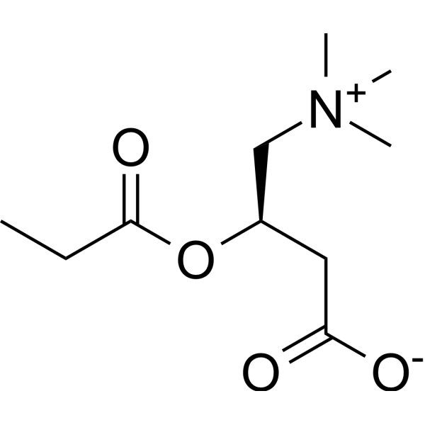
Propionyl-L-carnitine

Based on 1 Customer Validation
Propionyl-L-carnitine is an orally active L-carnitine derivative. Propionyl-L-carnitine has a high affinity for muscle L-carnitine transferase. Propionyl-L-carnitine increases Apoptosis, Bax, and reduces NF-κB, VCAM-1, MCP-1, and survivin. Propionyl-L-carnitine activates Src kinase, Akt, induces p-AMPK and nitric oxide synthesis. Propionyl-L-carnitine alleviates cardiovascular disease, obesity, and colitis.
For research use only. We do not sell to patients.
- Purity : 95.0%
- CAS No.: 20064-19-1
- Formula: C10H19NO4
- Molecular Weight:217.26
-
Storage:Powder -20°C, 3 years , 4°C, 2 years ; In solvent -80°C, 6 months , -20°C, 1 month

To place orders, for customer services and technical support, please contact: MedChemExpress USA
Tel: 609-228-6898 E-mail: [email protected] [email protected]


-
 Biological Activity
-
Chemical Information
-
Solvent & Solubility
- Protocols
- Purity & Documentation
- References
-
 Help & FAQs




Biological Activity
Description
IC50 & Target
IC50: muscular carnitine transferase[1]
In Vitro
Propionyl-L-carnitine (0-100 μM; 0-8 h) induces eNOS activation and nitric oxide synthesis in endothelial cells HAEC via PI3 and Akt kinases[2].
Propionyl-L-carnitine (100 μM; 6-24 h) induces IκB-α mRNA, and inhibits p65 and p50 in intimal cells[3].
MedChemExpress (MCE) has not independently confirmed the accuracy of these methods. They are for reference only. Further protocols information, click here.
-
Cell Line:HAEC
-
Concentration:25, 50, 75, 100 μM
-
Incubation Time:2, 4, 6, 8 h
-
Result:Increased eNOS phosphorylation and activity in a concentration- and time-dependent manner.
Increased eNOS activity by 350% at 50 μM.
Induced Akt activation.
Activated Src kinase.
Induced phosphorylation of AMPK.
In Vivo
Propionyl-L-carnitine (30-120 mg/kg/day, p.o., drinking water) enhances wound healing and counteracts microvascular endothelial cell dysfunction in rats[4].
Propionyl-L-carnitine (3.6 mg/mL, infused into the renal artery at the rate of 0.4 mL/min) prevents renal function deterioration due to ischemia/reperfusion in male Sprague-Dawley rats[5].
Propionyl-L-carnitine (200 mg/kg/day, p.o., 4 weeks) corrects metabolic and cardiovascular alterations in diet-induced obese mice and improves liver respiratory chain activity[6].
Propionyl-L-carnitine (25 mg/kg, intrarectal, twice daily, for 1 week; 120 mg/kg, p.o., once daily, for 1 week) reduces intestinal damage and microvascular dysfunction in rat TNBS-induced acute and reactivated colitis[7].
Propionyl-L-carnitine (200 mg/kg, p.o., drinking water) increases NO synthesis by enhancing eNOS expression in spontaneously hypertensive rats[8].
Propionyl-L-carnitine (500 mg/kg, i.p., 10 successive days) prevents the progression of Cisplatin (HY-17394)-induced cardiomyopathy in a carnitine-depleted rat model[9].
Propionyl-L-carnitine (200 mg/kg per d, p.o., 20 weeks) reduces body weight and hyperinsulinaemia in obese Zucker rats[10].
MedChemExpress (MCE) has not independently confirmed the accuracy of these methods. They are for reference only.
-
Animal Model:Male Wistar rats (200-225 g, 13-14 weeks old), skin flap model and full-thickness skin wound model[4]
-
Dosage:For the skin flap model, 100 mg/kg (dissolved in drinking water); for the full-thickness skin wound model, 30 mg/kg, 60 mg/kg, 120 mg/kg (dissolved in water).
-
Administration:Oral gavage (p.o.), drinking water
-
Result:Showed a faster blood flow recovery of the flap, an improved viability, reduced ROS production, increased NO level and iNOS expression (In the skin flap model).
Reduced the necrotic skin area after 8 days (in the skin flap model).
Had faster re-epithelialization, increased CD31+ capillaries, iNOS, VEGF and PlGF expression, and reduced Nox4+ microvessels (in the full-thickness skin wound model).
Clinical Trial
| NCT Number | Sponsor | Condition | Start Date |
Phase

|
|---|---|---|---|---|
| NCT01329991 | Plexxikon| | 2011-05 | PHASE1 |


Chemical Information
-
CAS No. 20064-19-1
-
Appearance Solid
-
Molecular Weight 217.26
-
Formula C10H19NO4
-
Color White to off-white
-
Shipping
Room temperature in continental US; may vary elsewhere.
-
Storage
Powder -20°C 3 years 4°C 2 years In solvent -80°C 6 months -20°C 1 month


Solvent & Solubility
In Vitro:
H2O : 125 mg/mL (575.35 mM; Need ultrasonic)
Please refer to the solubility information to select the appropriate solvent. Once prepared, please aliquot and store the solution to prevent product inactivation from repeated freeze-thaw cycles.
Storage method and period of stock solution: -80°C, 6 months; -20°C, 1 month. When stored at -80°C, please use it within 6 months. When stored at -20°C, please use it within 1 month.
* Note: If you choose water as the stock solution, please dilute it to the working solution, then filter and sterilize it with a 0.22 μm filter before use.
Please refer to the solubility information to select the appropriate solvent. Once prepared, please aliquot and store the solution to prevent product inactivation from repeated freeze-thaw cycles.
Storage method and period of stock solution: -80°C, 6 months; -20°C, 1 month. When stored at -80°C, please use it within 6 months. When stored at -20°C, please use it within 1 month.
* Note: If you choose water as the stock solution, please dilute it to the working solution, then filter and sterilize it with a 0.22 μm filter before use.
Concentration (start) × Volume (start) = Concentration (final) × Volume (final)

Protocols
-
Apoptosis
Apoptosis, also called programmed cell death, is generally characterized by distinct morphological characteristics.
-
TUNEL staining for apoptotic DNA fragmentation
TUNEL staining detects DNA strand breaks by using terminal deoxynucleotidyl transferase to add labeled nucleotides to exposed 3′-OH DNA termini, generating either microscopic staining in fixed cells or tissue sections, or fluorescence/cytometric signal in cell suspensions. TUNEL positivity reflects DNA fragmentation but should not be interpreted alone as definitive apoptosis, because TUNEL can also label necrotic, autolytic, mechanically damaged, or DNA-repair-associated DNA breaks.
-
Annexin V plus membrane-impermeant dye apoptosis staining
Annexin V-based apoptosis assays rely on the detection of phosphatidylserine (PS) externalization from the inner leaflet of the plasma membrane to the outer leaflet, an early biochemical hallmark of apoptosis. Fluorescently labeled Annexin V binds PS in a calcium-dependent manner, enabling identification of early apoptotic cells by flow cytometry or fluorescence microscopy. When combined with a membrane-impermeant DNA-binding dye (e. g. , propidium iodide), this approach allows discrimination between viable (Annexin V−/dye−), early apoptotic (Annexin V+/dye−), and late apoptotic or necrotic (Annexin V+/dye+) cell populations by assessing membrane integrity and PS exposure.
-
DSS-Induced Colitis
Dextran sulfate sodium (DSS)-induced colitis is generated by administering DSS in mouse drinking water, producing epithelial injury, barrier disruption, weight loss, diarrhea, fecal blood, colon shortening, histologic mucosal damage, and inflammatory mediator changes; the model is mainly used to study acute or chronic intestinal inflammation resembling selected features of ulcerative colitis. DSS injury is interpreted through clinical and tissue readouts rather than a single molecular endpoint: daily body weight, stool consistency, and bleeding are combined into a disease activity index, while colon length, histology, cytokines, myeloperoxidase activity, intestinal permeability, and tight-junction markers provide complementary measures of inflammation and barrier damage.
-
TNBS-Induced Colitis
TNBS-induced colitis is produced by intrarectal delivery of 2,4,6-trinitrobenzene sulfonic acid in ethanol, where ethanol disrupts the mucosal barrier and TNBS haptenates colonic proteins, generating immune-mediated colonic inflammation with weight loss, diarrhea, ulceration, transmural injury, inflammatory-cell infiltration, and cytokine responses. The model is used as an experimental intestinal inflammation model with Crohn’s disease–like features, especially when Th1-type responses, IL-12–dependent inflammation, chronic relapsing inflammation, or fibrosis-related endpoints are studied.
-
Apoptosis Solutions
Apoptosis is a regulated, generally non-lytic cell-death pathway that removes unwanted, damaged, infected, or abnormal cells through coordinated morphological changes, caspase activation, DNA fragmentation, and membrane remodeling. The intrinsic apoptosis pathway is controlled mainly by mitochondrial outer membrane permeabilization, BCL-2 family proteins, cytochrome c release, apoptosome formation, caspase-9 activation, and downstream executioner caspase-3/7 activation. The extrinsic apoptosis pathway is initiated by death receptors such as Fas, TNFR, and TRAIL receptors, which recruit adaptor proteins and activate caspase-8 before engaging executioner caspases or mitochondrial amplification through BID cleavage. Apoptosis is linked to many phenotypes, including cancer cell killing, tissue homeostasis, immune regulation, neurodegeneration, infection response, and treatment-induced cytotoxicity; unresolved questions include how apoptosis interacts with necroptosis, pyroptosis, ferroptos
-
Research Protocol for Cardiovascular Diseases
Cardiovascular disease can be modeled as maladaptive cardiac remodeling, where ischemic injury or pressure overload activates inflammatory signaling, fibroblast activation, extracellular-matrix deposition, cardiomyocyte hypertrophy, vascular remodeling, and progressive ventricular dysfunction. The TGF-β/SMAD axis is a central profibrotic pathway after myocardial injury and pressure overload, while innate immune and cytokine pathways regulate leukocyte recruitment, scar formation, and adverse remodeling. Key unresolved questions include which inflammatory signals are reparative versus harmful, when fibrosis is protective versus maladaptive, and whether pathway inhibition improves function without weakening necessary infarct healing or compensatory remodeling.
-
Research Protocol for Inflammation-related Diseases
The NLRP3 inflammasome is a cytosolic innate immune signaling platform that integrates priming signals and danger-signal activation to promote caspase-1 activation, maturation of IL-1β and IL-18, and gasdermin D-mediated pyroptotic cell death. The core experimental logic is to determine whether inflammatory disease phenotypes are driven by increased NLRP3 expression, ASC-containing inflammasome assembly, caspase-1 cleavage, GSDMD cleavage, and extracellular release of IL-1β/IL-18 rather than by nonspecific cell injury alone. The pathway is strongly linked to inflammation-related disease phenotypes because monosodium urate crystals activate NALP3/NLRP3 inflammasome signaling in gout-like crystal inflammation, cholesterol crystals activate NLRP3 inflammasomes in atherogenesis models, and DSS-induced intestinal inflammation has been reported to involve NLRP3 inflammasome activity. However, experimental colitis studies also show context-dependent protective effects of NLRP3 inflammasome co
-
Research Protocol for Metabolic Diseases
AMP-activated protein kinase, AMPK, is a conserved cellular energy sensor that responds to reduced cellular energy status and coordinates metabolism by increasing ATP-generating catabolic pathways while suppressing ATP-consuming anabolic processes. In metabolic disease research, the AMPK pathway is experimentally relevant because it regulates hepatic lipid synthesis, fatty acid oxidation, glucose production, skeletal-muscle glucose disposal, mTORC1-linked biosynthesis, autophagy, mitochondrial homeostasis, and whole-body energy balance. The central pathway logic is that energy stress, metformin, exercise-like stimulation, or direct AMPK activators increase AMPKα Thr172 phosphorylation and downstream substrate phosphorylation, including ACC and RAPTOR. Phosphorylation of ACC suppresses lipogenesis and supports fatty acid oxidation, whereas phosphorylation of RAPTOR suppresses mTORC1 signaling and links cellular energy status to growth and protein synthesis control. The pathway is linked


Purity & Documentation
-
Data Sheet (281 KB)
-
SDS (393 KB)
- English - EN (393 KB)
- Français - FR (393 KB)
- Deutsch - DE (393 KB)
- Norwegian - NO (393 KB)
- Español - ES (393 KB)
- Swedish - SV (393 KB)
- Italian - IT (393 KB)
- Korean - KR (393 KB)
- Portuguese - PT (393 KB)
-
Handling Instructions (2659 KB)
References
[1]. Victor Kamoen, et al. Propionyl-L-carnitine for intermittent claudication. Cochrane Database Syst Rev. 2021 Dec 26;12(12):CD010117. [Content Brief]
[2]. Ning WH, et al. Propionyl-L-carnitine induces eNOS activation and nitric oxide synthesis in endothelial cells via PI3 and Akt kinases. Vascul Pharmacol. 2013 Sep-Oct;59(3-4):76-82. [Content Brief]
[3]. Orlandi A, et al. Propionyl-L-carnitine reduces proliferation and potentiates Bax-related apoptosis of aortic intimal smooth muscle cells by modulating nuclear factor-kappaB activity. J Biol Chem. 2007 Feb 16;282(7):4932-4942. [Content Brief]
[4]. Scioli MG, et al. Propionyl-L-Carnitine Enhances Wound Healing and Counteracts Microvascular Endothelial Cell Dysfunction. PLoS One. 2015 Oct 16;10(10):e0140697. [Content Brief]
[5]. Mister M, et al. Propionyl-L-carnitine prevents renal function deterioration due to ischemia/reperfusion. Kidney Int. 2002 Mar;61(3):1064-78. [Content Brief]
[6]. Mingorance C, et al. Propionyl-L-carnitine corrects metabolic and cardiovascular alterations in diet-induced obese mice and improves liver respiratory chain activity. PLoS One. 2012;7(3):e34268. [Content Brief]
[7]. Scioli MG, et al. Propionyl-L-Carnitine is Efficacious in Ulcerative Colitis Through its Action on the Immune Function and Microvasculature. Clin Transl Gastroenterol. 2014 Mar 20;5(3):e55. [Content Brief]
[8]. Alvarez de Sotomayor M, et al. Effect of L-carnitine and propionyl-L-carnitine on endothelial function of small mesenteric arteries from SHR. J Vasc Res. 2007;44(5):354-64. [Content Brief]
[9]. Al-Majed AA, et al. Propionyl-L-carnitine prevents the progression of cisplatin-induced cardiomyopathy in a carnitine-depleted rat model. Pharmacol Res. 2006 Mar;53(3):278-86. [Content Brief]
[10]. Mingorance C, et al. Oral supplementation of propionyl-l-carnitine reduces body weight and hyperinsulinaemia in obese Zucker rats. Br J Nutr. 2009 Oct;102(8):1145-53. [Content Brief]


Complete Stock Solution Preparation Table
Please refer to the solubility information to select the appropriate solvent. Once prepared, please aliquot and store the solution to prevent product inactivation from repeated freeze-thaw cycles.
Storage method and period of stock solution: -80°C, 6 months; -20°C, 1 month. When stored at -80°C, please use it within 6 months. When stored at -20°C, please use it within 1 month.
| Optional Solvent | Concentration Solvent Mass | 1 mg | 5 mg | 10 mg | 25 mg |
|---|---|---|---|---|---|
| H2O | 1 mM | 4.6028 mL | 23.0139 mL | 46.0278 mL | 115.0695 mL |
| 5 mM | 0.9206 mL | 4.6028 mL | 9.2056 mL | 23.0139 mL | |
| 10 mM | 0.4603 mL | 2.3014 mL | 4.6028 mL | 11.5070 mL | |
| 15 mM | 0.3069 mL | 1.5343 mL | 3.0685 mL | 7.6713 mL | |
| 20 mM | 0.2301 mL | 1.1507 mL | 2.3014 mL | 5.7535 mL | |
| 25 mM | 0.1841 mL | 0.9206 mL | 1.8411 mL | 4.6028 mL | |
| 30 mM | 0.1534 mL | 0.7671 mL | 1.5343 mL | 3.8357 mL | |
| 40 mM | 0.1151 mL | 0.5753 mL | 1.1507 mL | 2.8767 mL | |
| 50 mM | 0.0921 mL | 0.4603 mL | 0.9206 mL | 2.3014 mL | |
| 60 mM | 0.0767 mL | 0.3836 mL | 0.7671 mL | 1.9178 mL | |
| 80 mM | 0.0575 mL | 0.2877 mL | 0.5753 mL | 1.4384 mL | |
| 100 mM | 0.0460 mL | 0.2301 mL | 0.4603 mL | 1.1507 mL |
* Note: If you choose water as the stock solution, please dilute it to the working solution, then filter and sterilize it with a 0.22 μm filter before use.


Propionyl-L-carnitine Related Classifications
HY-121705 Related Classifications
 Powered by Bioz