



Voacamine
Based on 1 publication(s) in Google Scholar
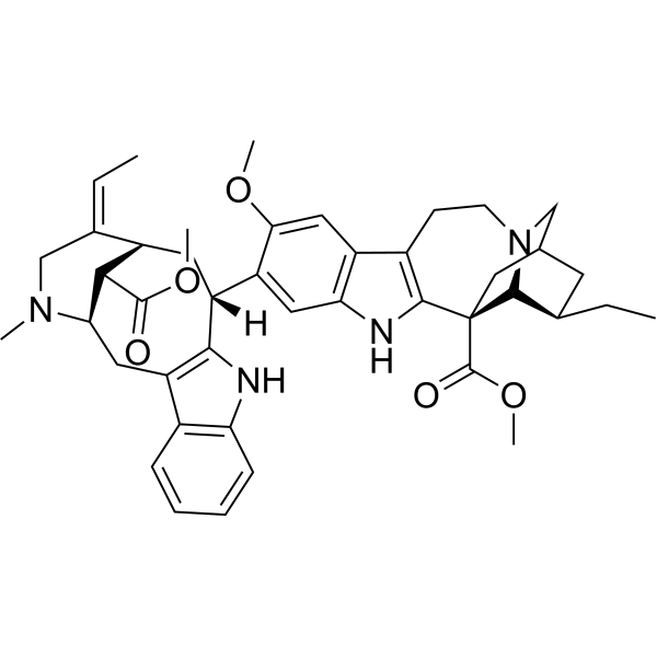
Voacamine is an indole alkaloid with cannabinoid 1 (CB1) antagonistic activity. Voacamine can inhibit nuclear translocation. Voacamine is effective in enhancing the effect of Doxorubicin (HY-15142A) as it interferes with the P-glycoprotein (P-gp) function. Voacamine promotes apoptosis-independent autophagic cell death in human osteosarcoma cells. Voacamine activates mitochondrial-associated apoptosis signaling pathway and inhibition of PI3K/Akt/mTOR signaling pathway to suppress breast cancer progression. Voacamine inhibits EGFR to exert oncogenic activity against colorectal cancer.
For research use only. We do not sell to patients.
- Purity : 99.64%
- CAS No.: 3371-85-5
- Formula: C43H52N4O5
- Molecular Weight:704.90
-
Storage:
4°C, protect from light
* In solvent : -80°C, 6 months; -20°C, 1 month (protect from light)

To place orders, for customer services and technical support, please contact: MedChemExpress USA
Tel: 609-228-6898 E-mail: [email protected] [email protected]


-
 Biological Activity
-
Chemical Information
-
Solvent & Solubility
- Protocols
- Purity & Documentation
- References
-
 Help & FAQs


-
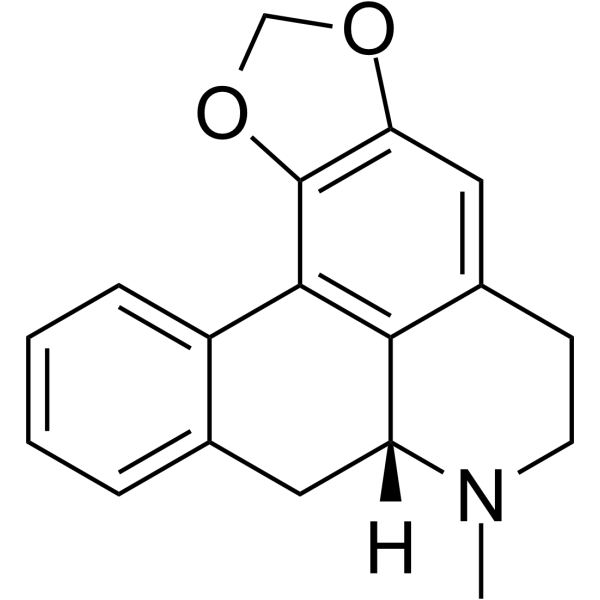
HY-121793
RoemerineEndogenous Metabolite -
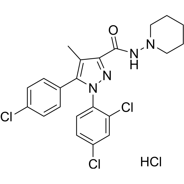
HY-14137
Rimonabant HydrochlorideCB1 Antagonist -
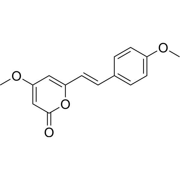
HY-N0919
YangoninCannabinoid Receptor Inhibitor -
HY-14275
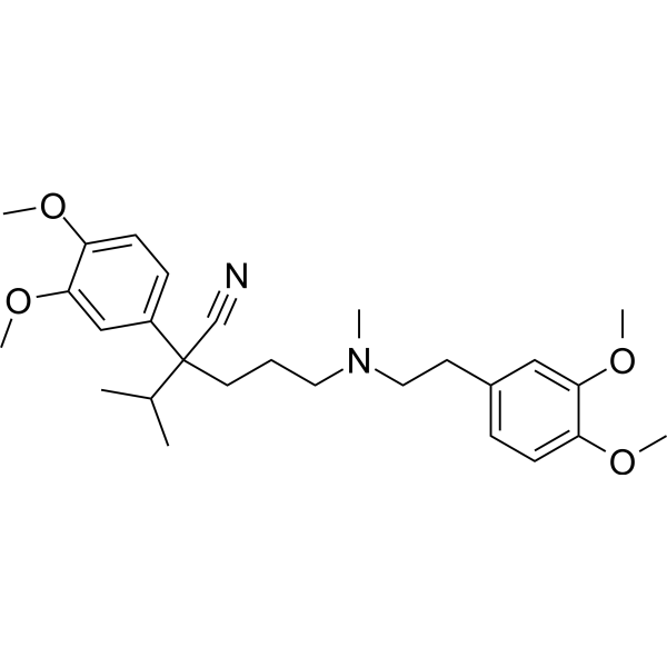
VerapamilCalcium Channel Blocker -
HY-17387
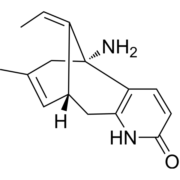
(-)-Huperzine AAChE/NMDA Receptor Inhibitor -
HY-101388
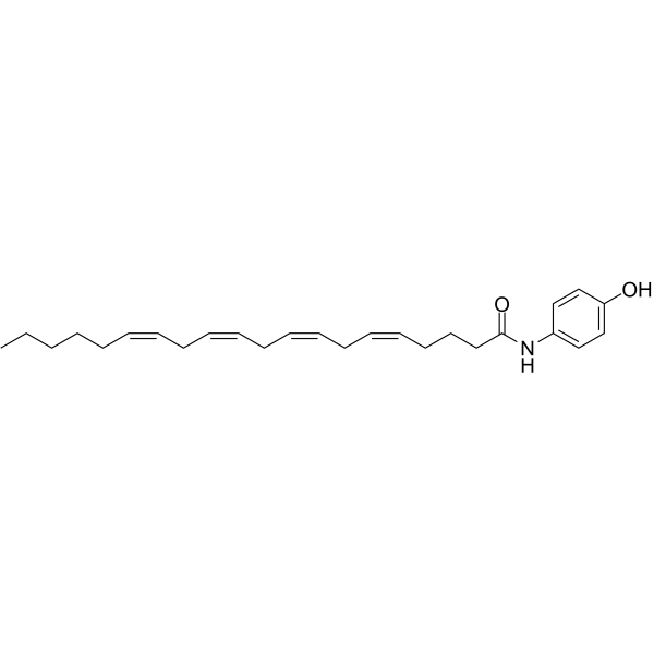
AM404Anandamide Inhibitor -
HY-112340
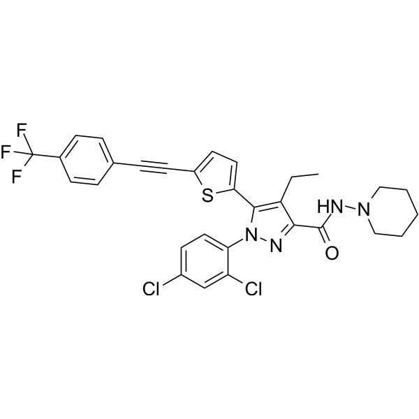
CB1 antagonist 4CB1 Receptor Antagonist -
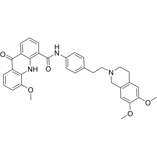
HY-50879
ElacridarP-gp And BCRP Dual-Inhibitor -
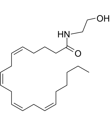
HY-10863
AnandamideCB1/2 Agonist -
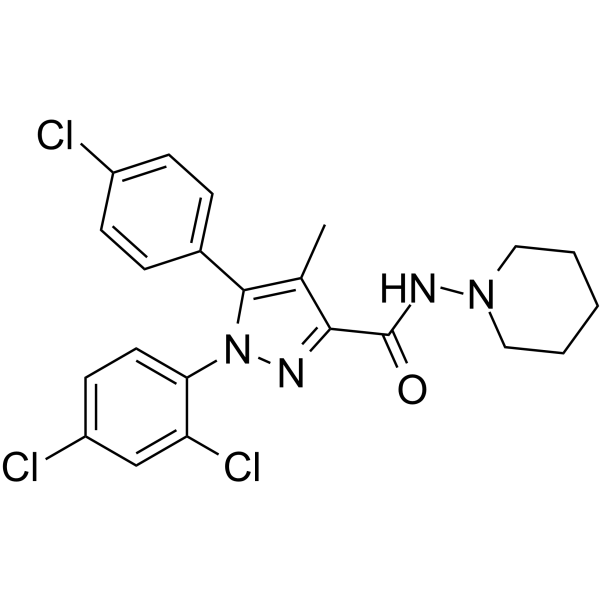
HY-14136
Rimonabant -
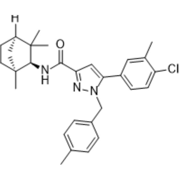
HY-13439
SR144528CB2 Receptor Antagonist -
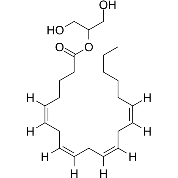
HY-W011051
2-Arachidonoylglycerol
Publications Citing Use of MedChemExpress (MCE) Voacamine
MoreAll EGFR Isoforms
More

Biological Activity
Description
In Vitro
Voacamine (1.4 μM) induces a slight increase in the apoptotic fraction of 23.6% in A2780 cells[2].
Voacamine (Compound VOA) (0-18 μM, 24-48 h) has antiproliferative activities against breast cancer cell lines (MCF-7 and 4T1)[3].
Voacamine (0-10 μM, 24-48 h) has anti-migratory and proapoptotic effect on MCF-7 and 4T1 cells[3].
Voacamine (1.5-5 μM) significantly diminishes the G0/G1 and G2/M phase of MCF-7 and 4T1 cells[3].
Voacamine (2.5-9 μM) collapses MMP and induces ROS production in MCF-7 and 4T1 cells[3].
Voacamine (0-8 μM) induces inhibition of p-PI3K/p-Akt/p-mTOR pathway and interacts with PI3K protein via molecular docking simulation[3].
Voacamine (1-10 μM, 24-72 h) results in a dose- and time-dependent reduction in the cell survival in U-2 osteosarcoma cells[4].
Voacamine (5 μM, 24 h) shows a higher number of cells in G1 phase than control and abundant formation of autophagosomes in U-2 osteosarcoma cells[4].
Voacamine (0-10 μM, 24-48 h) substantially restrains the viability of CRC cell lines (CT26 and HCT116) [5].
Voacamine (0-10 μM) induces apoptisis via EGFR/PI3K/Akt signaling pathway in CT26 and HCT116 cells[5].
MedChemExpress (MCE) has not independently confirmed the accuracy of these methods. They are for reference only. Further protocols information, click here.
-
Cell Line:MCF-7 and 4T1 cells
-
Concentration:0, 3, 6, 9, 12, 15, 18 μM (for MCF-7); 0, 2.5, 5, 7.5, 10, 12.5, 15 μM (for 4T1)
-
Incubation Time:24 and 48 h
-
Result:Exhibited low toxicities against human normal breast epithelial cells.
Had the most potent cytotoxic effect on breast cancer cells for 48 h with an IC50 of 0.99 and 1.42 μM for MCF-7 and 4T1 cells, respectively.
Reduced colony formation of MCF-7 and 4T1 cells.
In Vivo
Voacamine (15-30 mg/kg, i.p., 2 w) suppresses the progression of xenograft colorectal tumor in BALB/c mice injected with CT26 cells[5].
MedChemExpress (MCE) has not independently confirmed the accuracy of these methods. They are for reference only.
-
Animal Model:BALB/c mice with 4T1 cells induced xenograft tumor[3]
-
Dosage:15 and 30 mg/kg
-
Administration:Subcutaneous injection (s.c.)
-
Result:Increased body weights of mice whilst reduced the tumor volume and weight with 30 mg/kg dose.
Significantly downregulated levels of p-PI3K in a dose-dependent manner.


Chemical Information
-
CAS No. 3371-85-5
-
Appearance Solid
-
Molecular Weight 704.90
-
Formula C43H52N4O5
-
Color White to off-white
-
SMILES
O=C(OC)[C@]1([C@@H]2[N@](CC(C[C@H]2CC)C1)CC3)C4=C3C(C=C5OC)=C(N4)C=C5[C@](C[C@@H]6[C@H](C(OC)=O)[C@H](N(C)C/C6=C/C)C7)([H])C8=C7C9=C(N8)C=CC=C9
-
Structure Classification
-
Initial Source
-
Shipping
Room temperature in continental US; may vary elsewhere.
-
Storage
4°C, protect from light
* In solvent : -80°C, 6 months; -20°C, 1 month (protect from light)


Publications (1)
-
Journal Impact Factor
-
Most Recent
-
RSC Adv
A multi-stage computational pipeline and in vitro validation for the discovery of small-molecule translation inhibitors targeting the bacterial ribosome. [Abstract]2026 Apr 7;16(20):18359-18373. PMID: 41953617


Solvent & Solubility
In Vitro:
DMSO : ≥ 100 mg/mL (141.86 mM; Hygroscopic DMSO has a significant impact on the solubility of product, please use newly opened DMSO)
* "≥" means soluble, but saturation unknown.
Please refer to the solubility information to select the appropriate solvent. Once prepared, please aliquot and store the solution to prevent product inactivation from repeated freeze-thaw cycles.
Storage method and period of stock solution: -80°C, 6 months; -20°C, 1 month (protect from light). When stored at -80°C, please use it within 6 months. When stored at -20°C, please use it within 1 month.
Please refer to the solubility information to select the appropriate solvent. Once prepared, please aliquot and store the solution to prevent product inactivation from repeated freeze-thaw cycles.
Storage method and period of stock solution: -80°C, 6 months; -20°C, 1 month (protect from light). When stored at -80°C, please use it within 6 months. When stored at -20°C, please use it within 1 month.
Concentration (start) × Volume (start) = Concentration (final) × Volume (final)
In Vivo:
Select the appropriate dissolution method based on your experimental animal and administration route.
- For the following dissolution methods, please ensure to first prepare a clear stock solution using an In Vitro approach and then sequentially add co-solvents:
- To ensure reliable experimental results, the clarified stock solution can be appropriately stored based on storage conditions. As for the working solution for In Vivo experiments, it is recommended to prepare freshly and use it on the same day.
- The percentages shown for the solvents indicate their volumetric ratio in the final prepared solution. If precipitation or phase separation occurs during preparation, heat and/or sonication can be used to aid dissolution.
Add each solvent one by one: 10% DMSO 40% PEG300 5% Tween-80 45% Saline
Solubility: ≥ 2.5 mg/mL (3.55 mM); Clear solution
This protocol yields a clear solution of ≥ 2.5 mg/mL (saturation unknown).
Taking 1 mL working solution as an example, add 100 μL DMSO stock solution (25.0 mg/mL) to 400 μL PEG300, and mix evenly; then add 50 μL Tween-80 and mix evenly; then add 450 μL Saline to adjust the volume to 1 mL.
Preparation of Saline: Dissolve 0.9 g sodium chloride in ddH₂O and dilute to 100 mL to obtain a clear Saline solution.
Add each solvent one by one: 10% DMSO 90% (20% SBE-β-CD in Saline)
Solubility: 2.5 mg/mL (3.55 mM); Suspended solution; Need ultrasonic
This protocol yields a suspended solution of 2.5 mg/mL. Suspended solution can be used for oral and intraperitoneal injection.
Taking 1 mL working solution as an example, add 100 μL DMSO stock solution (25.0 mg/mL) to 900 μL 20% SBE-β-CD in Saline, and mix evenly.
Preparation of 20% SBE-β-CD in Saline (4°C, storage for one week): 2 g SBE-β-CD powder is dissolved in 10 mL Saline, completely dissolve until clear.
In Vivo Dissolution Calculator
Please enter the basic information of animal experiments:
-
-
-
-
Recommended: Prepare an additional quantity of animals to account for potential losses during experiments.
Please enter your animal formula composition:
-
%DMSO +
Recommended: Keep the proportion of DMSO in working solution below 2% if your animal is weak.
-
%+
-
+%Tween-80 + +
-
%Saline +
The co-solvents required include: DMSO, . All of co-solvents are available by MedChemExpress (MCE). , Tween 80. All of co-solvents are available by MedChemExpress (MCE).
Working solution concentration: 0.22 mg/mL
Method for preparing stock solution: mg drug dissolved in μL DMSO. Stock solution concentration: mg/mL. * In solvent : -80°C, 6 months; -20°C, 1 month (protect from light)
1. Take μL DMSO stock solution;
2. Add μL .
μL , mix evenly;
3. Then add μL Tween 80, mix evenly;
4. Then add μL
Please ensure that the stock solution in the first step is dissolved to a clear state, and add co-solvents in sequence. You can use ultrasonic heating (ultrasonic cleaner, recommended frequency 20-40 kHz), vortexing, etc. to assist dissolution.

Protocols
-
Cytoplasmic-Nuclear Fractionated Protein Extraction
Cytoplasmic-nuclear fractionated protein extraction separates soluble cytoplasmic proteins from nuclear-enriched proteins by mild plasma-membrane permeabilization, differential centrifugation, washing of nuclei, and extraction of nuclear proteins for downstream immunoblotting or related molecular analysis. The readout is the relative abundance of a protein in cytoplasmic and nuclear fractions, commonly assessed by western blotting together with compartment markers such as tubulin or pyruvate kinase for cytoplasm and lamin, nucleoporin, hnRNP, H2AX, or Lamin B for nuclear fractions.
-
Apoptosis
Apoptosis, also called programmed cell death, is generally characterized by distinct morphological characteristics.
-
TUNEL staining for apoptotic DNA fragmentation
TUNEL staining detects DNA strand breaks by using terminal deoxynucleotidyl transferase to add labeled nucleotides to exposed 3′-OH DNA termini, generating either microscopic staining in fixed cells or tissue sections, or fluorescence/cytometric signal in cell suspensions. TUNEL positivity reflects DNA fragmentation but should not be interpreted alone as definitive apoptosis, because TUNEL can also label necrotic, autolytic, mechanically damaged, or DNA-repair-associated DNA breaks.
-
Cell Cytotoxicity Assay
Cytotoxicity assays are usually based on the assessment of cell membrane damage, which can also be indirectly detected by measuring cell viability. Detection methods include MTT assay, CKK-8 assay, LDH assay and ATP assay, etc.
-
Mammalian live/dead viability and cytotoxicity staining
Live/dead viability and cytotoxicity staining assays are based on the simultaneous detection of intracellular esterase activity in metabolically active (viable) cells and membrane integrity loss in non-viable cells. In commonly used dual-staining approaches, membrane-permeant fluorogenic substrates are converted by intracellular esterases into fluorescent products in live cells, while impermeant DNA-binding dyes selectively enter cells with compromised plasma membranes and label nucleic acids in dead or dying cells, enabling discrimination between viable and non-viable populations by fluorescence microscopy or flow cytometry.
-
Annexin V plus membrane-impermeant dye apoptosis staining
Annexin V-based apoptosis assays rely on the detection of phosphatidylserine (PS) externalization from the inner leaflet of the plasma membrane to the outer leaflet, an early biochemical hallmark of apoptosis. Fluorescently labeled Annexin V binds PS in a calcium-dependent manner, enabling identification of early apoptotic cells by flow cytometry or fluorescence microscopy. When combined with a membrane-impermeant DNA-binding dye (e. g. , propidium iodide), this approach allows discrimination between viable (Annexin V−/dye−), early apoptotic (Annexin V+/dye−), and late apoptotic or necrotic (Annexin V+/dye+) cell populations by assessing membrane integrity and PS exposure.
-
Autophagy
Autophagy is a process in which eukaryotic cells use lysosomes to degrade their own cytoplasmic proteins and damaged organelles under the regulation of autophagy related gene (Atg). Microtubule-associated proteins light chain 3 (LC3) is recognized as autophagy marker, which transfers from cytoplasmic LC3 (LC3-I) to membrane type (LC3-II). LC3-II/I ratio could be detected by Western Blot and fluorescence microscopy.
-
Apoptosis Solutions
Apoptosis is a regulated, generally non-lytic cell-death pathway that removes unwanted, damaged, infected, or abnormal cells through coordinated morphological changes, caspase activation, DNA fragmentation, and membrane remodeling. The intrinsic apoptosis pathway is controlled mainly by mitochondrial outer membrane permeabilization, BCL-2 family proteins, cytochrome c release, apoptosome formation, caspase-9 activation, and downstream executioner caspase-3/7 activation. The extrinsic apoptosis pathway is initiated by death receptors such as Fas, TNFR, and TRAIL receptors, which recruit adaptor proteins and activate caspase-8 before engaging executioner caspases or mitochondrial amplification through BID cleavage. Apoptosis is linked to many phenotypes, including cancer cell killing, tissue homeostasis, immune regulation, neurodegeneration, infection response, and treatment-induced cytotoxicity; unresolved questions include how apoptosis interacts with necroptosis, pyroptosis, ferroptos
-
Breast Cancer Modeling
Breast cancer is a heterogeneous cancer, and it has been distinguished into four subtypes: luminal A, luminal B, HER2-positive and basal-like. Molecular mutations, epigenetic alterations, hormone exposure and immune microenvironment are related to the progression of breast cancer.


Purity & Documentation
-
Data Sheet (285 KB)
-
SDS (252 KB)
- English - EN (252 KB)
- Français - FR (252 KB)
- Deutsch - DE (252 KB)
- Norwegian - NO (252 KB)
- Español - ES (252 KB)
- Swedish - SV (252 KB)
- Italian - IT (252 KB)
- Korean - KR (252 KB)
- Portuguese - PT (252 KB)
-
Handling Instructions (2659 KB)
References
[1]. Kitajima M, et al. Discovery of indole alkaloids with cannabinoid CB1 receptor antagonistic activity. Bioorg Med Chem Lett. 2011 Apr 1;21(7):1962-4. [Content Brief]
[2]. Pellegrini, E., et al., (2022). A natural product, voacamine, sensitizes paclitaxel-resistant human ovarian cancer cells. Toxicology and applied pharmacology, 434, 115816. [Content Brief]
[3]. Zuo, Y., et al., (2022). Activation of mitochondrial-associated apoptosis signaling pathway and inhibition of PI3K/Akt/mTOR signaling pathway by voacamine suppress breast cancer progression. Phytomedicine : international journal of phytotherapy and phytopharmacology, 99, 154015. [Content Brief]
[4]. Meschini, S., et al., (2008). The plant alkaloid voacamine induces apoptosis-independent autophagic cell death on both sensitive and multidrug resistant human osteosarcoma cells. Autophagy, 4(8), 1020–1033. [Content Brief]
[5]. Chen, Y., et al., (2022). Voacamine is a novel inhibitor of EGFR exerting oncogenic activity against colorectal cancer through the mitochondrial pathway. Pharmacological research, 184, 106415. [Content Brief]


Complete Stock Solution Preparation Table
Please refer to the solubility information to select the appropriate solvent. Once prepared, please aliquot and store the solution to prevent product inactivation from repeated freeze-thaw cycles.
Storage method and period of stock solution: -80°C, 6 months; -20°C, 1 month (protect from light). When stored at -80°C, please use it within 6 months. When stored at -20°C, please use it within 1 month.
| Optional Solvent | Concentration Solvent Mass | 1 mg | 5 mg | 10 mg | 25 mg |
|---|---|---|---|---|---|
| DMSO | 1 mM | 1.4186 mL | 7.0932 mL | 14.1864 mL | 35.4660 mL |
| 5 mM | 0.2837 mL | 1.4186 mL | 2.8373 mL | 7.0932 mL | |
| 10 mM | 0.1419 mL | 0.7093 mL | 1.4186 mL | 3.5466 mL | |
| 15 mM | 0.0946 mL | 0.4729 mL | 0.9458 mL | 2.3644 mL | |
| 20 mM | 0.0709 mL | 0.3547 mL | 0.7093 mL | 1.7733 mL | |
| 25 mM | 0.0567 mL | 0.2837 mL | 0.5675 mL | 1.4186 mL | |
| 30 mM | 0.0473 mL | 0.2364 mL | 0.4729 mL | 1.1822 mL | |
| 40 mM | 0.0355 mL | 0.1773 mL | 0.3547 mL | 0.8867 mL | |
| 50 mM | 0.0284 mL | 0.1419 mL | 0.2837 mL | 0.7093 mL | |
| 60 mM | 0.0236 mL | 0.1182 mL | 0.2364 mL | 0.5911 mL | |
| 80 mM | 0.0177 mL | 0.0887 mL | 0.1773 mL | 0.4433 mL | |
| 100 mM | 0.0142 mL | 0.0709 mL | 0.1419 mL | 0.3547 mL |


Voacamine Related Classifications
HY-N6932 Related Classifications
 Powered by Bioz