



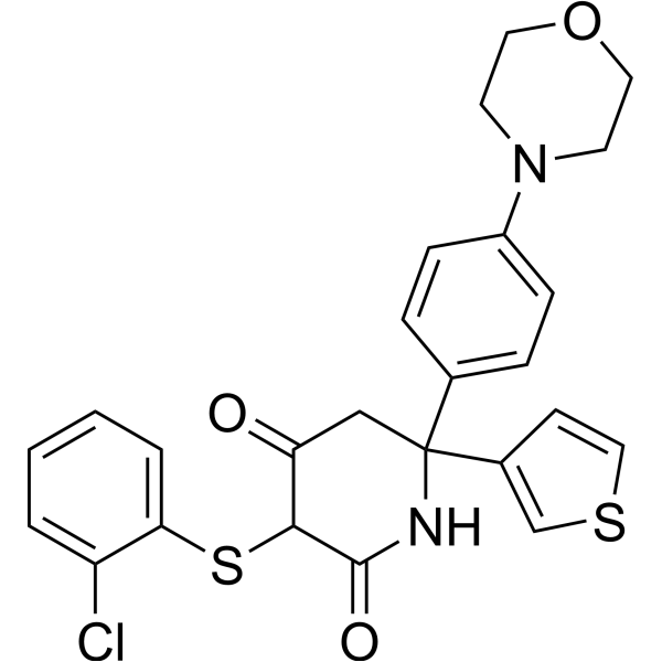
GNE-140 racemate
Based on 9 publication(s) in Google Scholar
GNE-140 racemate is a racemic mixture of (R)-GNE-140 (HY-100742A) and (S)-GNE-140 (HY-100742B), and also a tautomer of GNE-140 (HY-118241). GNE-140 racemate is an orally active lactate dehydrogenase inhibitor, with an IC50 of 3 nM against human LDHA and an IC50 of 5 nM against LDHB. GNE-140 racemate reduces the phosphorylation level and total protein expression of p38 MAPK, and inhibits EGF-induced AKT phosphorylation. GNE-140 racemate decreases lactate production, regulates glycolysis, the pentose phosphate pathway and nucleotide biosynthesis, increases extracellular pH, inhibits cell proliferation, migration and invasion, and induces caspase-3 activation and ROS accumulation. GNE-140 racemate can be used in research related to breast cancer, cystic fibrosis and pancreatic cancer.
For research use only. We do not sell to patients.
- Purity : 99.25%
- CAS No.: 1802977-61-2
- Formula: C25H23ClN2O3S2
- Molecular Weight:499.04
-
Storage:Powder -20°C, 3 years , 4°C, 2 years ; In solvent -80°C, 2 years , -20°C, 1 year

To place orders, for customer services and technical support, please contact: MedChemExpress USA
Tel: 609-228-6898 E-mail: [email protected] [email protected]


-
 Biological Activity
-
Chemical Information
-
Solvent & Solubility
- Protocols
- Purity & Documentation
- References
-
 Help & FAQs


-
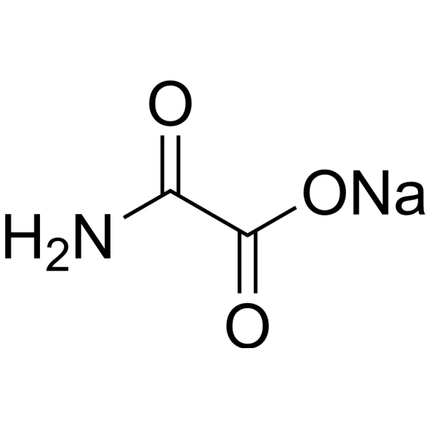
HY-W013032A
Oxamic acid sodiumLDH-A Inhibitor -
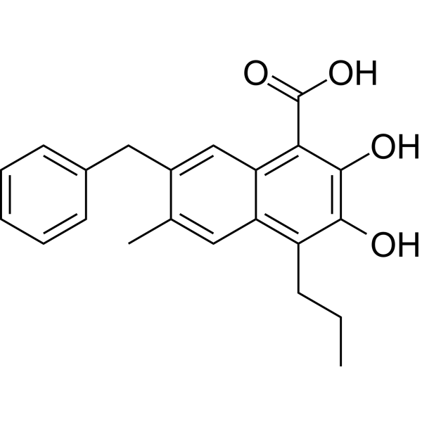
HY-16214
FX-11LDHA Inhibitor -
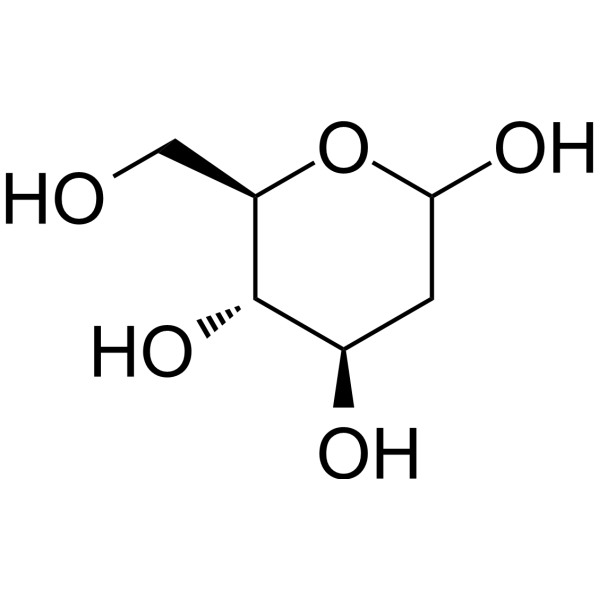
HY-13966
2-Deoxy-D-glucoseGlycolysis Inhibitor -
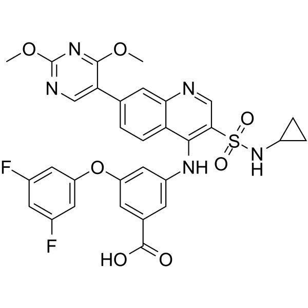
HY-100681
GSK2837808ALDHA Inhibitor -
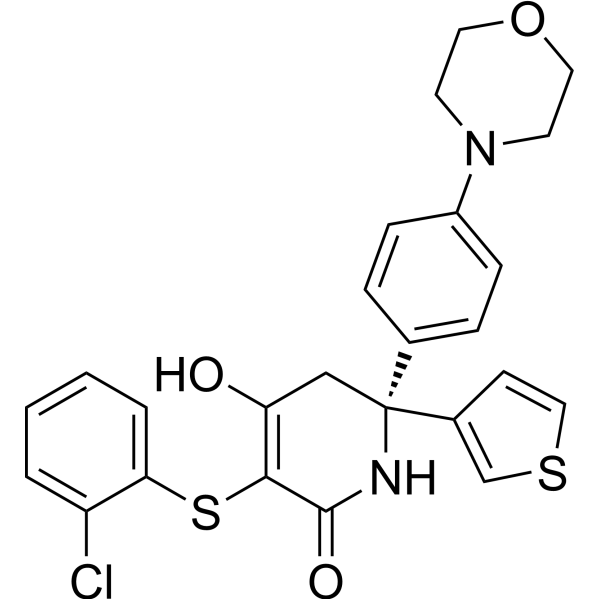
HY-100742A
(R)-GNE-140LDHA Inhibitor -
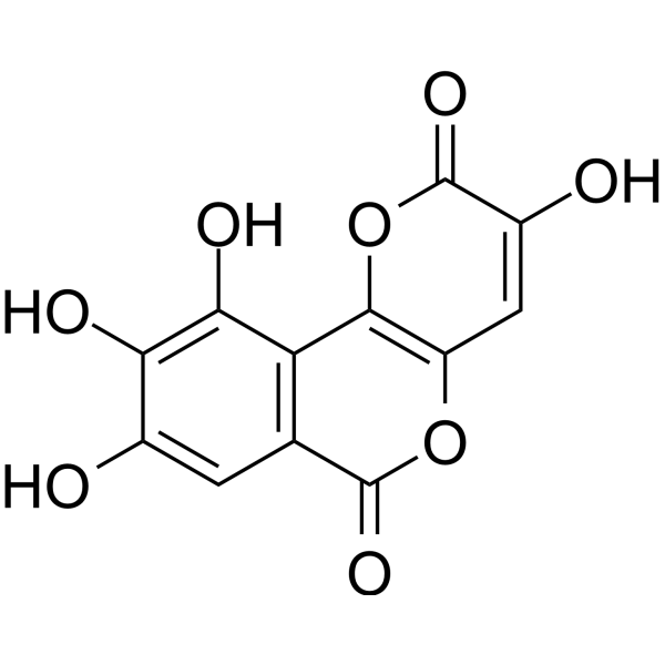
HY-W040118
GalloflavinLactate Dehydrogenase Inhibitor -

HY-N6782
OligomycinH+-ATP-Synthase Inhibitor -
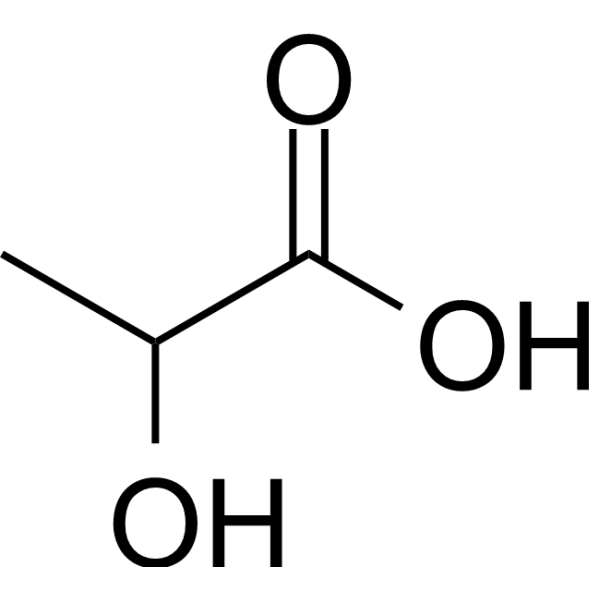
HY-B2227
Lactic acidGlycolysis Production -
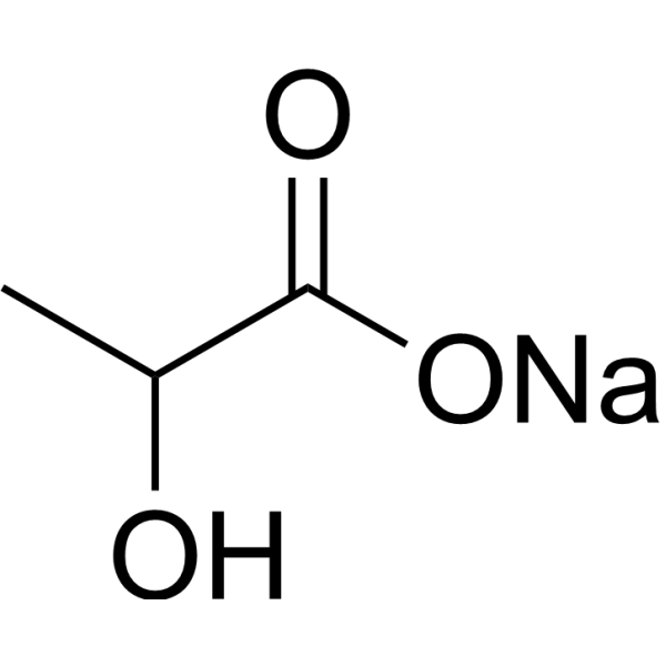
HY-B2227B
Lactic acid sodium (60% w/w in water)Glycolysis Production -

HY-B0215
AcetylcysteineMucolytic Agent -
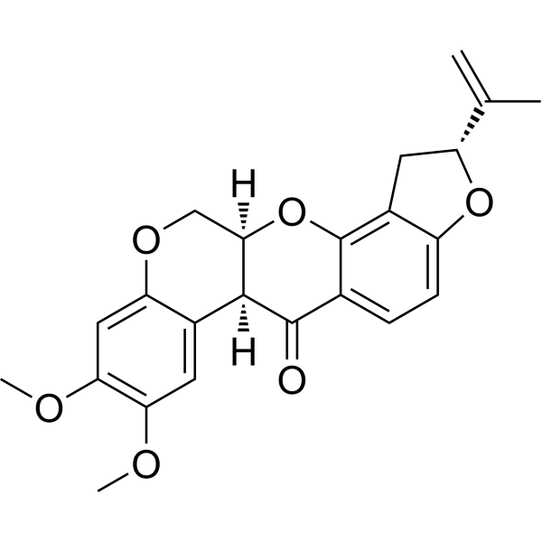
HY-B1756
RotenoneMitochondrial Complex I Inhibitor -
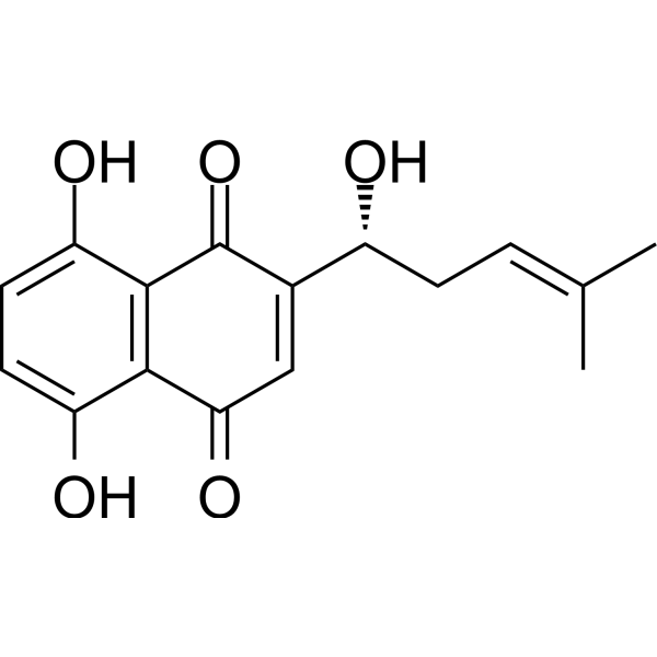
HY-N0822
ShikoninChloride Channel Inhibitor
Publications Citing Use of MedChemExpress (MCE) GNE-140 racemate
More- Gastroenterology. 2024 May;166(5):772-786.e14. [Abstract]
- Nat Commun. 2023 Jul 14;14(1):4129. [Abstract]
- Int J Biol Macromol. 2025 Sep 10:147598. [Abstract]
- Int J Biol Macromol. 2025 Apr 10;309(Pt 3):142858. [Abstract]
- J Anim Sci Biotechnol. 2025 Dec 18;16(1):174. [Abstract]
- Cell Mol Life Sci. 2019 Apr;76(8):1579-1593. [Abstract]
- Biochim Biophys Acta Mol Basis Dis. 2022 Dec 1;1868(12):166550. [Abstract]
- Am J Physiol Heart Circ Physiol. 2021 Jun 1;320(6):H2222-H2239. [Abstract]
- bioRxiv. 2024 June 27.
All EGFR Isoforms
MoreAll Caspase Isoforms
More

Biological Activity
Description
|
LDHA 3 nM (IC50) |
LDHB 5 nM (IC50) |
p38 MAPK |
Akt |
Caspase-3 |
Cellular Effect
|
Cell Line

|
Type | Value | Description | References |
|---|---|---|---|---|
| MIA PaCa-2 | IC50 |
0.67 μM
|
Reduction of cellular lactate levels in human MIA PaCa-2 pancreatic cancer cells after 6 hours of exposure.
Reduction of cellular lactate levels in human MIA PaCa-2 pancreatic cancer cells after 6 hours of exposure.
|
27479743 |
| MIA PaCa-2 | IC50 |
0.43 μM
|
Inhibition of proliferation in human MIA PaCa-2 pancreatic cancer cells.
Inhibition of proliferation in human MIA PaCa-2 pancreatic cancer cells.
|
27479743 |
| MIA PaCa-2 | IC50 |
0.47 μM
|
Reduction of glucose uptake in human MIA PaCa-2 pancreatic cancer cells after 6 hours of exposure.
Reduction of glucose uptake in human MIA PaCa-2 pancreatic cancer cells after 6 hours of exposure.
|
27479743 |
In Vitro
GNE-140 (200-300 μM) racemate inhibits intracellular LDH activity in pII ER- breast cancer cells[1].
GNE-140 (200-300 μM) racemate reduces extracellular lactate levels in ER− breast cancer cells[1].
GNE-140 (0.001-100 μM; 6 h) racemate inhibits LDHA activity in MIA PaCa-2 human pancreatic cancer cells, reducing lactate levels with an IC50 of 0.67 μM and increasing pyruvate levels after 6 hours of treatment[3].
GNE-140 (2 μM; 24 h) racemate alters global metabolism in MIA PaCa-2 human pancreatic cancer cells treated with 2 μM for 24 hours, increasing levels of glycolytic and pentose phosphate pathway metabolites without substantially affecting oxidative phosphorylation-associated metabolites[3].
GNE-140 (0.001-100 μM; 6 h) racemate reduces glucose uptake in MIA PaCa-2 human pancreatic cancer cells with an IC50 of 0.47 μM after 6 hours of treatment[3].
GNE-140 (2 μM; 6 h) racemate redirects glucose carbon flux away from lactate production and toward pyruvate, alanine, and pentose phosphate pathway metabolites in MIA PaCa-2 human pancreatic cancer cells treated with 2 μM for 6 hours, without increasing glutamine-dependent TCA cycle flux[3].
GNE-140 (72 h) racemate synergizes with AMPK inhibitor, S6K inhibitor, and Phenformin (HY-16397) to reduce viability and shift metabolism away from oxidative phosphorylation in MIA PaCa-2 human pancreatic cancer cells[3].
GNE-140 (200-300 μM; 4 days) inhibits proliferation of pII ER− breast cancer cells after 4 days of incubation at concentrations of 200 μM and 300 μM, with no effect seen after 1 day of incubation[1].
GNE-140 (200-300 μM; 24 h) racemate inhibits motility of pII ER- breast cancer cells after 24 h of incubation at concentrations of 200 μM and 300 μM[1].
GNE-140 (200-300 μM; 48 h) racemate inhibits invasion of pII ER- breast cancer cells after 48 h of incubation at concentrations of 200 μM and 300 μM[1].
GNE-140 (100-300 μM; 4 days) racemate inhibits proliferation of YS1.2 ER+ breast cancer cells after 4 days of incubation at concentrations of 100 μM, 200 μM, and 300 μM, with no effect seen after 1 day of incubation[1].
GNE-140 (200-300 μM; 24 h) racemate inhibits motility of YS1.2 ER+ breast cancer cells after 24 h of incubation at concentrations of 200 μM and 300 μM[1].
GNE-140 (200 μM; 30 min pre-incubation prior to 30 min EGF stimulation) racemate reduces EGF-induced phosphorylation of p38 MAPK and AKT, and lowers total p38 MAPK levels, in pII ER- breast cancer cells without altering EGF-induced phosphorylation of ERK1/2, Src, or NF-κB[1].
GNE-140 (0.001-100 μM) racemate inhibits proliferation of MIA PaCa-2 human pancreatic cancer cells with an IC50 of 0.43 μM, with potency dependent on cellular LDHA expression levels[3].
GNE-140 (72 h) racemate inhibits proliferation of 13% (4/30) of tested human pancreatic cancer cell lines with IC50 values below 5 μM, with sensitive lines relying more on glycolysis (lower baseline OCR) than resistant lines that rely on oxidative phosphorylation[3].
GNE-140 (200-300 μM; 4 days) racemate inhibits proliferation of MCF10A normal breast epithelial cells after 4 days of incubation at concentrations of 200 μM and 300 μM[1].
GNE-140 (1 μM; 72 h) racemate increases the extracellular pH of cultured IB3-1 CF lung epithelial cells[2].
GNE-140 racemate potently inhibits purified human LDHA, LDHB, and LDHC enzymes with nanomolar potency, shows no appreciable inhibition of MDH-1/MDH-2, and is highly selective against a broad panel of kinases[3].
GNE-140 (2 μM; 1-5 days) racemate induces caspase-3 activation starting at 1-2 days and cell death starting at 3 days in MIA PaCa-2 human pancreatic cancer cells treated with 2 μM, and sustained inhibition for more than 2 days is required to drive irreversible cell death[3].
GNE-140 (10 μM; 0-48 h) racemate increases reactive oxygen species levels in a time-dependent manner in MIA PaCa-2 human pancreatic cancer cells treated with 10 μM[3].
MedChemExpress (MCE) has not independently confirmed the accuracy of these methods. They are for reference only. Further protocols information, click here.
-
Cell Line:pII ER- breast cancer cells
-
Concentration:0.01-300 μM (4-day incubation); 200-300 μM (1-day incubation)
-
Incubation Time:4 days (for 0.01-300 μM); 1 day (for 200-300 μM)
-
Result:Inhibited pII cell viability on day 4 at concentrations of 200 μM and 300 μM.
Showed no effect on day 1 at 200 μM and 300 μM.
-
Cell Line:pII ER- breast cancer cells
-
Concentration:200-300 μM
-
Incubation Time:24 h
-
Result:Inhibited pII cell motility, as measured by reduced scratch closure, compared to untreated controls.
-
Cell Line:pII ER- breast cancer cells
-
Concentration:200-300 μM
-
Incubation Time:48 h
-
Result:Inhibited pII cell invasion compared to untreated controls.
-
Cell Line:YS1.2 ER+ breast cancer cells
-
Concentration:0.01-300 μM (4-day incubation); 200-300 μM (1-day incubation)
-
Incubation Time:4 days (for 0.01-300 μM); 1 day (for 200-300 μM)
-
Result:Inhibited YS1.2 cell viability on day 4 at concentrations of 100 μM, 200 μM, and 300 μM.
Showed no effect on day 1 at 200 μM and 300 μM.
-
Cell Line:YS1.2 ER+ breast cancer cells
-
Concentration:200-300 μM
-
Incubation Time:24 h
-
Result:Inhibited YS1.2 cell motility, as measured by reduced scratch closure, compared to untreated controls.
-
Cell Line:MCF10A normal breast epithelial cells
-
Concentration:0.01-300 μM
-
Incubation Time:4 days
-
Result:Inhibited MCF10A cell viability on day 4 at concentrations of 200 μM and 300 μM compared to untreated controls.
-
Cell Line:pII ER- breast cancer cells
-
Concentration:200 μM
-
Incubation Time:30 min pre-incubation prior to 30 min EGF stimulation
-
Result:Reduced EGF-induced phosphorylation of p38 MAPK and AKT compared to EGF-only treated cells.
Reduced total p38 MAPK protein levels compared to EGF-only treated cells.
Did not affect EGF-induced phosphorylation of ERK1/2, Src, or NF-κB.
In Vivo
MedChemExpress (MCE) has not independently confirmed the accuracy of these methods. They are for reference only.
-
Animal Model:NCR nu/nu (female, immunocompromised, subcutaneous implantation of human MIA PaCa-2-SHT2 cells)[3]
-
Dosage:100 mg/kg (7-day metabolite measurement; 21-day tumor growth assessment); 200 mg/kg (7-day metabolite measurement; 21-day tumor growth assessment); 400 mg/kg (7-day metabolite measurement; 21-day tumor growth assessment)
-
Administration:p.o.; BID; 7 days (metabolite measurement); p.o.; BID; 21 days (tumor growth assessment)
-
Result:Showed dose-dependent reduction in tumor lactate levels 1 h after final dose of 100, 200, or 400 mg/kg.
Showed dose-dependent increases in tumor pyruvate and glycerol-3-phosphate levels 1 h after final dose of 100, 200, or 400 mg/kg.
Detected no significant changes in tumor lactate, pyruvate, or glycerol-3-phosphate levels 6 h after final dose of 100, 200, or 400 mg/kg.
Resulted in no tumor growth inhibition compared to vehicle control after 21 days of treatment with 100, 200, or 400 mg/kg.


Chemical Information
-
CAS No. 1802977-61-2
-
Appearance Solid
-
Molecular Weight 499.04
-
Formula C25H23ClN2O3S2
-
Color White to off-white
-
SMILES
O=C(C1SC2=CC=CC=C2Cl)NC(C3=CSC=C3)(C4=CC=C(N5CCOCC5)C=C4)CC1=O
-
Shipping
Room temperature in continental US; may vary elsewhere.
-
Storage
Powder -20°C 3 years 4°C 2 years In solvent -80°C 2 years -20°C 1 year


Publications (9)
-
Journal Impact Factor
-
Most Recent
-
Gastroenterology
2024 May;166(5):772-786.e14. PMID: 38272100 -
Nat Commun
2023 Jul 14;14(1):4129. PMID: 37452018 -
Int J Biol Macromol
Targeting and reversing tumor acidic microenvironment with bacterial cellulose-based micelles to enhance immune infiltration. [Abstract]2025 Sep 10:147598. PMID: 40939806 -
Int J Biol Macromol
Histone lactylation regulates early embryonic development through m6A methyltransferase METTL3 in goats. [Abstract]2025 Apr 10;309(Pt 3):142858. PMID: 40216110 -
J Anim Sci Biotechnol
Glutamine relieves feed restriction-induced ruminal epithelial function damage through histone lysine lactylation in yaks. [Abstract]2025 Dec 18;16(1):174. PMID: 41408320 -
Cell Mol Life Sci
Impairment of CFTR activity in cultured epithelial cells upregulates the expression and activity of LDH resulting in lactic acid hypersecretion. [Abstract]2019 Apr;76(8):1579-1593. PMID: 30599064 -
Biochim Biophys Acta Mol Basis Dis
Integrated application of transcriptome and metabolomics reveals potential therapeutic targets for the polarization of atherosclerotic macrophages. [Abstract]2022 Dec 1;1868(12):166550. PMID: 36150660 -
Am J Physiol Heart Circ Physiol
Periostin promotes arterial calcification through PPARγ-related glucose metabolism reprogramming. [Abstract]2021 Jun 1;320(6):H2222-H2239. PMID: 33834866 -


Solvent & Solubility
In Vitro:
DMSO : 20 mg/mL (40.08 mM; Need ultrasonic; Hygroscopic DMSO has a significant impact on the solubility of product, please use newly opened DMSO)
Please refer to the solubility information to select the appropriate solvent. Once prepared, please aliquot and store the solution to prevent product inactivation from repeated freeze-thaw cycles.
Storage method and period of stock solution: -80°C, 2 years; -20°C, 1 year. When stored at -80°C, please use it within 2 years. When stored at -20°C, please use it within 1 year.
Please refer to the solubility information to select the appropriate solvent. Once prepared, please aliquot and store the solution to prevent product inactivation from repeated freeze-thaw cycles.
Storage method and period of stock solution: -80°C, 2 years; -20°C, 1 year. When stored at -80°C, please use it within 2 years. When stored at -20°C, please use it within 1 year.
Concentration (start) × Volume (start) = Concentration (final) × Volume (final)

Protocols
-
RNA extraction experimental
By lysing cells, releasing RNA, and removing impurities such as proteins and DNA, high-purity RNA products are finally obtained. The commonly used traditional method is the guanidine isothiocyanate/phenol/chloroform method (Trizol), which is suitable for a variety of animal materials including animal tissues, microorganisms, cultured cells, etc., and most plant materials.
-
Collagen: Sirius Red Staining
Sirius Red or picrosirius red staining is a histochemical method for visualizing collagen-rich extracellular matrix in tissue sections, and collagen fibers are detected as red-stained structures under bright-field microscopy with enhanced birefringence under polarized light. Picrosirius red is useful for assessing total collagen organization, distribution, and fibrosis burden, but polarized color should not be interpreted as a definitive collagen type I versus type III readout because color is affected by fiber orientation, thickness, and packing.
-
Cell migration
Cell migration is a method that plays an important role in wound healing, cell differentiation, embryonic development, etc.
-
Somatic Cell Culture
A method of simulating the in vivo environment in vitro to maintain the cell growth, differentation and main functions.
-
Connective Tissue: Masson's Trichrome/Collagen Trichrome Staining
Masson’s Trichrome (collagen/trichrome staining) is a histological technique that differentially stains tissue compartments using sequential acidic dyes to distinguish collagen from muscle and cytoplasmic components based on dye affinity and tissue permeability differences, enabling visualization of fibrosis and connective tissue architecture in histological sections. The classical formulation typically uses Weigert's iron hematoxylin for nuclear staining, Biebrich scarlet-acid fuchsin for cytoplasm and muscle, and aniline blue (or light green variants) for collagen, producing a characteristic blue/green collagen signal contrasted against red cytoplasm and dark nuclei. The staining principle relies on selective displacement of smaller dye molecules by larger anionic dyes in collagen-rich regions under controlled acidified conditions, which enhances collagen-specific dye retention. This property makes the method widely used for fibrosis assessment in organs such as heart, liver, lung, a
-
Breast Cancer Modeling
Breast cancer is a heterogeneous cancer, and it has been distinguished into four subtypes: luminal A, luminal B, HER2-positive and basal-like. Molecular mutations, epigenetic alterations, hormone exposure and immune microenvironment are related to the progression of breast cancer.
-
Cell Cytotoxicity Assay
Cytotoxicity assays are usually based on the assessment of cell membrane damage, which can also be indirectly detected by measuring cell viability. Detection methods include MTT assay, CKK-8 assay, LDH assay and ATP assay, etc.
-
Protein Extraction
Protein extraction uses physical, chemical or biological methods, such as ultrasonic disruption, salting out, cell lysis, electrophoresis, etc., to destroy the cell membrane structure and to separate the proteins from different components according to their characteristics.
-
Apoptosis Solutions
Apoptosis is a regulated, generally non-lytic cell-death pathway that removes unwanted, damaged, infected, or abnormal cells through coordinated morphological changes, caspase activation, DNA fragmentation, and membrane remodeling. The intrinsic apoptosis pathway is controlled mainly by mitochondrial outer membrane permeabilization, BCL-2 family proteins, cytochrome c release, apoptosome formation, caspase-9 activation, and downstream executioner caspase-3/7 activation. The extrinsic apoptosis pathway is initiated by death receptors such as Fas, TNFR, and TRAIL receptors, which recruit adaptor proteins and activate caspase-8 before engaging executioner caspases or mitochondrial amplification through BID cleavage. Apoptosis is linked to many phenotypes, including cancer cell killing, tissue homeostasis, immune regulation, neurodegeneration, infection response, and treatment-induced cytotoxicity; unresolved questions include how apoptosis interacts with necroptosis, pyroptosis, ferroptos
-
Cell invasion
Cell invasion is the ability of cells to migrate from one area to another via the extracellular matrix. Cell invasion is the response of normal and cancer cells to chemical and mechanical stimuli. Before migrating to a new region, the extracellular matrix is degraded by proteases within the cell. Cell invasion often occurs during wound repair, vascularization and inflammation, abnormal tissue invasion, and tumor cell metastasis.
-
MTT Cell Proliferation Assay
The MTT assay is a colorimetric endpoint assay for estimating viable cell number, cell growth, cytotoxicity, or cell activation in cultured mammalian cells. Living cells reduce the yellow tetrazolium salt MTT into purple/blue formazan, while dead cells do not generate the same signal; the resulting color can be quantified with a multiwell spectrophotometer. MTT reduction is commonly interpreted as a readout of metabolic activity that often correlates with viable cell number, but it should not be treated as a direct cell-counting method unless the assay is optimized for the cell type and experimental condition. Studies show that MTT reduction can involve mitochondrial and non-mitochondrial reducing systems, and formazan may accumulate in intracellular lipid droplets rather than simply marking mitochondria.
-
Western Blot
Western blotting (WB) is a commonly used experimental method in molecular biology, biochemistry, and immunogenetics for identifying and quantifying target proteins. It combines gel electrophoresis with immunoassay, enabling researchers to analyze protein expression, post-translational modifications, and molecular weight.
-
BrdU Incorporation Assay
Bromodeoxyuridine (BrdU) incorporation assay is based on the principle that BrdU, a thymidine analog, is incorporated into newly synthesized DNA during the S phase of the cell cycle, thereby serving as a marker of DNA replication and cellular proliferation. Incorporated BrdU can be detected using anti-BrdU antibodies following DNA denaturation, enabling visualization or quantification of proliferating cells through immunochemical detection methods such as immunofluorescence or immunohistochemistry.
-
ROS/oxidative-stress fluorescent staining
ROS/oxidative-stress fluorescent staining uses cell-permeant fluorogenic probes that become fluorescent after oxidation inside cells or tissues; commonly used examples include DCFH-DA/DCFDA for broad cellular oxidant detection, DHE for superoxide-related signal detection, MitoSOX for mitochondrial superoxide-related signal detection, and CellROX probes for oxidative-stress-associated fluorescence readouts. The assay detects probe oxidation rather than a single ROS species unless the probe and analysis method have been chemically validated for that species. DCFH-DA enters cells, is deacetylated by intracellular esterases to DCFH, and produces fluorescent DCF after oxidation, so the readout is used as an operational measure of total cellular oxidative stress rather than a species-specific ROS measurement. DHE and MitoSOX can report superoxide-related oxidation, but red fluorescence alone can include non-specific ethidium-like oxidation products; HPLC or optimized spectral approaches are
-
CFSE Dye Dilution Proliferation Assay
The CFSE (carboxyfluorescein diacetate succinimidyl ester) dye dilution proliferation assay is based on the covalent labeling of intracellular proteins by a cell-permeant fluorescent dye that becomes fluorescent upon intracellular ester cleavage and then is stably retained within cells. As labeled cells divide, the dye is partitioned equally between daughter cells, resulting in a stepwise halving of fluorescence intensity that can be quantified by flow cytometry to determine the number of cell divisions undergone by each cell population. This fluorescence dilution approach enables quantitative tracking of lymphocyte proliferation at the single-cell level over multiple rounds of division. CFSE-based proliferation analysis has been widely applied to measure antigen-driven lymphocyte expansion in vitro, where discrete fluorescence peaks correspond to successive cell divisions and allow reconstruction of proliferative history within heterogeneous populations.
-
Dye-dilution cell tracking and proliferation staining
Dye-dilution cell tracking assays quantify cell proliferation by covalently labeling intracellular proteins with a stable fluorescent dye that is equally partitioned between daughter cells during mitosis, resulting in stepwise halving of fluorescence intensity with each cell division as measured by flow cytometry histograms. Carboxyfluorescein diacetate succinimidyl ester (CFSE) is a prototypical dye that diffuses into cells, is enzymatically converted into a fluorescent compound, and then covalently binds intracellular amine groups, producing long-lived fluorescence suitable for tracking multiple rounds of division in vitro and in vivo. Successive generations of dividing cells form discrete peaks of decreasing fluorescence intensity, enabling estimation of proliferation history, precursor frequency, and division index within heterogeneous populations. Alternative dyes such as CellTrace Violet (CTV) and far-red membrane dyes (e. g. , PKH26) follow the same dilution principle but differ
-
CCK-8/WST-8 Cell Proliferation Assay
The CCK-8/WST-8 assay is based on the reduction of the water-soluble tetrazolium salt WST-8 to a water-soluble formazan product by cellular dehydrogenases in metabolically active cells, where the generated formazan amount is proportional to the number of living cells and is quantified by measuring absorbance in the visible range, providing a colorimetric readout for cell viability and proliferation assessment. This class of tetrazolium-based assays improves upon earlier MTT-based systems by producing a water-soluble formazan, eliminating the need for organic solubilization steps and enabling direct spectrophotometric measurement in culture medium.
-
Protocol for Cell Cycle
Cell-cycle analysis by flow cytometry measures DNA content in single cells to estimate the fraction of cells in G0/G1, S, and G2/M phases. Propidium iodide intercalates into DNA, and after RNA removal with RNase, fluorescence intensity reflects cellular DNA content: 2N cells are assigned to G0/G1, cells between 2N and 4N to S phase, and 4N cells to G2/M. DNA-content analysis alone cannot reliably separate G0 from G1 or G2 from M. Ki-67 can distinguish quiescent G0 cells from cycling cells, EdU or BrdU incorporation marks active DNA synthesis in S phase, and phospho-histone H3 staining identifies mitotic cells within the 4N population.
-
Cell Counting-Based Growth Curve Assay
Cell counting-based growth curve assays quantify cell proliferation by directly measuring changes in viable cell number over time using manual or automated counting methods such as hemocytometer-based counting or instrument-assisted cell enumeration, enabling construction of growth curves that reflect population expansion dynamics in response to culture conditions. A widely used approach is trypan blue exclusion with hemocytometer counting, where membrane-compromised (non-viable) cells take up the dye, allowing discrimination between viable and non-viable cells while simultaneously enabling total cell number quantification. Repeated sampling across time points allows estimation of proliferation rate, growth phases, and comparative growth kinetics between experimental conditions.
-
Colony Formation (Clonogenic) Assay
The clonogenic (colony formation) assay measures the ability of a single cell to retain reproductive viability and form a macroscopic colony, typically defined as a cluster derived from one progenitor cell after a defined growth period. This assay is widely used to evaluate cell survival after exposure to ionizing radiation or cytotoxic treatments and is considered a standard method in radiation biology for generating dose-response relationships of reproductive cell death. Colony formation reflects long-term proliferative capacity rather than short-term metabolic activity, and survival is quantified by comparing treated versus untreated conditions based on colony number and derived survival fractions.
-
Protocol For Protein Expression And Purification
Recombinant protein expression in Escherichia coli followed by purification of a His-tagged soluble protein by immobilized metal affinity chromatography (IMAC), with optional MBP fusion and TEV tag removal when the construct includes these elements. The biological readout is production of the encoded target protein, detected as an inducible band at the expected molecular mass by SDS-PAGE and quantified by total protein assay or chromatographic absorbance; the purification readout is enrichment of the target protein in elution fractions after selective binding of polyhistidine residues to immobilized Ni2+/metal-chelate resin and elution by imidazole-containing buffer. Expression is driven by an inducible bacterial expression system, commonly T7/lac-based, in which IPTG or lactose/auto-induction activates transcription and translation of the cloned gene; lower induction temperature, lower inducer concentration, induction timing, and solubility-enhancing fusion tags can influence the frac
-
Cell Viability Determination by MTT Colorimetric Assay
The following protocol uses the MTT colorimetric assay as a classic literature-established method for assessing cell viability/metabolic activity in cultured mammalian cells. MTT[3-(4,5-dimethylthiazol-2-yl)-2,5-diphenyltetrazolium bromide] is reduced by metabolically active cells to a colored formazan product; the amount of formazan is quantified spectrophotometrically and provides an indirect measure of metabolically active viable cells. Importantly, MTT reduction reflects cellular oxidoreductase/metabolic activity rather than an absolute direct count of living cells, so changes in cellular metabolism can alter the signal independently of cell number.
-
Kinase activity and phosphorylation assays
Kinase activity assays measure the ability of kinases to transfer phosphate groups from ATP to specific substrates, while phosphorylation assays detect the presence and levels of phosphorylated proteins. Common methods include radiolabeled ATP incorporation (e. g. ,), ADP release detection via bioluminescence (e. g. ,[3]), enzyme-linked immunosorbent assays (ELISA) for phospho-specific epitopes (e. g. ,[6]), and microtiter-based formats for high-throughput screening (e. g. ,[8]). The ADP-Glo assay quantifies kinase activity by measuring ADP produced during phosphorylation using a luciferase-based system. Radiometric assays involve autoradiography or scintillation counting after incorporation of 32P-labeled ATP into substrate proteins. ELISA-based approaches rely on phospho-specific antibodies to detect activated kinases in cell lysates or purified samples.
-
Fibrosis/Collagen Morphometry
Fibrosis and collagen morphometry is based on the quantitative visualization of fibrillar collagen deposition in tissue sections using histochemical stains such as Sirius Red (Picrosirius Red) or Masson's trichrome, followed by image-based or polarization-enhanced analysis to estimate collagen proportional area as a surrogate of extracellular matrix accumulation during fibrotic remodeling. Sirius Red combined with polarized light microscopy enhances detection of collagen fibers due to birefringence properties, enabling more specific visualization of collagen type I and III fibrils compared to conventional bright-field histology, while whole-section or region-restricted digital morphometry reduces field-selection bias in fibrosis assessment. Alternative quantitative approaches include second harmonic generation (SHG) and two-photon excited fluorescence microscopy, which enable label-free detection of fibrillar collagen and have been validated against histological staining and biochemica
-
EdU Incorporation Assay (Click Chemistry-Based DNA Synthesis Measurement)
The EdU incorporation assay measures DNA synthesis by adding the thymidine analog 5-ethynyl-2′-deoxyuridine to cells or tissues, where it is incorporated into newly synthesized DNA during S phase. Incorporated EdU is detected by copper-catalyzed azide-alkyne cycloaddition, in which a fluorescent azide covalently reacts with the ethynyl group on EdU, allowing S-phase cells to be detected by fluorescence microscopy, flow cytometry, or high-content imaging. EdU detection does not require DNA denaturation or anti-BrdU antibody access, which preserves sample structure and improves compatibility with immunostaining and multiparameter cytometry compared with BrdU-based detection. EdU can be cytotoxic in a cell-type- and exposure-dependent manner, so pulse duration, concentration, and continuous-labeling designs should be validated for each cell type.
-
Ki-67 Immunostaining Proliferation Assay
Ki-67 immunostaining measures the growth fraction of a cell population by detecting Ki-67, a nuclear antigen present in proliferating cells and absent in quiescent G0 cells. The readout is the percentage of Ki-67-positive nuclei among total counted cells, commonly called the Ki-67 labeling index or proliferation index.
-
PCNA Immunodetection Proliferation Assay
PCNA immunodetection measures proliferative activity by detecting proliferating cell nuclear antigen, a nuclear protein associated with DNA polymerase δ function and DNA replication. The assay readout is the proportion of PCNA-positive nuclei among total counted cells, but PCNA labeling is not identical to BrdU labeling because PCNA can mark late G1/early S-associated replication competence and may persist beyond active DNA synthesis depending on fixation and extraction conditions.
-
Protocol for Cell Counting and Cell Density Analysis
Cell counting and cell-density analysis estimate the number of cells in a known volume or field area. Manual hemocytometer counting uses a chamber of defined geometry to convert counted cells into cells/mL, while automated counters and image-analysis workflows detect cell objects from optical, brightfield, fluorescence, impedance, or digital-image features. Trypan blue viability counting is based on dye exclusion: viable cells with intact membranes exclude dye, while non-viable cells with compromised membranes stain blue. The readout is total cell density, viable-cell density, dead-cell density, and percent viability. Cell density can also be estimated from microscopy images by counting objects per image area, from flow cytometry using calibrated volume or reference particles, or from in situ microscopy in bioreactors after calibration against reference methods such as hemocytometer or flow cytometry.
-
Protocol for Kinase activity and phosphorylation assays
Kinase activity assays measure transfer of phosphate from ATP to a protein or peptide substrate, generating phosphorylated substrate, ADP, or incorporated radiolabeled phosphate as the readout; phosphorylation assays measure site-specific phosphorylation in cells or tissues as a proxy for kinase-pathway activation, inhibition, or substrate regulation. Phosphorylation can be detected by phospho-specific Western blot, immunoprecipitation kinase assay, phospho-immunofluorescence, phospho-flow cytometry, luminescent ADP detection, radiolabeled ATP incorporation, or reporter-based pathway assays, and these readouts can be applied to cancer cells, primary neurons, mouse tumors, organoids, inflammatory macrophages, ferroptosis studies, and mitophagy studies when the kinase target is biologically relevant.


Purity & Documentation
-
Data Sheet (312 KB)
-
SDS (393 KB)
- English - EN (393 KB)
- Français - FR (393 KB)
- Deutsch - DE (393 KB)
- Norwegian - NO (393 KB)
- Español - ES (393 KB)
- Swedish - SV (393 KB)
- Italian - IT (393 KB)
- Korean - KR (393 KB)
- Portuguese - PT (393 KB)
-
Handling Instructions (2659 KB)
References
[1]. Khajah MA, et al. The effect of lactate dehydrogenase inhibitors on proliferation, motility and invasion of breast cancer cells highlights a new role for lactate. Molecular medicine reports. 2024 Jan;29(1):12. [Content Brief]
[2]. Valdivieso ÁG, et al. Impairment of CFTR activity in cultured epithelial cells upregulates the expression and activity of LDH resulting in lactic acid hypersecretion. Cellular and molecular life sciences : CMLS. 2019 Apr;76(8):1579-1593. [Content Brief]
[3]. Boudreau A, et al. Metabolic plasticity underpins innate and acquired resistance to LDHA inhibition. Nature chemical biology. 2016 Oct;12(10):779-86. [Content Brief]
[4]. Ji JJ, et al. Kallistatin/Serpina3c inhibits cardiac fibrosis after myocardial infarction by regulating glycolysis via Nr4a1 activation. Biochimica et biophysica acta. Molecular basis of disease. 2022 Sep 01;1868(9):166441. [Content Brief]


Complete Stock Solution Preparation Table
Please refer to the solubility information to select the appropriate solvent. Once prepared, please aliquot and store the solution to prevent product inactivation from repeated freeze-thaw cycles.
Storage method and period of stock solution: -80°C, 2 years; -20°C, 1 year. When stored at -80°C, please use it within 2 years. When stored at -20°C, please use it within 1 year.
| Optional Solvent | Concentration Solvent Mass | 1 mg | 5 mg | 10 mg | 25 mg |
|---|---|---|---|---|---|
| DMSO | 1 mM | 2.0038 mL | 10.0192 mL | 20.0385 mL | 50.0962 mL |
| 5 mM | 0.4008 mL | 2.0038 mL | 4.0077 mL | 10.0192 mL | |
| 10 mM | 0.2004 mL | 1.0019 mL | 2.0038 mL | 5.0096 mL | |
| 15 mM | 0.1336 mL | 0.6679 mL | 1.3359 mL | 3.3397 mL | |
| 20 mM | 0.1002 mL | 0.5010 mL | 1.0019 mL | 2.5048 mL | |
| 25 mM | 0.0802 mL | 0.4008 mL | 0.8015 mL | 2.0038 mL | |
| 30 mM | 0.0668 mL | 0.3340 mL | 0.6679 mL | 1.6699 mL | |
| 40 mM | 0.0501 mL | 0.2505 mL | 0.5010 mL | 1.2524 mL |


GNE-140 racemate Related Classifications
HY-100742 Related Classifications
 Powered by Bioz