



Gestodene

Based on 1 Customer Validation
Gestodene (SHB 331; WL 70) is an orally active synthetic progestogen compound of the 19-nortestosterone class. Gestodene binds with high affinity to the progesterone receptor, inhibits 5α-reductase, binds to androgen and aldosterone receptors, and inactivates CYP3A. Gestodene acts as a positive allosteric modulator of PAR1, enhances PAR1-mediated signaling pathways, and is capable of increasing the activation potency of PAR1-AP toward PAR1, thereby promoting ERK1/2 phosphorylation, receptor internalization, cell morphological changes, and human platelet aggregation. Gestodene and its A-ring reduced metabolites promote the proliferation, differentiation, and mineralization of neonatal rat osteoblasts. Gestodene itself has no binding capacity for estrogen receptors, and this osteogenic activity depends on intracellular metabolism to generate A-ring reduced products with estrogen-like agonistic activity. Gestodene is useful for research on diseases related to progesterone secretion and diseases related to thrombosis.
For research use only. We do not sell to patients.
- Purity : 99.89%
- CAS No.: 60282-87-3
- Formula: C21H26O2
- Molecular Weight:310.43
-
Storage:Powder -20°C, 3 years ; In solvent -80°C, 6 months , -20°C, 1 month

To place orders, for customer services and technical support, please contact: MedChemExpress USA
Tel: 609-228-6898 E-mail: [email protected] [email protected]


-
 Biological Activity
-
Chemical Information
-
Solvent & Solubility
- Protocols
- Purity & Documentation
- References
-
 Help & FAQs


-
HY-B0111
DrospirenoneProgesterone Receptor Agonist -
HY-B0257
LevonorgestrelProgesterone Inhibitor -
HY-B0216
EthinylestradiolEndogenous Metabolite -
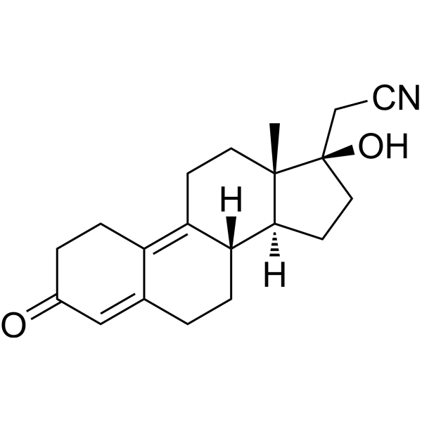
HY-B0084
DienogestProgesterone Receptor Agonist -
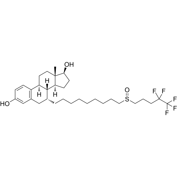
HY-13636
FulvestrantEstrogen Receptor Antagonist -
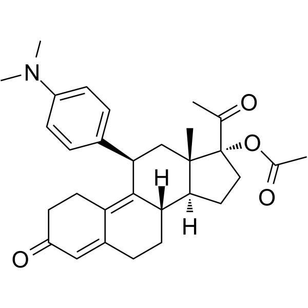
HY-16508
Ulipristal acetateSelective Progesterone Receptor Modulator -
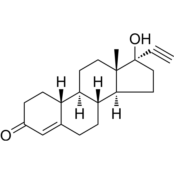
HY-B0554
NorethindroneProgestin -
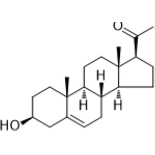
HY-B0151
PregnenoloneCB1 Inhibitor -
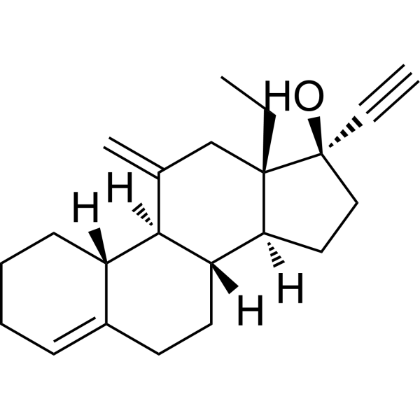
HY-12516
DesogestrelProgesterone Receptor Agonist -
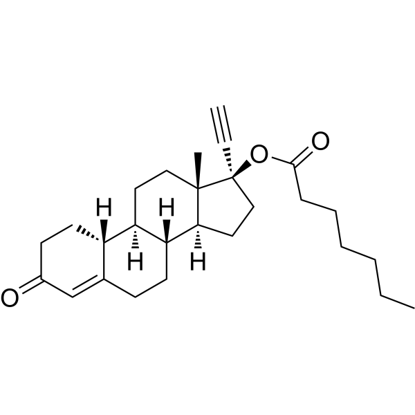
HY-A0285
Norethisterone enanthateParenteral Progestogen -
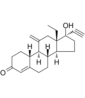
HY-B0652
EtonogestrelProgesterone/ERR Agonist -
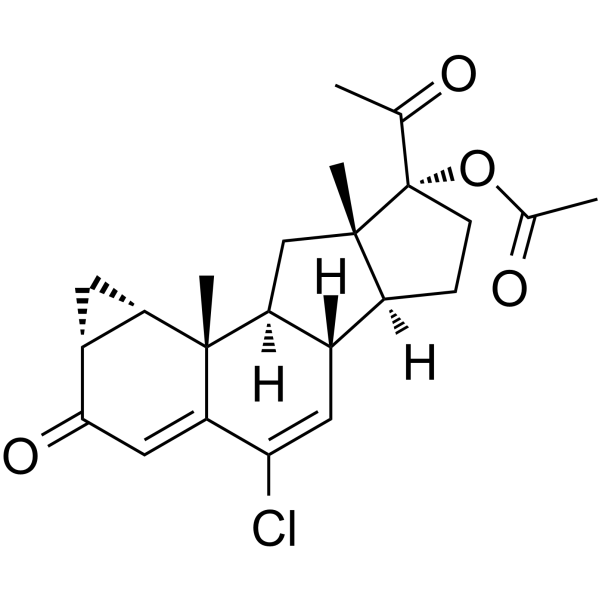
HY-13604
Cyproterone acetateAndrogen Receptor Antagonist


Biological Activity
Description
|
ERK1 |
ERK2 |
MMP-1 |
MMP-2 |
MMP-3 |
MMP-10 |
CYP3A |
Cellular Effect
|
Cell Line

|
Type | Value | Description | References |
|---|---|---|---|---|
| HT-29 | EC50 |
8.15 μM
|
Potency of gestodene to enhance PAR1-AP-induced intracellular calcium increase in human HT29 cells pre-incubated for 10 min and measured by Fluo-4 NW calcium assay.
Potency of gestodene to enhance PAR1-AP-induced intracellular calcium increase in human HT29 cells pre-incubated for 10 min and measured by Fluo-4 NW calcium assay.
|
39130626 |
| HT-29 | EC50 |
4.54 μM
|
Potency of gestodene to enhance thrombin-induced intracellular calcium increase in human HT29 cells pre-incubated for 10 min and measured by Fluo-4 NW calcium assay.
Potency of gestodene to enhance thrombin-induced intracellular calcium increase in human HT29 cells pre-incubated for 10 min and measured by Fluo-4 NW calcium assay.
|
39130626 |
In Vitro
Gestodene exhibits a high relative binding affinity for the progesterone receptor, shows low to moderate binding affinity for the androgen receptor, and has weak binding capacity for the estrogen receptor[1].
Gestodene inhibits the hydroxylation of ethinyl estradiol in a concentration-dependent manner in human liver microsomes and also inhibits the oxidative metabolism of Cyclosporine and Diazepam[1].
Gestodene (GDN) (1-500 nM; 1-25 days) stimulates the proliferation of neonatal rat calvarial osteoblasts, increases alkaline phosphatase activity in neonatal rat calvarial osteoblasts, and increases cell-associated osteocalcin content in neonatal rat calvarial osteoblasts; the 5α-reductase inhibitor Finasteride (HY-13635) and the aldosterone ketosteroid dehydrogenase inhibitor Flufenamic acid (HY-B1221) inhibit the Gestodene-induced increase in alkaline phosphatase activity[2].
Gestodene (50-500 nM; 25 days) increases cell-associated calcium content in neonatal rat calvarial osteoblasts[2].
Gestodene (1-250 nM; 18 h) shows no binding affinity for estrogen receptors in the cytosolic fraction of neonatal rat calvarial osteoblasts[2].
Gestodene is extensively metabolized to A-ring reduced derivatives (5α gestodene, 3α,5α gestodene, and 3β,5α gestodene) in rat anterior pituitary, hypothalamus, and ventral prostate homogenates[3].
Gestodene (3 μM; 3 days; 4-16 h; 30 min) enhances PAR1-AP-induced proliferation, migration, and ERK1/2 phosphorylation in A2058 cells, HaCaT cells, and HDF cells in a PAR1-dependent manner; this enhancing effect is blocked by Vorapaxar (HY-10119)[4].
Gestodene acts as a positive allosteric modulator of PAR1 in HT29 cells, enhancing PAR1-AP- and thrombin-induced intracellular calcium levels with EC50 values of 8.15 μM and 4.54 μM, respectively, without affecting PAR2 or PAR4 activity[5].
Gestodene (10 μM) enhances PAR1-AP-induced intracellular calcium levels in MEG-01 cells, reducing the EC50 of PAR1-AP from 6.58 μM to 3.41 μM[5].
Gestodene (10 μM; 10 min) enhances PAR1-AP-induced ERK1/2 phosphorylation in MEG-01 cells through a PAR1-dependent mechanism[5].
Gestodene (10 μM) enhances PAR1-AP-induced PAR1 internalization in HT29 cells, and this effect is blocked by Vorapaxar[5].
Gestodene (10 μM; 30 min) enhances PAR1-AP-induced morphological changes (decreased circularity) in MEG-01 cells, and this effect is blocked by Vorapaxar[5].
Gestodene (GDN) (10-100 nM; 6 h) significantly increases insulin 2 and glucokinase gene expression in isolated rat pancreatic islets[8].
Gestodene (100 nM; 6 h) significantly enhances glucose-stimulated insulin secretion in isolated rat pancreatic islets cultured in 11 mM D-glucose; moreover, in the presence of steroid-metabolizing enzyme inhibitors, the Gestodene-induced increase in insulin 2 and glucokinase gene expression in rat pancreatic islets is suppressed, indicating that the stimulatory effect of Gestodene is exerted through enzyme-generated tetrahydro-reduced metabolites[8].
Gestodene (10 min) acts as a selective positive allosteric modulator of PAR1 in HT29 cells, with no effect on PAR2 or PAR4; it enhances PAR1-AP-induced calcium signaling in A2058 cells in a PAR1-dependent manner and shifts the dose-response curve to the left (EC50 decreases from 3.1 to 0.8 μM)[4].
Gestodene (3 μM; 24 h) enhances PAR1-AP-induced expression of MMP-1, MMP-2, MMP-3, MMP-10, and fibronectin in HaCaT cells as well as COL1A1 expression in NIH-3T3 cells through a PAR1-dependent signaling pathway[4].
Gestodene (GES) (10-10-10-6 M; 7 days) stimulates the growth of T47D-A breast tumor subclone cells but does not stimulate the growth of T47D-S breast tumor subclone cells[7].
Gestodene (10-10-10-6 M; 7 days) abolishes the estradiol-induced cell proliferation effect in T47D-S human breast tumor subclone cells; the proliferation effect induced in T47D-A cells is partially inhibited by antiestrogens (4-Hydroxytamoxifen, ICI 164384) and is not antagonized by antiprogestins (RU 38486 (HY-13683), Org 31710)[7].
MedChemExpress (MCE) has not independently confirmed the accuracy of these methods. They are for reference only. Further protocols information, click here.
-
Cell Line:Primary calvarial osteoblasts isolated from neonatal Wistar rats
-
Concentration:1, 10, 50, 100, 500 nM
-
Incubation Time:15 days
-
Result:Showed a significant increase in AP activity at 50 nM and above when compared to vehicle alone.
-
Cell Line:Primary calvarial osteoblasts isolated from neonatal Wistar rats
-
Concentration:1, 10, 50, 100, 500 nM
-
Incubation Time:25 days
-
Result:Resulted in a significant increase in cell-associated OC content when compared to cells incubated with vehicle alone.
-
Cell Line:Primary calvarial osteoblasts isolated from neonatal Wistar rats
-
Concentration:500 nM (Gestodene)
250 µM (Finasteride)
250 µM (Flufenamic acid) -
Incubation Time:15 days
-
Result:The presence of finasteride resulted in a complete inhibition of the GDN-induced increase of the AP activity.
The presence of flufenamic acid also inhibited the GDN-induced increase of the AP activity.
-
Cell Line:A2058, HaCaT and HDF Cells
-
Concentration:3 μM
PAR1-AP (10 μM)
Vorapaxar (30 nM) -
Incubation Time:3 days
-
Result:Enhanced PAR1-AP-induced proliferation of A2058, HaCaT, and HDF cells in a PAR1-dependent manner; this enhancing effect can be blocked by Vorapaxar.
-
Cell Line:A2058, HaCaT and HDF Cells
-
Concentration:3 μM
PAR1-AP (10 μM)
Vorapaxar (30 nM) -
Incubation Time:24 h (A2058 Cells)
14 h (HaCaT Cells)
16 h (HDF Cells) -
Result:Enhanced PAR1-AP-induced migration of A2058, HaCaT, and HDF cells in a PAR1-dependent manner.
-
Cell Line:HaCaT and
NIH-3T3 Cells -
Concentration:3 μM
PAR1-AP (10 μM)
Vorapaxar (30 nM) -
Incubation Time:24 h
-
Result:Enhanced PAR1-AP-induced expression of MMP-1, MMP-2, MMP-3, MMP-10, fibronectin in HaCaT cells and COL1A1 in NIH-3T3 cells via PAR1-dependent signaling.
-
Cell Line:MEG-01 cells
-
Concentration:10 μM
PAR1-AP (30 μM)
Vorapaxar (1 μM) -
Incubation Time:10 min
-
Result:Enhanced PAR1-AP-induced ERK1/2 phosphorylation in MEG-01 cells through a PAR1-dependent mechanism.
-
Cell Line:T47D-A, T47D-S
-
Concentration:10-10 M; 10-8 M; 10-6 M
-
Incubation Time:7 days
-
Result:Stimulated the growth of T47D-A cells but not that of T47D-S cells.
In Vivo
Gestodene (100-600 ng/L; water exposure; renewed daily; 7-day pre-exposure followed by 45 days of continuous exposure) reduces the cumulative egg production per female in adult zebrafish, with no mortality observed, and alters sex steroid levels and mRNA expression of HPG and HPT axis genes in a concentration- and sex-specific manner[6].
MedChemExpress (MCE) has not independently confirmed the accuracy of these methods. They are for reference only.
-
Animal Model:ICR mice (male, 7 weeks old, 30-32 g)[4]
-
Dosage:2 mg/g vaseline, while vorapaxar was applied at a dose of 0.6 mg/vaseline
-
Administration:Topical; once daily; 8 consecutive days
-
Result:Achieved complete wound closure with 0% residual wound area by Day 8, compared to 22.02% residual wound area in the control group.
Exhibited extensive collagen fiber accumulation, evidenced by prominent blue staining throughout the wound area in Masson's trichrome staining.
-
Animal Model:Wild-type (sexually mature, 6-7 months old, both males and females)[6]
-
Dosage:100, 600 ng/L
-
Administration:waterborne immersion; renewed daily; 45 days
-
Result:Reduced female daily egg production, male body length, HSI (both sexes), male 11‑ketotestosterone, and male spermatocyte proportion following gestodene water‑borne exposure.
Altered body weight and female GSI at 600 ng/L gestodene exposure, with zero mortality across tested concentrations.
Modified mRNA levels of cyp19b, npr, mprα, gnih, fshr, pgrmc1, ar, esr1, vtg1, dio1 and dio3‑b in zebrafish brain‑pituitary complex, gonads and liver in concentration‑ and sex‑dependent manners after gestodene treatment.
Clinical Trial
| NCT Number | Sponsor | Condition | Start Date |
Phase
|
|---|---|---|---|---|
| NCT01329991 | Plexxikon| | 2011-05 | PHASE1 |


Chemical Information
-
CAS No. 60282-87-3
-
Appearance Solid
-
Molecular Weight 310.43
-
Formula C21H26O2
-
Color White to off-white
-
SMILES
CC[C@@]1([C@]2(O)C#C)[C@](C=C2)([H])[C@@](CCC3=CC4=O)([H])[C@]([C@@]3([H])CC4)([H])CC1
-
Synonyms
SHB 331; WL 70
-
Shipping
Room temperature in continental US; may vary elsewhere.
-
Storage
Powder -20°C 3 years In solvent -80°C 6 months -20°C 1 month


Solvent & Solubility
In Vitro:
DMSO : ≥ 100 mg/mL (322.13 mM; Hygroscopic DMSO has a significant impact on the solubility of product, please use newly opened DMSO)
* "≥" means soluble, but saturation unknown.
Please refer to the solubility information to select the appropriate solvent. Once prepared, please aliquot and store the solution to prevent product inactivation from repeated freeze-thaw cycles.
Storage method and period of stock solution: -80°C, 6 months; -20°C, 1 month. When stored at -80°C, please use it within 6 months. When stored at -20°C, please use it within 1 month.
Please refer to the solubility information to select the appropriate solvent. Once prepared, please aliquot and store the solution to prevent product inactivation from repeated freeze-thaw cycles.
Storage method and period of stock solution: -80°C, 6 months; -20°C, 1 month. When stored at -80°C, please use it within 6 months. When stored at -20°C, please use it within 1 month.
Concentration (start) × Volume (start) = Concentration (final) × Volume (final)
In Vivo:
Select the appropriate dissolution method based on your experimental animal and administration route.
- For the following dissolution methods, please ensure to first prepare a clear stock solution using an In Vitro approach and then sequentially add co-solvents:
- To ensure reliable experimental results, the clarified stock solution can be appropriately stored based on storage conditions. As for the working solution for In Vivo experiments, it is recommended to prepare freshly and use it on the same day.
- The percentages shown for the solvents indicate their volumetric ratio in the final prepared solution. If precipitation or phase separation occurs during preparation, heat and/or sonication can be used to aid dissolution.
Add each solvent one by one: 10% DMSO 40% PEG300 5% Tween-80 45% Saline
Solubility: ≥ 2.5 mg/mL (8.05 mM); Clear solution
This protocol yields a clear solution of ≥ 2.5 mg/mL (saturation unknown).
Taking 1 mL working solution as an example, add 100 μL DMSO stock solution (25.0 mg/mL) to 400 μL PEG300, and mix evenly; then add 50 μL Tween-80 and mix evenly; then add 450 μL Saline to adjust the volume to 1 mL.
Preparation of Saline: Dissolve 0.9 g sodium chloride in ddH₂O and dilute to 100 mL to obtain a clear Saline solution.
Add each solvent one by one: 10% DMSO 90% (20% SBE-β-CD in Saline)
Solubility: ≥ 2.5 mg/mL (8.05 mM); Clear solution
This protocol yields a clear solution of ≥ 2.5 mg/mL (saturation unknown).
Taking 1 mL working solution as an example, add 100 μL DMSO stock solution (25.0 mg/mL) to 900 μL 20% SBE-β-CD in Saline, and mix evenly.
Preparation of 20% SBE-β-CD in Saline (4°C, storage for one week): 2 g SBE-β-CD powder is dissolved in 10 mL Saline, completely dissolve until clear.
In Vivo Dissolution Calculator
Please enter the basic information of animal experiments:
-
-
-
-
Recommended: Prepare an additional quantity of animals to account for potential losses during experiments.
Please enter your animal formula composition:
-
%DMSO +
Recommended: Keep the proportion of DMSO in working solution below 2% if your animal is weak.
-
%+
-
+%Tween-80 + +
-
%Saline +
The co-solvents required include: DMSO, . All of co-solvents are available by MedChemExpress (MCE). , Tween 80. All of co-solvents are available by MedChemExpress (MCE).
Working solution concentration: 0.22 mg/mL
Method for preparing stock solution: mg drug dissolved in μL DMSO. Stock solution concentration: mg/mL.
1. Take μL DMSO stock solution;
2. Add μL .
μL , mix evenly;
3. Then add μL Tween 80, mix evenly;
4. Then add μL
Please ensure that the stock solution in the first step is dissolved to a clear state, and add co-solvents in sequence. You can use ultrasonic heating (ultrasonic cleaner, recommended frequency 20-40 kHz), vortexing, etc. to assist dissolution.

Protocols
-
Kinase activity and phosphorylation assays
Kinase activity assays measure the ability of kinases to transfer phosphate groups from ATP to specific substrates, while phosphorylation assays detect the presence and levels of phosphorylated proteins. Common methods include radiolabeled ATP incorporation (e. g. ,), ADP release detection via bioluminescence (e. g. ,[3]), enzyme-linked immunosorbent assays (ELISA) for phospho-specific epitopes (e. g. ,[6]), and microtiter-based formats for high-throughput screening (e. g. ,[8]). The ADP-Glo assay quantifies kinase activity by measuring ADP produced during phosphorylation using a luciferase-based system. Radiometric assays involve autoradiography or scintillation counting after incorporation of 32P-labeled ATP into substrate proteins. ELISA-based approaches rely on phospho-specific antibodies to detect activated kinases in cell lysates or purified samples.
-
Western Blot
Western blotting (WB) is a commonly used experimental method in molecular biology, biochemistry, and immunogenetics for identifying and quantifying target proteins. It combines gel electrophoresis with immunoassay, enabling researchers to analyze protein expression, post-translational modifications, and molecular weight.
-
Mesenchymal stromal/stem cell osteogenic differentiation
Mesenchymal stromal/stem cells can be induced toward an osteoblast-like lineage in vitro by culture in osteogenic medium containing dexamethasone, ascorbic acid or ascorbate-2-phosphate, and β-glycerophosphate; the differentiation process is commonly evaluated by alkaline phosphatase activity, osteogenic marker expression, collagenous matrix formation, and calcium-rich matrix mineralization. The main readouts are alkaline phosphatase activity as an early osteogenic marker and Alizarin Red S staining as a calcium-deposit readout for mineralized extracellular matrix; Alizarin Red S can be inspected microscopically or extracted and measured colorimetrically at 405 nm.
-
Research Protocol for Endocrine Diseases
Endocrine diseases often arise from disrupted hormone production, hormone signaling, or target-tissue responsiveness; for diabetes-focused endocrine disease models, insulin signaling regulates glucose uptake, hepatic glucose output, lipid metabolism, and β-cell compensation. Type 2 diabetes develops through interacting defects in insulin resistance, β-cell dysfunction, adipose inflammation, hepatic glucose overproduction, altered incretin signaling, and ectopic lipid metabolism. A major unresolved question is whether endocrine dysfunction is driven primarily by target-tissue insulin resistance, intrinsic β-cell failure, immune/inflammatory stress, or combined multi-organ failure that differs by disease stage.
-
Protocol for Kinase activity and phosphorylation assays
Kinase activity assays measure transfer of phosphate from ATP to a protein or peptide substrate, generating phosphorylated substrate, ADP, or incorporated radiolabeled phosphate as the readout; phosphorylation assays measure site-specific phosphorylation in cells or tissues as a proxy for kinase-pathway activation, inhibition, or substrate regulation. Phosphorylation can be detected by phospho-specific Western blot, immunoprecipitation kinase assay, phospho-immunofluorescence, phospho-flow cytometry, luminescent ADP detection, radiolabeled ATP incorporation, or reporter-based pathway assays, and these readouts can be applied to cancer cells, primary neurons, mouse tumors, organoids, inflammatory macrophages, ferroptosis studies, and mitophagy studies when the kinase target is biologically relevant.
-
Cell Viability Determination by MTT Colorimetric Assay
The following protocol uses the MTT colorimetric assay as a classic literature-established method for assessing cell viability/metabolic activity in cultured mammalian cells. MTT[3-(4,5-dimethylthiazol-2-yl)-2,5-diphenyltetrazolium bromide] is reduced by metabolically active cells to a colored formazan product; the amount of formazan is quantified spectrophotometrically and provides an indirect measure of metabolically active viable cells. Importantly, MTT reduction reflects cellular oxidoreductase/metabolic activity rather than an absolute direct count of living cells, so changes in cellular metabolism can alter the signal independently of cell number.


Purity & Documentation
-
Data Sheet (304 KB)
-
SDS (418 KB)
- English - EN (418 KB)
- Français - FR (418 KB)
- Deutsch - DE (418 KB)
- Norwegian - NO (418 KB)
- Español - ES (418 KB)
- Swedish - SV (418 KB)
- Italian - IT (418 KB)
- Korean - KR (418 KB)
- Portuguese - PT (418 KB)
-
Handling Instructions (2659 KB)
References


Complete Stock Solution Preparation Table
Please refer to the solubility information to select the appropriate solvent. Once prepared, please aliquot and store the solution to prevent product inactivation from repeated freeze-thaw cycles.
Storage method and period of stock solution: -80°C, 6 months; -20°C, 1 month. When stored at -80°C, please use it within 6 months. When stored at -20°C, please use it within 1 month.
| Optional Solvent | Concentration Solvent Mass | 1 mg | 5 mg | 10 mg | 25 mg |
|---|---|---|---|---|---|
| DMSO | 1 mM | 3.2213 mL | 16.1067 mL | 32.2134 mL | 80.5335 mL |
| 5 mM | 0.6443 mL | 3.2213 mL | 6.4427 mL | 16.1067 mL | |
| 10 mM | 0.3221 mL | 1.6107 mL | 3.2213 mL | 8.0533 mL | |
| 15 mM | 0.2148 mL | 1.0738 mL | 2.1476 mL | 5.3689 mL | |
| 20 mM | 0.1611 mL | 0.8053 mL | 1.6107 mL | 4.0267 mL | |
| 25 mM | 0.1289 mL | 0.6443 mL | 1.2885 mL | 3.2213 mL | |
| 30 mM | 0.1074 mL | 0.5369 mL | 1.0738 mL | 2.6844 mL | |
| 40 mM | 0.0805 mL | 0.4027 mL | 0.8053 mL | 2.0133 mL | |
| 50 mM | 0.0644 mL | 0.3221 mL | 0.6443 mL | 1.6107 mL | |
| 60 mM | 0.0537 mL | 0.2684 mL | 0.5369 mL | 1.3422 mL | |
| 80 mM | 0.0403 mL | 0.2013 mL | 0.4027 mL | 1.0067 mL | |
| 100 mM | 0.0322 mL | 0.1611 mL | 0.3221 mL | 0.8053 mL |


Gestodene Related Classifications
HY-B0110 Related Classifications
 Powered by Bioz
Keywords
- Gestodene
- 60282-87-3
- SHB 331
- WL 70
- SHB331
- SHB-331
- WL70
- WL 70
- WL-70
- Progesterone Receptor
- ERK
- Cytochrome P450
- Protease Activated Receptor (PAR)
- 5 alpha Reductase
- MMP
- Drug Intermediate
- T47D-A cells
- neonatal rat calvarial osteoblasts
- CYP3A
- MCF-7 breast cancer cells
- progesterone receptors
- HT29 cells
- PAR1-selective positive allosteric modulator
- 5α-reductase
- SHBG
- A2058 cells
- Inhibitor
- inhibitor
- inhibit