



IRF1-IN-1
Based on 1 publication(s) in Google Scholar
IRF1-IN-1 (Compound I-2) is an IRF1 inhibitor. IRF1-IN-1 decreases the recruitment of IRF1 to the promoter of CASP1. IRF1-IN-1 inhibits cell death signaling pathway (i.e., cleavage of Caspase 1, GSDMD, IL-1 and PARP1). IRF1-IN-1 has a protective effect on ionizing radiation-induced inflammatory skin injury.
Nur für Forschungszwecke. Wir verkaufen nicht an Patienten.
- Reinheit : 99.72%
- CAS. Nr.: 701225-07-2
- Formel: C22H24N4O4S
- Molecular Weight:440.52
-
Speicherung:
4°C, sealed storage, away from moisture
* In solvent : -80°C, 6 months; -20°C, 1 month (sealed storage, away from moisture)


To place orders, for customer services and technical support, please contact: MedChemExpress USA
Tel: 609-228-6898 E-mail: [email protected] [email protected]


-
 Biologische Aktivität
-
Chemical Information
-
Lösungsmittel & Löslichkeit
- Protokoll
- Reinheit & Dokumentation
- Verweise
-
 Help & FAQs


-

HY-171007
IRF1-IN-2IRF1 Inhibitor -
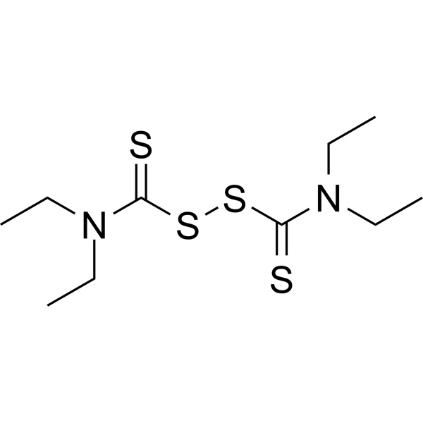
HY-B0240
DisulfiramALDH1 Inhibitor -
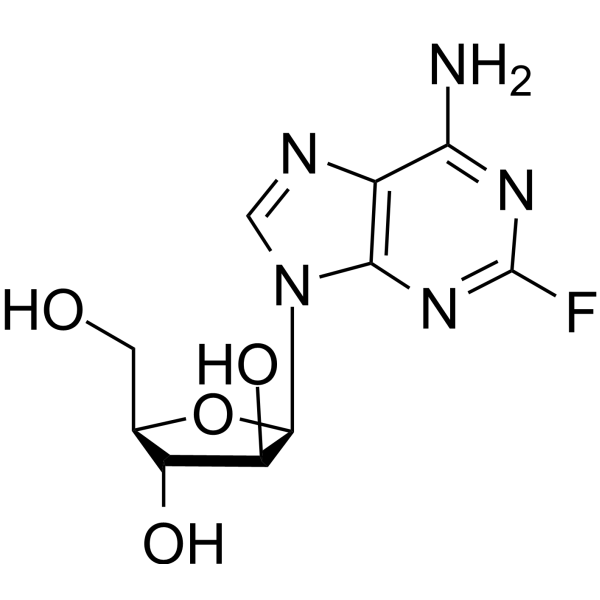
HY-B0069
FludarabineDNA Synthesis Inhibitor -
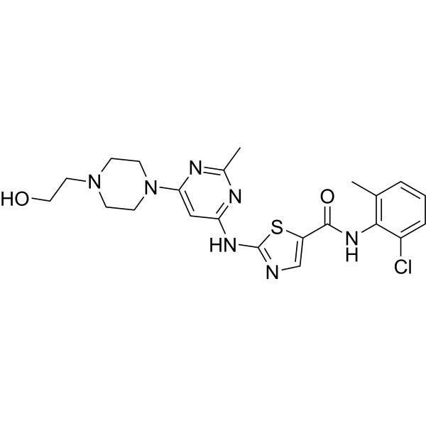
HY-10181
DasatinibSrc/Bcr-Abl Inhibitor -
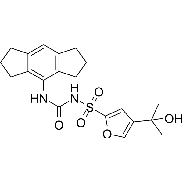
HY-12815
MCC950NLRP3 Inhibitor -
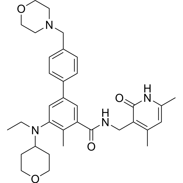
HY-13803
TazemetostatEZH2 Inhibitor -
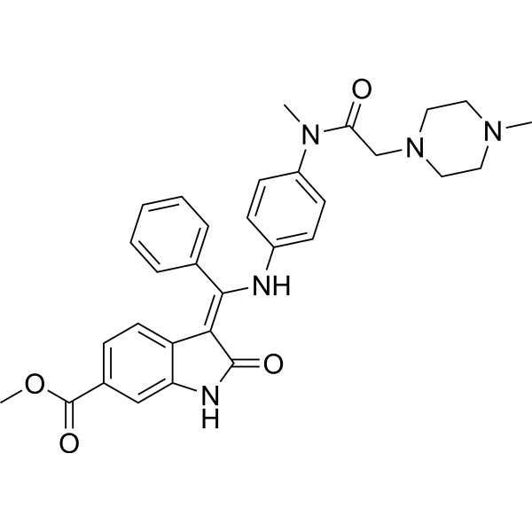
HY-50904
NintedanibPDGFR/VEGFR/FGFR Inhibitor -
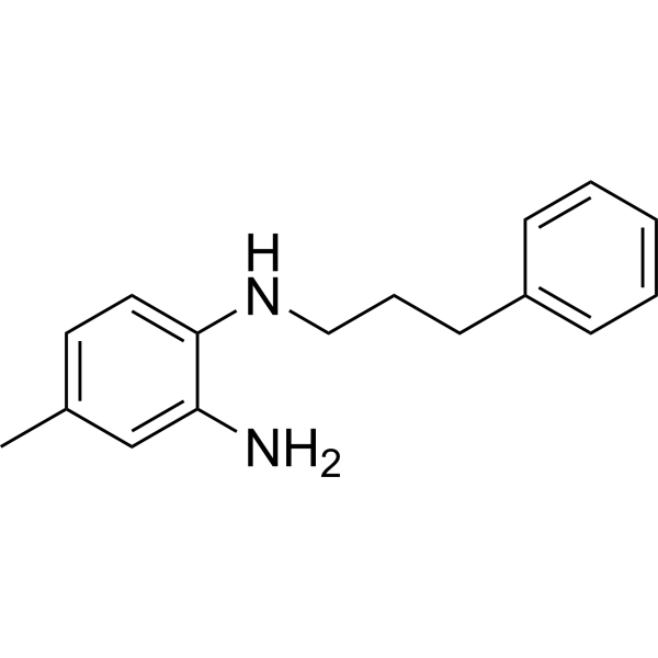
HY-13982
JSH-23NF-κB Inhibitor -
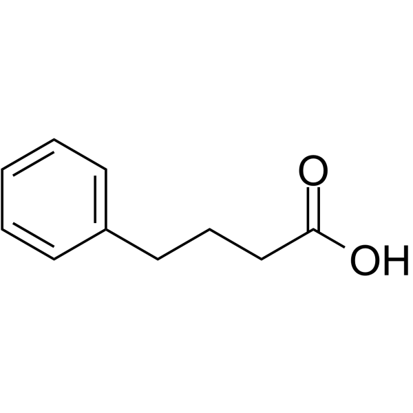
HY-A0281
4-Phenylbutyric acidHDAC Inhibitor -
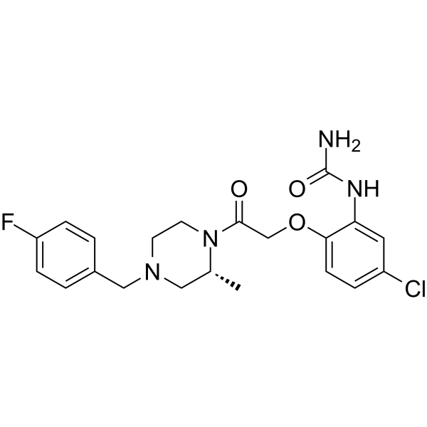
HY-12080
BX471CCR1 Antagonist -
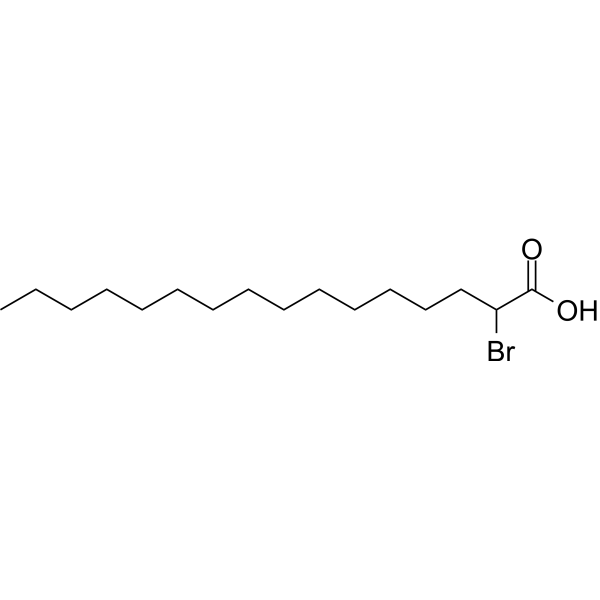
HY-111770
2-Bromohexadecanoic acidPyroptosis Inhibitor -
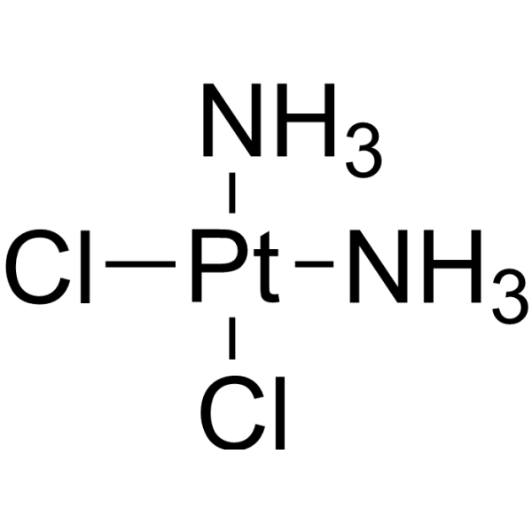
HY-17394
CisplatinDNA Alkylator
Publications Citing Use of MedChemExpress (MCE) IRF1-IN-1
MoreAlle Caspase Isoform-spezifische Produkte anzeigen
More

Biologische Aktivität
Beschreibung
IC50 & Target
[1]|
PARP-1 |
IL-1 |
Caspase-1 |
In Vitro
IRF1-IN-1 pretreatment (20 μM, 12 h) decreases the recruitment of IRF1 to the promoter of CASP1 in irradiated HaCaT cells (20 Gy)[1].
IRF1-IN-1 (20 μM, 24 h) attenuates the NSP-10 plasmid transfection-induced IRF1 activation in HELF, HaCaT and WS1 cells[1].
IRF1-IN-1 (50 μM, 24 h) treatment reduces the transcriptional activity of IRF1 in HELF cells 96 h after SARS-CoV-2 pseudovirus infection[1].
IRF1-IN-1 decreases radiation (20 Gy)-induced cell death in K150 cells[1].
IRF1-IN-1 maintains mitochondrial activity and ROS production in skin cells in the early stage after irradiation[1].
IRF1-IN-1 significantly inhibits the cell death signaling pathway, i.e., suppresses cleavage of Caspase 1, GSDMD, IL-1 and PARP1[1].
MedChemExpress (MCE) has not independently confirmed the accuracy of these methods. They are for reference only.
In Vivo
MedChemExpress (MCE) has not independently confirmed the accuracy of these methods. They are for reference only.
-
Animal Model:Radiogenic skin injury mice (intraperitoneal injection of pentobarbital sodium (1%, 30 mg/kg), 35 Gy at the dose rate of 1000 cGy/min by a 6-MeV electron beam)[1]
-
Dosage:100 μg/d
-
Administration:Subcutaneous injection (s.c.), one every other day before irradiation, pretreatment
-
Result:Showed a significant reduction in acute skin inflammatory manifestations, such as erythema and exudation and accelerated the healing process.
Showed protective effects on the function and structural integrity of radiation-induced lesions to the claws.


Chemical Information
-
CAS. Nr. 701225-07-2
-
Appearance Solid
-
Molecular Weight 440.52
-
Formel C22H24N4O4S
-
Color White to off-white
-
SMILES
O=C1C2=C(N=CN1CCC(NC3=CC=C(S(=O)(N4CCCCC4)=O)C=C3)=O)C=CC=C2
-
Versand
Room temperature in continental US; may vary elsewhere.
-
Speicherung
4°C, sealed storage, away from moisture
* In solvent : -80°C, 6 months; -20°C, 1 month (sealed storage, away from moisture)


Publications (1)
-
Journal Impact Factor
-
Most Recent
-
J Inflamm Res
Integrated Multi-Omics Analysis Reveals IRF1-Driven Microglial PANoptosis via ZBP1 in Spinal Cord Injury. [Abstract]2026 Mar 16:19:574990. PMID: 41867458


Lösungsmittel & Löslichkeit
In Vitro:
DMSO : 83.33 mg/mL (189.16 mM; Need ultrasonic; Hygroscopic DMSO has a significant impact on the solubility of product, please use newly opened DMSO)
Please refer to the solubility information to select the appropriate solvent. Once prepared, please aliquot and store the solution to prevent product inactivation from repeated freeze-thaw cycles.
Storage method and period of stock solution: -80°C, 6 months; -20°C, 1 month (sealed storage, away from moisture). When stored at -80°C, please use it within 6 months. When stored at -20°C, please use it within 1 month.
Please refer to the solubility information to select the appropriate solvent. Once prepared, please aliquot and store the solution to prevent product inactivation from repeated freeze-thaw cycles.
Storage method and period of stock solution: -80°C, 6 months; -20°C, 1 month (sealed storage, away from moisture). When stored at -80°C, please use it within 6 months. When stored at -20°C, please use it within 1 month.
Konzentration (Stammlösung) × Volumen (Stammlösung) = Konzentration (Ziellösung) × Volumen (Ziellösung)
In Vivo:
Select the appropriate dissolution method based on your experimental animal and administration route.
- For the following dissolution methods, please ensure to first prepare a clear stock solution using an In Vitro approach and then sequentially add co-solvents:
- To ensure reliable experimental results, the clarified stock solution can be appropriately stored based on storage conditions. As for the working solution for In Vivo experiments, it is recommended to prepare freshly and use it on the same day.
- The percentages shown for the solvents indicate their volumetric ratio in the final prepared solution. If precipitation or phase separation occurs during preparation, heat and/or sonication can be used to aid dissolution.
Add each solvent one by one: 10% DMSO 40% PEG300 5% Tween-80 45% Saline
Solubility: ≥ 8 mg/mL (18.16 mM); Clear solution
This protocol yields a clear solution of ≥ 8 mg/mL (saturation unknown).
Taking 1 mL working solution as an example, add 100 μL DMSO stock solution (80.0 mg/mL) to 400 μL PEG300, and mix evenly; then add 50 μL Tween-80 and mix evenly; then add 450 μL Saline to adjust the volume to 1 mL.
Preparation of Saline: Dissolve 0.9 g sodium chloride in ddH₂O and dilute to 100 mL to obtain a clear Saline solution.
Add each solvent one by one: 10% DMSO 90% (20% SBE-β-CD in Saline)
Solubility: ≥ 8 mg/mL (18.16 mM); Clear solution
This protocol yields a clear solution of ≥ 8 mg/mL (saturation unknown).
Taking 1 mL working solution as an example, add 100 μL DMSO stock solution (80.0 mg/mL) to 900 μL 20% SBE-β-CD in Saline, and mix evenly.
Preparation of 20% SBE-β-CD in Saline (4°C, storage for one week): 2 g SBE-β-CD powder is dissolved in 10 mL Saline, completely dissolve until clear.
In Vivo Dissolution Calculator
Please enter the basic information of animal experiments:
-
-
-
-
Recommended: Prepare an additional quantity of animals to account for potential losses during experiments.
Please enter your animal formula composition:
-
%DMSO +
Recommended: Keep the proportion of DMSO in working solution below 2% if your animal is weak.
-
%+
-
+%Tween-80 + +
-
%Saline +
The co-solvents required include: DMSO, . All of co-solvents are available by MedChemExpress (MCE). , Tween 80. All of co-solvents are available by MedChemExpress (MCE).
Working solution concentration: 0.22 mg/mL
Method for preparing stock solution: mg drug dissolved in μL DMSO. Stock solution concentration: mg/mL. * In solvent : -80°C, 6 months; -20°C, 1 month (sealed storage, away from moisture)
1. Take μL DMSO stock solution;
2. Add μL .
μL , mix evenly;
3. Then add μL Tween 80, mix evenly;
4. Then add μL
Please ensure that the stock solution in the first step is dissolved to a clear state, and add co-solvents in sequence. You can use ultrasonic heating (ultrasonic cleaner, recommended frequency 20-40 kHz), vortexing, etc. to assist dissolution.


Reinheit & Dokumentation
-
Data Sheet (273 KB)
-
SDS (252 KB)
- English - EN (252 KB)
- Français - FR (252 KB)
- Deutsch - DE (252 KB)
- Norwegian - NO (252 KB)
- Español - ES (252 KB)
- Swedish - SV (252 KB)
- Italian - IT (252 KB)
- Korean - KR (252 KB)
- Portuguese - PT (252 KB)
-
Handling Instructions (2659 KB)
Verweise


Complete Stock Solution Preparation Table
Please refer to the solubility information to select the appropriate solvent. Once prepared, please aliquot and store the solution to prevent product inactivation from repeated freeze-thaw cycles.
Storage method and period of stock solution: -80°C, 6 months; -20°C, 1 month (sealed storage, away from moisture). When stored at -80°C, please use it within 6 months. When stored at -20°C, please use it within 1 month.
| Optional Solvent | Concentration Solvent Mass | 1 mg | 5 mg | 10 mg | 25 mg |
|---|---|---|---|---|---|
| DMSO | 1 mM | 2.2700 mL | 11.3502 mL | 22.7004 mL | 56.7511 mL |
| 5 mM | 0.4540 mL | 2.2700 mL | 4.5401 mL | 11.3502 mL | |
| 10 mM | 0.2270 mL | 1.1350 mL | 2.2700 mL | 5.6751 mL | |
| 15 mM | 0.1513 mL | 0.7567 mL | 1.5134 mL | 3.7834 mL | |
| 20 mM | 0.1135 mL | 0.5675 mL | 1.1350 mL | 2.8376 mL | |
| 25 mM | 0.0908 mL | 0.4540 mL | 0.9080 mL | 2.2700 mL | |
| 30 mM | 0.0757 mL | 0.3783 mL | 0.7567 mL | 1.8917 mL | |
| 40 mM | 0.0568 mL | 0.2838 mL | 0.5675 mL | 1.4188 mL | |
| 50 mM | 0.0454 mL | 0.2270 mL | 0.4540 mL | 1.1350 mL | |
| 60 mM | 0.0378 mL | 0.1892 mL | 0.3783 mL | 0.9459 mL | |
| 80 mM | 0.0284 mL | 0.1419 mL | 0.2838 mL | 0.7094 mL | |
| 100 mM | 0.0227 mL | 0.1135 mL | 0.2270 mL | 0.5675 mL |


IRF1-IN-1 Related Classifications
HY-171006 Related Classifications
 Powered by Bioz