



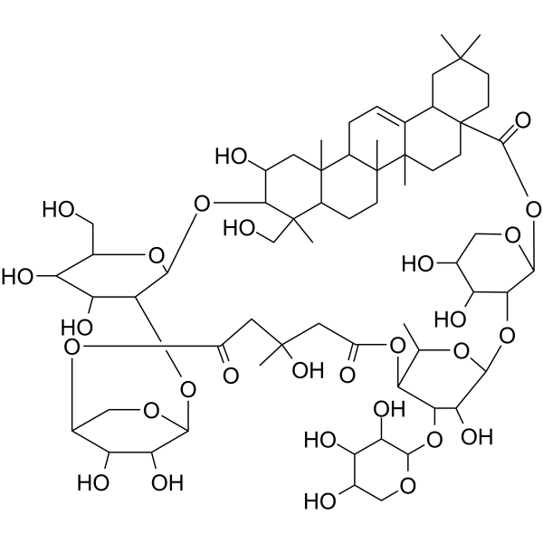
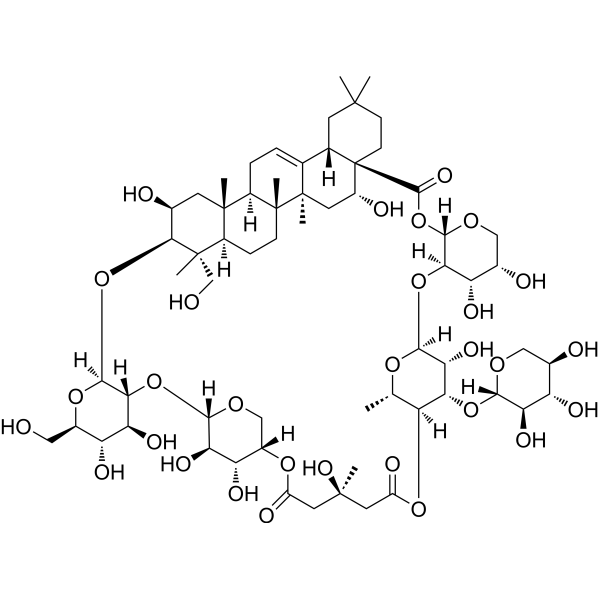
Tubeimoside I
Based on 5 publication(s) in Google Scholar
Tubeimoside I is an orally active HSPD1 inhibitor. Tubeimoside I inhibits NF-κB, MAPK, as well as regulates eNOS-VEGF. Tubeimoside I induces cytoprotective Autophagy via an Akt-mediated pathway. Tubeimoside I inhibits proinflammatory cytokine (IL-6 and IL-1β) production. Tubeimoside I exhibits anti-inflammatory activities. Tubeimoside I promotes angiogenesis and improves sepsis symptoms. Tubeimoside I is used in the research of inflammatory diseases, various cancers, sepsis and ischemic diseases.
For research use only. We do not sell to patients.
- Purity : 99.96%
- CAS No.: 102040-03-9
- Formula: C63H98O29
- Molecular Weight:1319.43
-
Storage:Powder -20°C, 3 years , 4°C, 2 years ; In solvent -80°C, 2 years , -20°C, 1 year

To place orders, for customer services and technical support, please contact: MedChemExpress USA
Tel: 609-228-6898 E-mail: [email protected] [email protected]


-
 Biological Activity
-
Chemical Information
-
Solvent & Solubility
- Protocols
- Purity & Documentation
- References
-
 Help & FAQs


-
HY-N0891
Tubeimoside IIAntiviral/Anticancer Agent -

HY-10219
RapamycinmTOR Inhibitor -
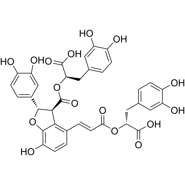
HY-N1362
Salvianolic acid BAntioxidant -
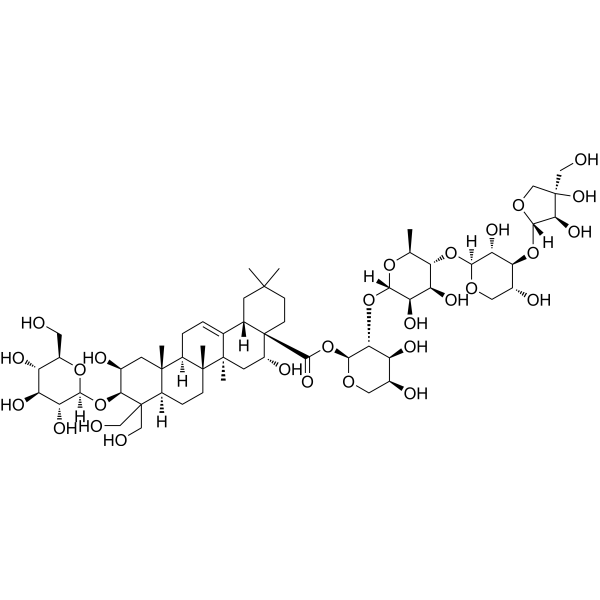
HY-N1411
Platycodin DAMPK Activator -
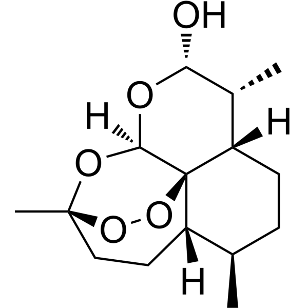
HY-N0176
DihydroartemisininAntimalarial Agent -
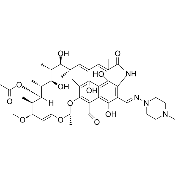
HY-B0272
RifampicinAntivirus Agent -
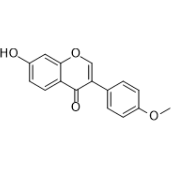
HY-N0183
FormononetinFGFR2 Inhibitor -
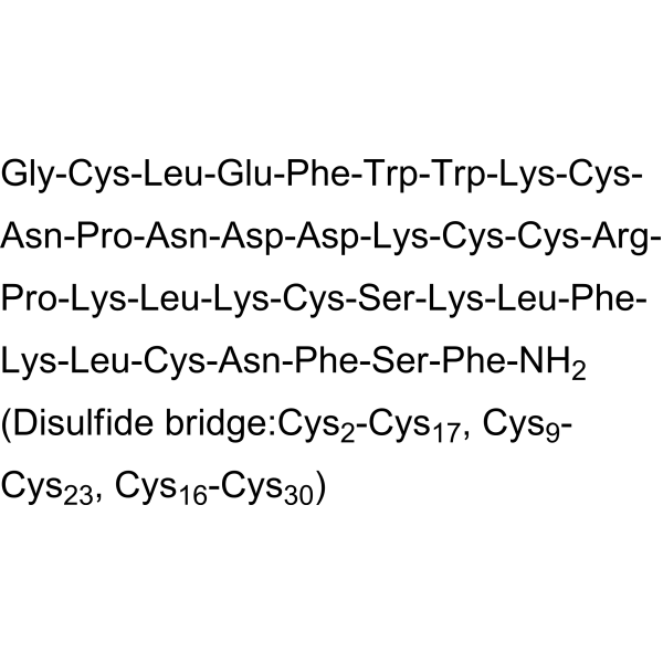
HY-P1410
GsMTx4MSCs Inhibitor -
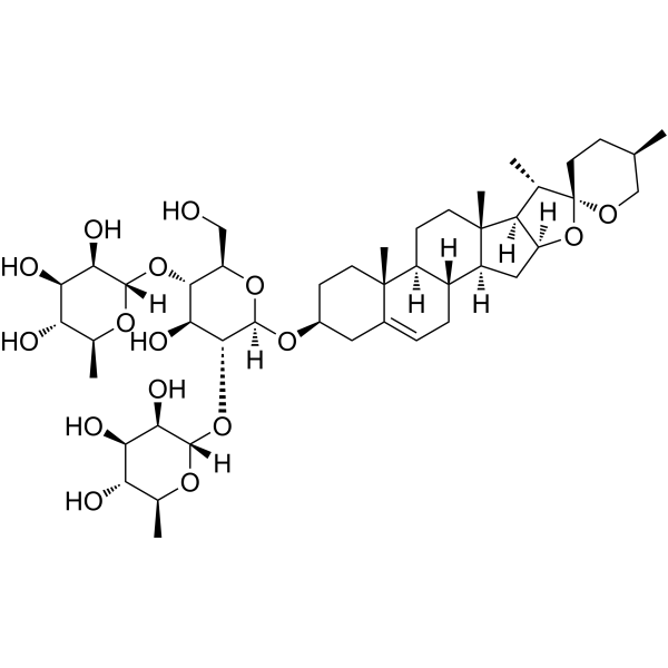
HY-N0124
DioscinAnticancer Agent -
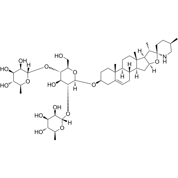
HY-N0069
SolamargineP-gp Inhibitor -
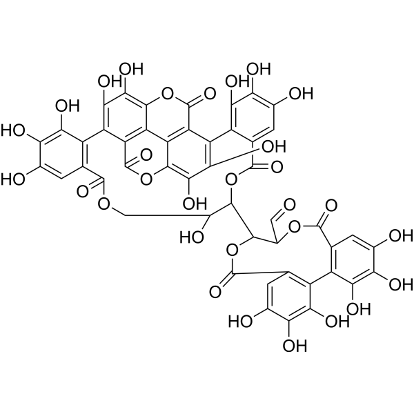
HY-N0063
Punicalagin3CLpro Inhibitor -
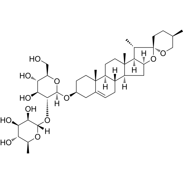
HY-N6940
Prosapogenin AAnticancer Agent
Publications Citing Use of MedChemExpress (MCE) Tubeimoside I
More Customer Validation & Images

-
Cell Proliferation/Viability Assay
-
Flow Cytometry
-
WB
-
IF
-
In Vivo Efficacy Study
All VEGFR Isoforms
More

Biological Activity
Description
Cellular Effect
|
Cell Line

|
Type | Value | Description | References |
|---|---|---|---|---|
| A549 | IC50 |
12.3 μM
Compound: TBM I
|
Antiproliferative activity against human A549 cells after 72 hrs by MTS assay
Antiproliferative activity against human A549 cells after 72 hrs by MTS assay
|
[PMID: 30439592] |
| DU-145 | IC50 |
10 μM
Compound: TBM I
|
Antiproliferative activity against human DU145 cells after 24 hrs by MTT assay
Antiproliferative activity against human DU145 cells after 24 hrs by MTT assay
|
[PMID: 30439592] |
| HeLa | IC50 |
17.1 μM
Compound: TBM I
|
Antiproliferative activity against human HeLa cells after 72 hrs by MTT assay
Antiproliferative activity against human HeLa cells after 72 hrs by MTT assay
|
[PMID: 30439592] |
| HeLa | IC50 |
25 μM
Compound: TBM I
|
Cytotoxicity against human HeLa cells after 24 hrs by MTT assay
Cytotoxicity against human HeLa cells after 24 hrs by MTT assay
|
[PMID: 30439592] |
| HeLa | IC50 |
27.1 μM
Compound: TBM I
|
Antiproliferative activity against human HeLa cells after 48 hrs by MTT assay
Antiproliferative activity against human HeLa cells after 48 hrs by MTT assay
|
[PMID: 30439592] |
| HeLa | IC50 |
34.8 μM
Compound: TBM I
|
Antiproliferative activity against human HeLa cells after 24 hrs by MTT assay
Antiproliferative activity against human HeLa cells after 24 hrs by MTT assay
|
[PMID: 30439592] |
| HepG2 | IC50 |
15.5 μM
Compound: TBM I
|
Antiproliferative activity against human HepG2 cells after 24 hrs by MTT assay
Antiproliferative activity against human HepG2 cells after 24 hrs by MTT assay
|
[PMID: 30439592] |
| HUVEC | IC50 |
17.9 μM
Compound: TBM I
|
Antiproliferative activity against HUVEC after 72 hrs
Antiproliferative activity against HUVEC after 72 hrs
|
[PMID: 30439592] |
| HUVEC | IC50 |
21.4 μM
Compound: TBM I
|
Antiproliferative activity against HUVEC after 48 hrs
Antiproliferative activity against HUVEC after 48 hrs
|
[PMID: 30439592] |
| HUVEC | IC50 |
24.2 μM
Compound: TBM I
|
Antiproliferative activity against HUVEC after 24 hrs
Antiproliferative activity against HUVEC after 24 hrs
|
[PMID: 30439592] |
| JEG-3 | IC50 |
8.5 μM
Compound: TBM I
|
Antiproliferative activity against human JEG3 cells after 24 hrs by MTT assay
Antiproliferative activity against human JEG3 cells after 24 hrs by MTT assay
|
[PMID: 30439592] |
| NCI-H1299 | IC50 |
10 μM
Compound: TBM I
|
Antiproliferative activity against human NCI-H1299 cells after 48 hrs by CCK-8 assay
Antiproliferative activity against human NCI-H1299 cells after 48 hrs by CCK-8 assay
|
[PMID: 30439592] |
| NCI-H460 | IC50 |
20 μM
Compound: TBM I
|
Antiproliferative activity against human NCI-H460 cells after 9 hrs by MTT assay
Antiproliferative activity against human NCI-H460 cells after 9 hrs by MTT assay
|
[PMID: 30439592] |
| NCI-H460 | IC50 |
23.3 μM
Compound: TBM I
|
Antiproliferative activity against human NCI-H460 cells after 24 hrs by WST-1 assay
Antiproliferative activity against human NCI-H460 cells after 24 hrs by WST-1 assay
|
[PMID: 30439592] |
| PC-3 | IC50 |
20 μM
Compound: TBM I
|
Antiproliferative activity against human PC3 cells after 24 hrs by MTT assay
Antiproliferative activity against human PC3 cells after 24 hrs by MTT assay
|
[PMID: 30439592] |
| PC-9 | IC50 |
10.2 μM
Compound: TBM I
|
Antiproliferative activity against human PC9 cells after 72 hrs by MTS assay
Antiproliferative activity against human PC9 cells after 72 hrs by MTS assay
|
[PMID: 30439592] |
| SK-OV-3 | IC50 |
16 μM
Compound: TBM I
|
Antiproliferative activity against human SKOV3 cells after 24 hrs by MTT assay
Antiproliferative activity against human SKOV3 cells after 24 hrs by MTT assay
|
[PMID: 30439592] |
| T-24 | IC50 |
20 μM
Compound: TBM I
|
Antiproliferative activity against human T24 cells after 24 hrs by MTT assay
Antiproliferative activity against human T24 cells after 24 hrs by MTT assay
|
[PMID: 30439592] |
In Vitro
Tubeimoside I (0.5-8 µM; 6-48 h) induces cytoprotective autophagy in human breast cancer cells (MDA-MB-231, MCF-7, T47D) via Akt-mediated pathway[1].
Tubeimoside I (20 µM; 24 h) inhibits the late stage of autophagic flux in lung cancer cells transfected with mCherry-GFP-LC3 tandem plasmids[2].
Tubeimoside I (0-30 µM; 24 h) markedly decreases cervical cancer (Hela and SiHa) cell viability in a dose-dependent manner[3].
Tubeimoside I (25 μM; 4-24 h) causes cell cycle arrest at the G2/M phase in HeLa cells in a dose- and time- dependent manner[4].
Tubeimoside I (3-15 μM; 12-24 h) induces oxidative stress-mediated apoptosis and G0/G1 phase arrest in human prostate carcinoma cells DU145 and PC3[5].
Tubeimoside I (15-30 μM; 24 h) induces apoptosis in HepG2 and L-02 cells[6].
Tubeimoside I (2-6 μM; 1 h) attenuates LPS-induced inflammation in RAW 264.7 cells, inhibiting the production of pro-inflammatory cytokines TNF-α, IL-6 and IL-1β[7].
Tubeimoside I (0.5-10 μM; 48 h) inhibits the viability of HCT-116 colon cancer cells in a dose-dependent manner[10].
MedChemExpress (MCE) has not independently confirmed the accuracy of these methods. They are for reference only.
-
Cell Line:MCF-7, MDA-MB-231, T47D
-
Concentration:0.5 μM, 1 μM, 2 μM, 4 μM, 8 μM
-
Incubation Time:6, 9, 12, 24, 36 h
-
Result:Induced apoptosis in a dose-dependent manner, as determined by increased cleaved PARP and caspase-3 levels.
Decreased the levels of Mcl-1, Bcl-xl, and Bcl-2 in a time- and dose- dependent manner.
In Vivo
Tubeimoside I (3 mg/kg; i.p.; daily; 16 days) inhibits the growth of HeLa-xenografted tumors in nude mice, as indicated by smaller tumor size, volume, and mass[3].
Tubeimoside I (1-4 mg/kg; i.p.; 1 h before LPS challenge) reduces lung injury, down-regulates the secretion of TNF-α, IL-6 and IL-1β, and inhibits the activation of NF-κB and MAPK in a murine model of LPS-induced acute lung injury[7].
Tubeimoside I (45-180 mg/kg; p.o.; daily; 21 consecutive days) attenuates inflammation and oxidative damage in a mice model of PM2.5‑induced pulmonary injury[8].
Tubeimoside I (4 mg/kg; i.p.; 1 h before cecal ligation and puncture (CLP)) improves survival of mice in sepsis by inhibiting inducible nitric oxide synthase expression[9].
Tubeimoside I (4 mg/kg; i.p.; every day; 28 days) improves recovery from hindlimb ischemia and increases capillary density in C57BL/6 mice[11].
MedChemExpress (MCE) has not independently confirmed the accuracy of these methods. They are for reference only.
-
Animal Model:5-week-old male BALB/c nude mice (weighing 18-22 g), lung cancer xenograft model established by subcutaneous injection of NCI-H1299 cells[2]
-
Dosage:1 mg/kg, 4 mg/kg
-
Administration:Intraperitoneal injection, 13 days
-
Result:Reduced the tumor volume and weight.
Significantly upregulated the expression levels of cleaved-PARP, cleaved-caspase 3, LC3-II, and p62 in the tumor tissues.
-
Animal Model:Female BALB/c nude mice (4-week-old) with HeLa cell xenograft model[3]
-
Dosage:3 mg/kg
-
Administration:Intraperitoneal injection, daily, for 16 days
-
Result:Decreased the expression of Ki67 in tumor tissues, indicating reduced proliferating ability.
Induced tumor cell apoptosis, as evidenced by the accumulation of cleaved-CASP3 and cleaved-PARP1 in tumor tissues.
Induced autophagosome accumulation in tumor cells, with increased LC3 II expression and enhanced LC3 staining in tumor tissues.
- Human pluripotent stem cell endothelial-cell differentiation
- Vascular/Branching Fractal Analysis
- LPS-Induced Endotoxemia/Systemic Inflammation
- Autophagy
- Endothelial Tube Formation Assay
- Research Protocol for Inflammation-related Diseases
- Lysosome and acidic-vesicle live-cell staining
- Macroautophagy Solutions
- Endothelial Cell Migration/Angiogenic Sprouting Assay
- Pyroptosis Solutions


Chemical Information
-
CAS No. 102040-03-9
-
Appearance Solid
-
Molecular Weight 1319.43
-
Formula C63H98O29
-
Color White to off-white
-
SMILES
C[C@@]12C([C@@]3([H])[C@](C(O[C@@H]4OC[C@H](O)[C@H](O)[C@H]4O[C@@H]5O[C@@H](C)[C@H](OC(C[C@](O)(C)CC6=O)=O)[C@@H](O[C@@H]7OC[C@@H](O)[C@H](O)[C@H]7O)[C@H]5O)=O)(CCC(C)(C)C3)CC2)=CC[C@@]8([H])[C@@]1(C)CC[C@]([C@](C)(CO)[C@H]9O[C@@H]%10O[C@H](CO)[C@@H](O)[C@H](O)[C@H]%10O[C@@H]%11OC[C@H](O6)[C@H](O)[C@H]%11O)([H])[C@]8(C)C[C@@H]9O
-
Synonyms
Tubeimoside-1; Lobatoside-H
-
Structure Classification
-
Initial Source
-
Shipping
Room temperature in continental US; may vary elsewhere.
-
Storage
Powder -20°C 3 years 4°C 2 years In solvent -80°C 2 years -20°C 1 year


Publications (5)
-
Journal Impact Factor
-
Most Recent
-
Pharmacol Res
Cardamonin retards progression of autosomal dominant polycystic kidney disease via inhibiting renal cyst growth and interstitial fibrosis. [Abstract]2020 May:155:104751. PMID: 32151678 -
Phytomedicine
Tubeimoside Ⅰ targets sodium/potassium ATPase alpha 1 subunit to selectively eliminate senescent cells and alleviate aging-associated abnormalities. [Abstract]2026 Jul:156:158202. PMID: 42035526 -
Transl Oncol
Tubeimoside I induces mitophagy by activating the PINK1/Parkin/Mfn2 signaling pathway in acute myeloid leukemia cells. [Abstract]2025 May:55:102355. PMID: 40112502
Tubeimoside I purchased from MedChemExpress. Usage Cited in: Transl Oncol. 2025 May:55:102355. [Abstract]
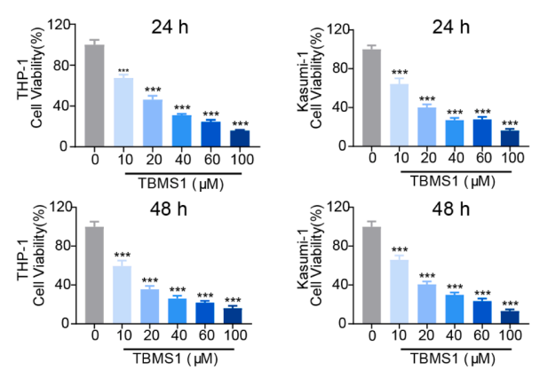
Cell viability was assessed after PBS and TBMS1 (Tubeimoside I) treatment for 24 and 48 hours.
Tubeimoside I purchased from MedChemExpress. Usage Cited in: Transl Oncol. 2025 May:55:102355. [Abstract]
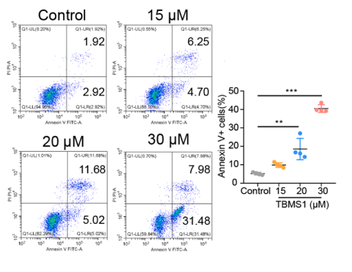
Annexin V-PI flow cytometry analysis of apoptosis in AML cells treated with TBMS1 (Tubeimoside I) for 24 hours.
Tubeimoside I purchased from MedChemExpress. Usage Cited in: Transl Oncol. 2025 May:55:102355. [Abstract]
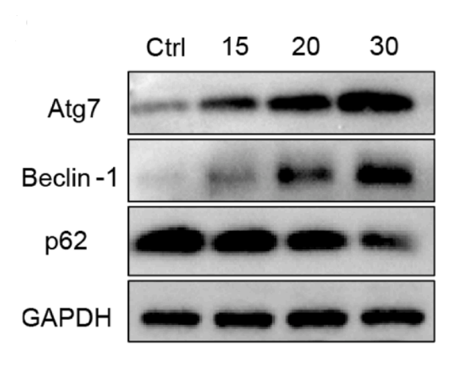
TBMS1 (Tubeimoside I) induces mitophagy in AML cells. The expression of autophagy-related proteins was detected by Western blot.
Tubeimoside I purchased from MedChemExpress. Usage Cited in: Transl Oncol. 2025 May:55:102355. [Abstract]
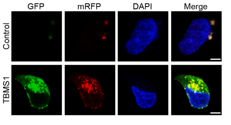
TBMS1 (Tubeimoside I) induces mitophagy in AML cells. LC3B protein expression was detected by confocal microscopy.
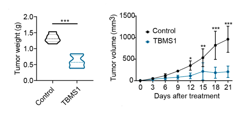
Tubeimoside I purchased from MedChemExpress. Usage Cited in: Transl Oncol. 2025 May:55:102355. [Abstract]
Tumor weight and volume after 21 days of treatment with saline and TBMS1 (Tubeimoside I: 5 mg/kg).
-
Biochem Biophys Res Commun
Engineered MAP30ER: A plant toxin-derived platform for EGFR-Targeted delivery of protein and chemotherapeutic payloads. [Abstract]2025 Aug 30:776:152184. PMID: 40517671 -
Oxid Med Cell Longev
2023 Jan 14:2023:9966355. PMID: 36691640


Solvent & Solubility
In Vitro:
DMSO : 50 mg/mL (37.90 mM; ultrasonic and warming and heat to 60°C; Hygroscopic DMSO has a significant impact on the solubility of product, please use newly opened DMSO)
Please refer to the solubility information to select the appropriate solvent. Once prepared, please aliquot and store the solution to prevent product inactivation from repeated freeze-thaw cycles.
Storage method and period of stock solution: -80°C, 2 years; -20°C, 1 year. When stored at -80°C, please use it within 2 years. When stored at -20°C, please use it within 1 year.
Please refer to the solubility information to select the appropriate solvent. Once prepared, please aliquot and store the solution to prevent product inactivation from repeated freeze-thaw cycles.
Storage method and period of stock solution: -80°C, 2 years; -20°C, 1 year. When stored at -80°C, please use it within 2 years. When stored at -20°C, please use it within 1 year.
Concentration (start) × Volume (start) = Concentration (final) × Volume (final)
In Vivo:
Select the appropriate dissolution method based on your experimental animal and administration route.
- For the following dissolution methods, please ensure to first prepare a clear stock solution using an In Vitro approach and then sequentially add co-solvents:
- To ensure reliable experimental results, the clarified stock solution can be appropriately stored based on storage conditions. As for the working solution for In Vivo experiments, it is recommended to prepare freshly and use it on the same day.
- The percentages shown for the solvents indicate their volumetric ratio in the final prepared solution. If precipitation or phase separation occurs during preparation, heat and/or sonication can be used to aid dissolution.
Add each solvent one by one: 10% DMSO 40% PEG300 5% Tween-80 45% Saline
Solubility: ≥ 2.5 mg/mL (1.89 mM); Clear solution
This protocol yields a clear solution of ≥ 2.5 mg/mL (saturation unknown).
Taking 1 mL working solution as an example, add 100 μL DMSO stock solution (25.0 mg/mL) to 400 μL PEG300, and mix evenly; then add 50 μL Tween-80 and mix evenly; then add 450 μL Saline to adjust the volume to 1 mL.
Preparation of Saline: Dissolve 0.9 g sodium chloride in ddH₂O and dilute to 100 mL to obtain a clear Saline solution.
Add each solvent one by one: 10% DMSO 90% (20% SBE-β-CD in Saline)
Solubility: ≥ 2.5 mg/mL (1.89 mM); Clear solution
This protocol yields a clear solution of ≥ 2.5 mg/mL (saturation unknown).
Taking 1 mL working solution as an example, add 100 μL DMSO stock solution (25.0 mg/mL) to 900 μL 20% SBE-β-CD in Saline, and mix evenly.
Preparation of 20% SBE-β-CD in Saline (4°C, storage for one week): 2 g SBE-β-CD powder is dissolved in 10 mL Saline, completely dissolve until clear.
In Vivo Dissolution Calculator
Please enter the basic information of animal experiments:
-
-
-
-
Recommended: Prepare an additional quantity of animals to account for potential losses during experiments.
Please enter your animal formula composition:
-
%DMSO +
Recommended: Keep the proportion of DMSO in working solution below 2% if your animal is weak.
-
%+
-
+%Tween-80 + +
-
%Saline +
The co-solvents required include: DMSO, . All of co-solvents are available by MedChemExpress (MCE). , Tween 80. All of co-solvents are available by MedChemExpress (MCE).
Working solution concentration: 0.22 mg/mL
Method for preparing stock solution: mg drug dissolved in μL DMSO. Stock solution concentration: mg/mL.
1. Take μL DMSO stock solution;
2. Add μL .
μL , mix evenly;
3. Then add μL Tween 80, mix evenly;
4. Then add μL
Please ensure that the stock solution in the first step is dissolved to a clear state, and add co-solvents in sequence. You can use ultrasonic heating (ultrasonic cleaner, recommended frequency 20-40 kHz), vortexing, etc. to assist dissolution.


Purity & Documentation
-
Data Sheet (298 KB)
-
SDS (393 KB)
- English - EN (393 KB)
- Français - FR (393 KB)
- Deutsch - DE (393 KB)
- Norwegian - NO (393 KB)
- Español - ES (393 KB)
- Swedish - SV (393 KB)
- Italian - IT (393 KB)
- Korean - KR (393 KB)
- Portuguese - PT (393 KB)
-
Handling Instructions (2659 KB)
References
[1]. Jiang SL, et al. Tubeimoside-1, a triterpenoid saponin, induces cytoprotective autophagy in human breast cancer cells in vitro via Akt-mediated pathway. Acta Pharmacol Sin. 2019 Jul;40(7):919-928. [Content Brief]
[2]. Wang K, et al. Tubeimoside I-induced lung cancer cell death and the underlying crosstalk between lysosomes and mitochondria. Cell Death Dis. 2020 Aug 26;11(8):708. [Content Brief]
[3]. Feng X, et al. Tubeimoside I induces accumulation of impaired autophagolysosome against cervical cancer cells by both initiating autophagy and inhibiting lysosomal function. Cell Death Dis. 2018 Nov 2;9(11):1117. [Content Brief]
[4]. Xu Y, et al. Multiple pathways were involved in tubeimoside-1-induced cytotoxicity of HeLa cells. J Proteomics. 2011 Dec 21;75(2):491-501. [Content Brief]
[5]. Yang JB, et al. Tubeimoside-1 induces oxidative stress-mediated apoptosis and G0/G1 phase arrest in human prostate carcinoma cells in vitro. Acta Pharmacol Sin. 2016 Jul;37(7):950-62. [Content Brief]
[6]. Wang Y, et al. Natural plant extract tubeimoside I promotes apoptosis-mediated cell death in cultured human hepatoma (HepG2) cells. Biol Pharm Bull. 2011;34(6):831-8. [Content Brief]
[7]. Wu Q, et al. Tubeimoside-1 attenuates LPS-induced inflammation in RAW 264.7 macrophages and mouse models. Immunopharmacol Immunotoxicol. 2013 Aug;35(4):514-23. [Content Brief]
[8]. Zhang JB, et al. Tubeimoside I attenuates inflammation and oxidative damage in a mice model of PM2.5-induced pulmonary injury. Exp Ther Med. 2018 Feb;15(2):1602-1607. [Content Brief]
[9]. Luo M, et al.Tubeimoside I improves survival of mice in sepsis by inhibiting inducible nitric oxide synthase expression. Biomed Pharmacother. 2020 Jun;126:110083. [Content Brief]
[10]. Wu T, et al. Tubeimoside-I, an inhibitor of HSPD1, enhances cytotoxicity of oxaliplatin by activating ER stress and MAPK signaling pathways in colorectal cancer. J Ethnopharmacol. 2025 Jan 10;336:118754. [Content Brief]
[11]. Yang X, et al. Tubeimoside I promotes angiogenesis via activation of eNOS-VEGF signaling pathway. J Ethnopharmacol. 2021 Mar 1;267:113642. [Content Brief]


Complete Stock Solution Preparation Table
Please refer to the solubility information to select the appropriate solvent. Once prepared, please aliquot and store the solution to prevent product inactivation from repeated freeze-thaw cycles.
Storage method and period of stock solution: -80°C, 2 years; -20°C, 1 year. When stored at -80°C, please use it within 2 years. When stored at -20°C, please use it within 1 year.
| Optional Solvent | Concentration Solvent Mass | 1 mg | 5 mg | 10 mg | 25 mg |
|---|---|---|---|---|---|
| DMSO | 1 mM | 0.7579 mL | 3.7895 mL | 7.5790 mL | 18.9476 mL |
| 5 mM | 0.1516 mL | 0.7579 mL | 1.5158 mL | 3.7895 mL | |
| 10 mM | 0.0758 mL | 0.3790 mL | 0.7579 mL | 1.8948 mL | |
| 15 mM | 0.0505 mL | 0.2526 mL | 0.5053 mL | 1.2632 mL | |
| 20 mM | 0.0379 mL | 0.1895 mL | 0.3790 mL | 0.9474 mL | |
| 25 mM | 0.0303 mL | 0.1516 mL | 0.3032 mL | 0.7579 mL | |
| 30 mM | 0.0253 mL | 0.1263 mL | 0.2526 mL | 0.6316 mL |


Tubeimoside I Related Classifications
HY-N0890 Related Classifications
 Powered by Bioz