



CAY10526
Based on 2 publication(s) in Google Scholar
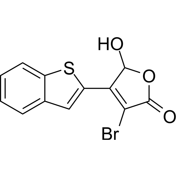
CAY10526 is an inhibitor of Y-box binding protein 1 (YB-1) and microsomal prostaglandin E2 synthase 1 (mPGES1). CAY10526 inhibits the production of PGE2 by suppressing YB-1 and mPGES1. CAY10526 induces cell apoptosis (apoptosis) and inhibits the JAK/STAT, TGF-β/Smad3 and PI3K/AKT signaling pathways. CAY10526 can be used in research related to melanoma, prostate cancer, esophageal adenocarcinoma, T-cell lymphoma, etc.
For research use only. We do not sell to patients.
- Purity : 99.12%
- CAS No.: 938069-71-7
- Formula: C12H7BrO3S
- Molecular Weight:311.16
-
Storage:
4°C, sealed storage, away from moisture and light
* In solvent : -80°C, 6 months; -20°C, 1 month (sealed storage, away from moisture and light)

To place orders, for customer services and technical support, please contact: MedChemExpress USA
Tel: 609-228-6898 E-mail: [email protected] [email protected]


-
 Biological Activity
-
Chemical Information
-
Solvent & Solubility
- Protocols
- Purity & Documentation
- References
-
 Help & FAQs


-
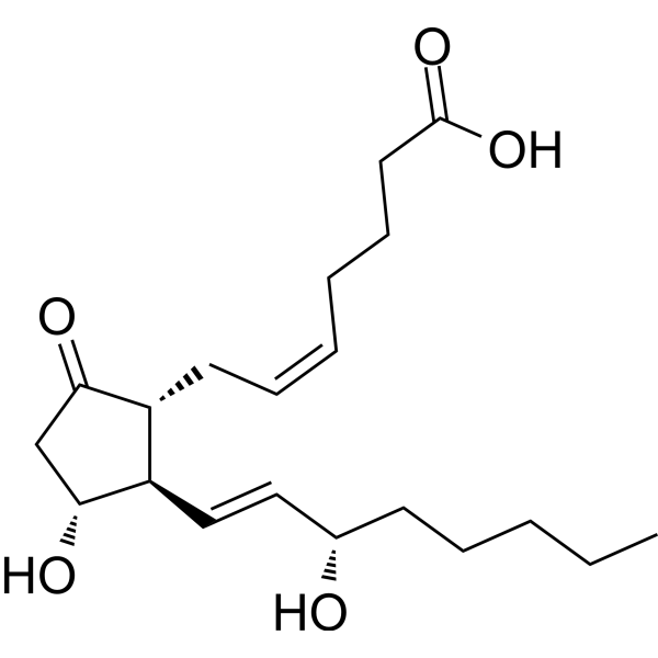
HY-101952
Prostaglandin E2Prostaglandin Receptor Agonist -
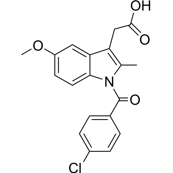
HY-14397
IndomethacinCOX1/2 Inhibitor -
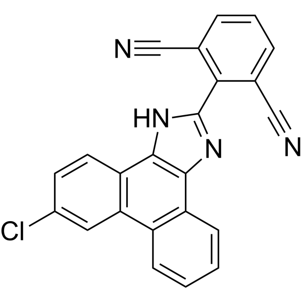
HY-13283
MF63MF63 Inhibitor -
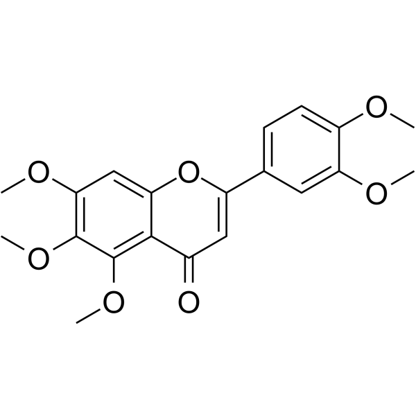
HY-N0297
SinensetinTNF Receptor Inhibitor -
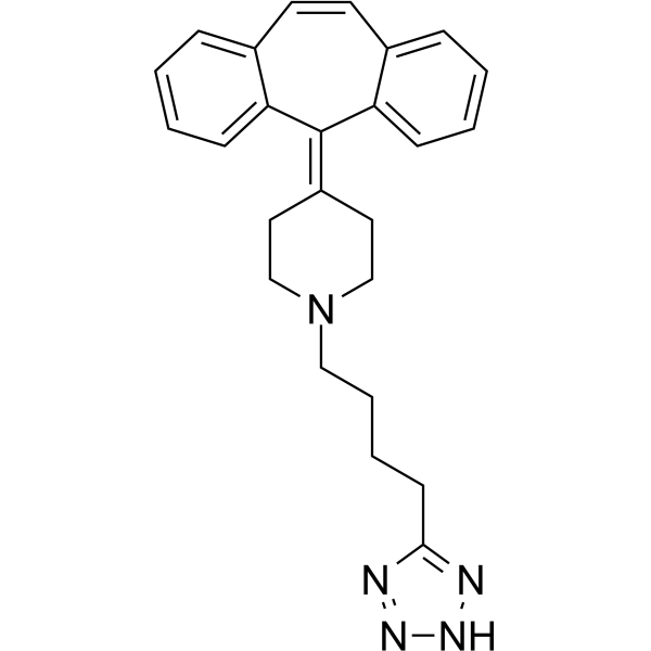
HY-13988
AT-56L-PGDS Inhibitor -
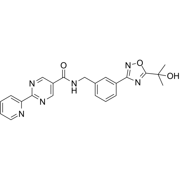
HY-12791
hPGDS-IN-1hPGDS Inhibitor -
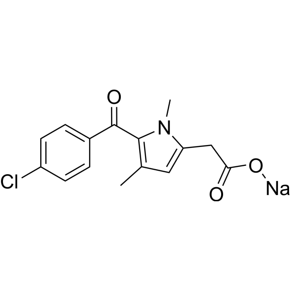
HY-B0890
Zomepirac sodium saltProstaglandin Synthetase Inhibitor -
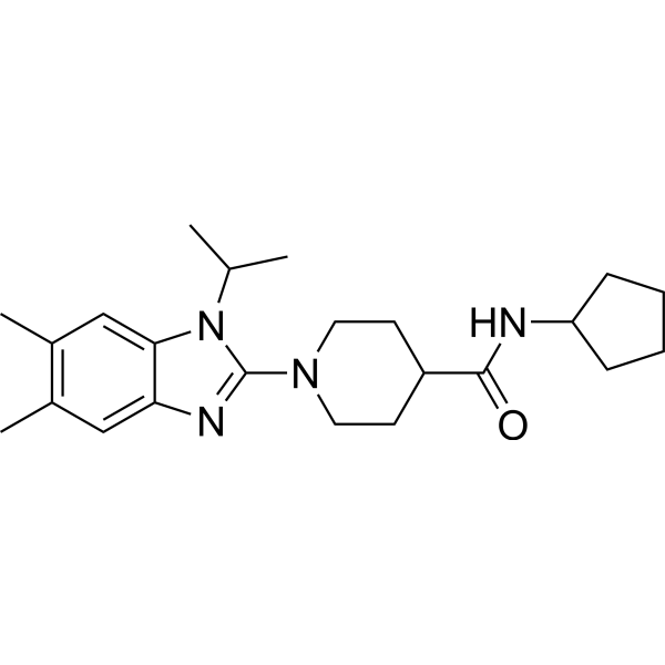
HY-118282
mPGES1-IN-7mPGES-1 Inhibitor -
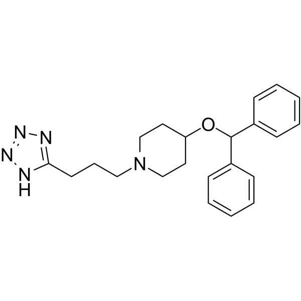
HY-108259
HQL-79Prostaglandin D Synthase Inhibitor -
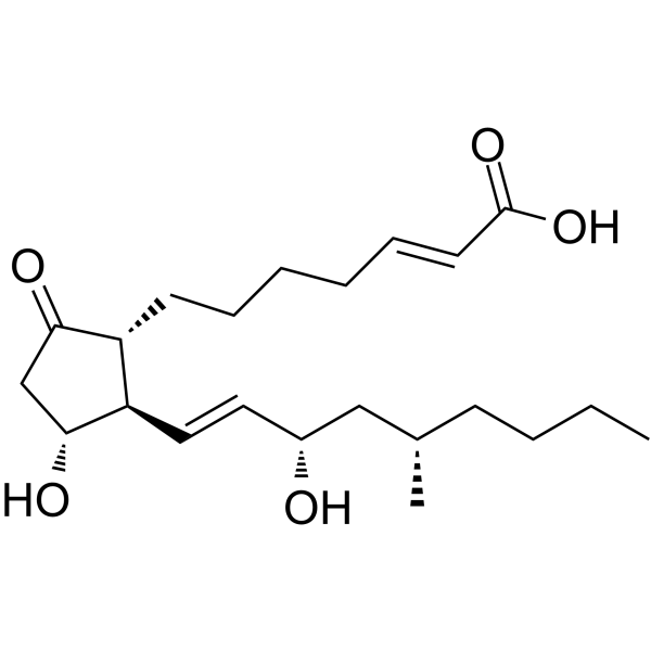
HY-B0683
LimaprostVasodilator Agent -
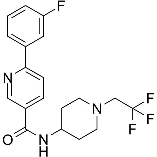
HY-10439
HPGDS inhibitor 1HPGDS Inhibitor -
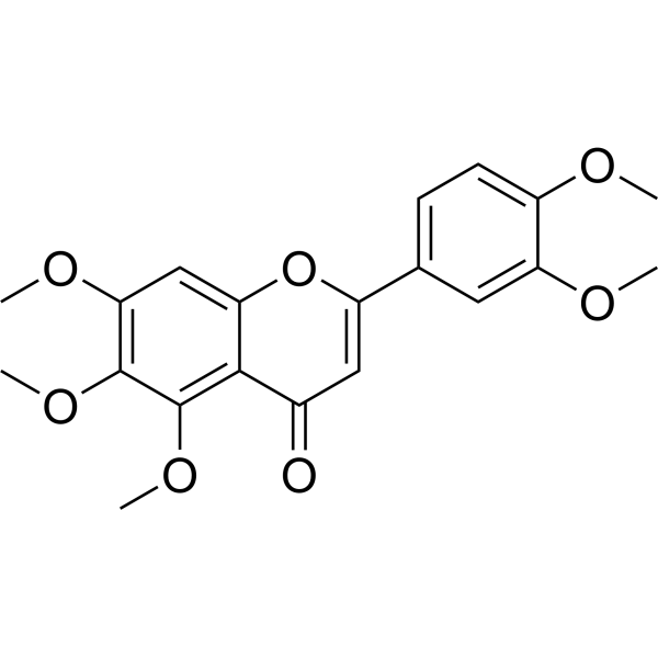
HY-N0297R
Sinensetin (Standard)
Publications Citing Use of MedChemExpress (MCE) CAY10526
More

Biological Activity
Description
Cellular Effect
|
Cell Line

|
Type | Value | Description | References |
|---|---|---|---|---|
| A549 | IC50 |
>10 μM
Compound: 1
|
Inhibition of mPGES-1-mediated PGE2 formation in IL-1beta-stimulated human A549 cells preincubated for 10 mins measured after 1 min by RP-HPLC analysis
Inhibition of mPGES-1-mediated PGE2 formation in IL-1beta-stimulated human A549 cells preincubated for 10 mins measured after 1 min by RP-HPLC analysis
|
[PMID: 22795900] |
| A549 | IC50 |
12.5 μM
Compound: CAY10526
|
Antiproliferative activity against human A549 cells assessed as inhibition of cell viability incubated for 48 hrs by MTT assay
Antiproliferative activity against human A549 cells assessed as inhibition of cell viability incubated for 48 hrs by MTT assay
|
[PMID: 32435385] |
| A549 | IC50 |
9.1 μM
Compound: CAY10526
|
Antiproliferative activity against human A549 cells assessed as inhibition of cell viability incubated for 72 hrs by MTT assay
Antiproliferative activity against human A549 cells assessed as inhibition of cell viability incubated for 72 hrs by MTT assay
|
[PMID: 32435385] |
| A549 | IC50 |
98.5 μM
Compound: CAY10526
|
Antiproliferative activity against human A549 cells assessed as inhibition of cell viability incubated for 24 hrs by MTT assay
Antiproliferative activity against human A549 cells assessed as inhibition of cell viability incubated for 24 hrs by MTT assay
|
[PMID: 32435385] |
| RAW264.7 | IC50 |
1.8 μM
Compound: 4e
|
Inhibition of PGE2 production in LPS-stimulated mouse RAW264.7 cells after 18 hrs
Inhibition of PGE2 production in LPS-stimulated mouse RAW264.7 cells after 18 hrs
|
[PMID: 17407277] |
| RAW264.7 | IC50 |
1.8 μM
Compound: 34
|
Inhibition of lipopolysaccharide-stimulated PGE2 production in mouse RAW264.7 cells
Inhibition of lipopolysaccharide-stimulated PGE2 production in mouse RAW264.7 cells
|
[PMID: 18459759] |
In Vitro
CAY10526 (5 μM; 1 h) potently inhibits NO donor-induced PGE2 production in human melanoma cell lines A375 and SB2[1].
CAY10526 (0-20 μM; 72 h) potently reduces the viability of human melanoma cell lines A375, SB2 and WM793 with high mPGES1 expression, with an IC50 < 5 μM; whereas in human melanoma cell lines SK-MEL-28, WM1361A with low mPGES1 expression and BJ fibroblasts, the activity of this compound is significantly attenuated (IC50 >10 μM)[1].
CAY10526 (5 μM; 48 h) regulates 69 proteins associated with cell death and survival, including reducing the phosphorylation level of YB-1, cyclin B1, and the anti-apoptotic proteins BCL-2/BCL-XL, while upregulating p21, the pro-apoptotic proteins BAX/BAK, and activated caspase-3 in A375 and SB2 human melanoma cells[1].
CAY10526 (1-50 μM; 12 h) inhibits the proliferation of DU-145 prostate cancer cells in a concentration-dependent manner. A significant reduction in cell viability is observed at concentrations of 10 μM and 20 μM, and the inhibitory effect reaches a plateau when the concentration exceeds 20 μM[2].
CAY10526 (10-20 μM; 12 h) downregulates mPGES-1 expression in DU-145 prostate cancer cells in a concentration-dependent manner in vitro; at a concentration of 10 μM, it maintains high expression levels of Beclin-1 compared with the complete medium control group, while at 20 μM, it significantly reduces the expression of both mPGES-1 and Beclin-1[2].
CAY10526 (1-50 μM; 18 h) potently inhibits leptin-induced PGE2 production in OE33 esophageal adenocarcinoma cells, achieving complete inhibition at concentrations of 10 μM and 50 μM[4].
CAY10526 (10 μM; 18 h) inhibits leptin-induced mPGES-1 protein expression in OE33 esophageal adenocarcinoma cells without altering COX-2 expression[4].
CAY10526 (1-50 μM; 24 h) inhibits leptin-induced proliferation in OE33 esophageal adenocarcinoma cells in a concentration-dependent manner, reaching maximum inhibitory activity at 10 μM and 50 μM, which is comparable to the activity of active COX-2 inhibitors at corresponding concentrations[4].
CAY10526 (1-50 μM; 48 h) reduces the number of viable serum-cultured OE33 esophageal adenocarcinoma cells in a concentration-dependent manner, with maximal activity observed at 10 μM[4].
CAY10526 (1-50 μM; 25 h) inhibits leptin-induced increase in the number of viable OE33 esophageal adenocarcinoma cells in a concentration-dependent manner, with its maximal activity achieved at 10 μM[4].
CAY10526 (10 μM; 25 h) inhibits leptin-induced proliferation in OE33 esophageal adenocarcinoma cells by targeting the upstream pathway of PGE2 production, while exogenous PGE2 restores this proliferative effect[4].
CAY10526 (10 μM; 25 h) inhibits IL-1β-induced PGE2 production and promotes PGI2 production in HUVECs, without affecting endothelial prostacyclin synthesis[4].
CAY10526 (25 h) inhibits thrombin-induced PGE2 production and promotes PGI2 production in human umbilical vein endothelial cells (HUVECs), without affecting endothelial prostacyclin synthesis[4].
CAY10526 (10-80 μM; 24-72 h) inhibits the proliferation of human T-cell lymphoma Hut78 cells, with an IC50 of 27.64 μM at 24 h, and suppresses cell viability in a dose-dependent manner after 72 h of incubation[5].
CAY10526 (10-80 μM; 24 h) reduces the expression of mPGES-1 protein in human T-cell lymphoma Hut78 cells in a dose-dependent manner, and exerts significant inhibitory effects at concentrations of 40 μM and 80 μM after 24 h[5].
CAY10526 (10-80 μM; 24 h) inhibits PGE2 synthesis in human T-cell lymphoma Hut78 cells in a dose-dependent manner, and exerts significant inhibitory effects at concentrations of 10, 20, 40 and 80 μM after 24 h[5].
CAY10526 (20-40 μM; 24 h) induces apoptosis in human T-cell lymphoma Hut78 cells in a dose-dependent manner, and the apoptosis rates at concentrations of 20 μM and 40 μM increase significantly after 24 h[5].
CAY10526 (20-40 μM; 24 h) upregulates the expression of activated caspase-3 in human T-cell lymphoma Hut78 cells in a dose-dependent manner, and the expression levels at 20 μM and 40 μM increase significantly after 24 h[5].
CAY10526 (20-40 μM; 24 h) dose-dependently inhibits the JAK/STAT signaling pathway in human T-cell lymphoma Hut78 cells by reducing the expression of JAK1, JAK2 and p-STAT3; after 24 h, the inhibitory effect is significant at concentrations of 20 μM and 40 μM, while the total STAT3 level remains unchanged[5].
CAY10526 (20-40 μM; 24 h) dose-dependently inhibits the PI3K/AKT signaling pathway in human T-cell lymphoma Hut78 cells by reducing the expression of PI3Kp110, PI3Kp85 and p-AKT; after 24 h of treatment, the inhibitory effect is significant at concentrations of 20 μM and 40 μM, while the total AKT level remains unchanged[5].
CAY10526 (20-40 μM; 24 h) inhibits the TGF-β/Smad3 signaling pathway in human T-cell lymphoma Hut78 cells in a dose-dependent manner by reducing the expression of TGF-β and p-Smad3; after 24 h of treatment, the inhibitory effect is significant at concentrations of 20 μM and 40 μM, while the total Smad3 level remains unchanged[5].
CAY10526 (20-40 μM; 24 h) reduces CyclinD1 expression in human T-cell lymphoma Hut78 cells in a dose-dependent manner, and exerts significant inhibitory effects at concentrations of 20 μM and 40 μM after 24 h[5].
MedChemExpress (MCE) has not independently confirmed the accuracy of these methods. They are for reference only. Further protocols information, click here.
-
Cell Line:A375, SB2, WM793, SK-MEL-28, WM1361A human melanoma cells, BJ normal fibroblasts
-
Concentration:0, 1, 5, 10 and 20 μM
-
Incubation Time:72 h
-
Result:Inhibited cell viability with an IC50 < 5 μM in mPGES1-high melanoma cell lines (A375, SB2, WM793).
Showed significantly weaker activity with an IC50 >10 μM in mPGES1-low melanoma cell lines (SK-MEL-28, WM1361A) and normal BJ fibroblasts.
-
Cell Line:DU-145 prostate cancer cells
-
Concentration:1, 10, 20 and 50 μM
-
Incubation Time:12 h
-
Result:Inhibited DU-145 cell proliferation in a concentration-dependent manner.
Exhibited optical density values of 0.34 at 1 μM, 0.22 at 10 μM, 0.08 at 20 μM, and 0.06 at 50 μM.
Reduced cell viability significantly at 10 μM and 20 μM compared to control.
Showed no significant difference in cell viability between 20 μM and 50 μM groups.
-
Cell Line:DU-145 prostate cancer cells
-
Concentration:10 and 20 μM
-
Incubation Time:12 h (serum-free conditions)
-
Result:Downregulated mPGES-1 protein expression in a concentration-dependent manner.
Reduced Beclin-1 expression at 10 μM compared to serum-free group, while levels remained higher than complete medium control group.
Significantly downregulated both mPGES-1 and Beclin-1 at 20 μM.
-
Cell Line:leptin-stimulated OE33 oesophageal adenocarcinoma cells
-
Concentration:1 μM, 10 μM, 50 μM
-
Incubation Time:60 min pretreatment, followed by 18 h leptin stimulation
-
Result:Caused a concentration-dependent reduction in leptin-driven, COX-2-dependent PGE2 production.
Abolished leptin-induced PGE2 production at 10 μM and 50 μM, achieving inhibition comparable to COX-2 inhibitors celecoxib and NS-398 at their active concentrations.
-
Cell Line:leptin-stimulated OE33 oesophageal adenocarcinoma cells
-
Concentration:10 μM
-
Incubation Time:60 min pretreatment, followed by 18 h leptin stimulation
-
Result:Completely blocked leptin-induced mPGES-1 protein expression.
Left COX-2 protein expression unaffected.
-
Cell Line:leptin-stimulated OE33 oesophageal adenocarcinoma cells
-
Concentration:1 μM, 10 μM, 50 μM
-
Incubation Time:60 min pretreatment, followed by 24 h leptin stimulation
-
Result:Caused a concentration-dependent decrease in leptin-stimulated OE33 cell proliferation.
Blocked leptin-induced proliferation at 10 μM and 50 μM with effectiveness matching COX-2 inhibitors celecoxib (1 μM) and NS-398 (10 μM).
-
Cell Line:serum-cultured OE33 oesophageal adenocarcinoma cells
-
Concentration:1 μM, 10 μM, 50 μM
-
Incubation Time:48 h
-
Result:Caused a concentration-dependent reduction in viable OE33 cell numbers, with no further reduction observed at concentrations above 10 μM.
Significantly reduced viable cell numbers at all tested concentrations compared to untreated controls.
-
Cell Line:leptin-stimulated OE33 oesophageal adenocarcinoma cells
-
Concentration:1 μM, 10 μM, 50 μM
-
Incubation Time:60 min pretreatment, followed by 24 h leptin stimulation
-
Result:Blunted the leptin-stimulated increase in viable OE33 cell numbers in a concentration-dependent manner, with no additional effect observed at concentrations above 10 μM.
Significantly reduced leptin-stimulated viable cell numbers at all tested concentrations compared to leptin-only controls.
-
Cell Line:leptin-stimulated OE33 oesophageal adenocarcinoma cells (PGE2-rescue experiment)
-
Concentration:10 μM
-
Incubation Time:60 min pretreatment, followed by 24 h stimulation with leptin, PGE2, or leptin + PGE2
-
Result:Allowed exogenous PGE2 to stimulate OE33 cell proliferation even in its presence.
Saw the inhibitory effect on leptin-stimulated proliferation reversed by added PGE2.
-
Cell Line:IL-1β-stimulated human umbilical vein endothelial cells (HUVECs)
-
Concentration:10 μM
-
Incubation Time:60 min pretreatment, followed by 24 h IL-1β stimulation
-
Result:Effectively blocked IL-1β-driven PGE2 production in HUVECs.
Significantly enhanced IL-1β-stimulated PGI2 production, unlike COX-2 inhibitors celecoxib and NS-398 which abolished production of both PGE2 and PGI2.
-
Cell Line:thrombin-stimulated human umbilical vein endothelial cells (HUVECs)
-
Concentration:10 μM
-
Incubation Time:60 min pretreatment, followed by 24 h thrombin stimulation
-
Result:Blocked thrombin-stimulated PGE2 production in HUVECs.
Significantly enhanced thrombin-stimulated PGI2 production, unlike COX-2 inhibitors celecoxib and NS-398 which abolished production of both PGE2 and PGI2.
-
Cell Line:human T-cell lymphoma Hut78 cells
-
Concentration:10, 20, 40 and 80 μM
-
Incubation Time:24 h
-
Result:Down-regulated mPGES-1 protein expression in Hut78 cells in a dose-dependent manner.
Caused statistically significant reductions at 40 μM and 80 μM compared to untreated cells.
-
Cell Line:human T-cell lymphoma Hut78 cells
-
Concentration:20 and 40 μM
-
Incubation Time:24 h
-
Result:Significantly increased the apoptosis rate of Hut78 cells in a dose-dependent manner.
Caused statistically significant elevations at 20 μM and 40 μM compared to untreated cells.
-
Cell Line:human T-cell lymphoma Hut78 cells
-
Concentration:20 and 40 μM
-
Incubation Time:24 h
-
Result:Increased the expression of cleaved caspase-3 in Hut78 cells in a dose-dependent manner.
Caused statistically significant elevations at 20 μM and 40 μM compared to untreated cells.\nDecreased the expression of JAK1, JAK2, and p-STAT3 in Hut78 cells in a dose-dependent manner.
Caused statistically significant reductions at 20 μM and 40 μM compared to untreated cells.
Left total STAT3 expression unchanged.\nDecreased the expression of PI3Kp110, PI3Kp85, and p-AKT in Hut78 cells in a dose-dependent manner.
Caused statistically significant reductions at 20 μM and 40 μM compared to untreated cells.
Left total AKT expression unchanged.\nDecreased the expression of TGF-β and p-Smad3 in Hut78 cells in a dose-dependent manner.
Caused statistically significant reductions at 20 μM and 40 μM compared to untreated cells.
Left total Smad3 expression unchanged.\nDecreased CyclinD1 expression in Hut78 cells in a dose-dependent manner.
Caused statistically significant reductions at 20 μM and 40 μM compared to untreated cells.
In Vivo
CAY10526 (2.5 mg/kg; i.p.; daily; 7 days) either during obstruction or starting at obstruction release normalizes PGE2 levels and improves or restores colon motility and smooth muscle contractility in post-BO rats[3].
MedChemExpress (MCE) has not independently confirmed the accuracy of these methods. They are for reference only.
-
Animal Model:athymic nu/nu (male, 6-8 weeks old, subcutaneous xenograft model)[1]
-
Dosage:25 mg/kg; 50 mg/kg
-
Administration:i.p.; daily; 15 days
-
Result:Reduced tumor volume by 43% and tumor weight by 48% relative to vehicle controls at 50 mg/kg dose.
Decreased phosphorylation of Y-box binding protein 1 (p-YB-1) and increased levels of cleaved caspase 3 in tumor tissues at 50 mg/kg dose.
-
Animal Model:Sprague-Dawley (male, 225-275 g, partial colon obstruction induced with obstruction band, released after 7 days to create post-bowel obstruction state)[3]
-
Dosage:2.5 mg/kg (starting on obstruction release); 2.5 mg/kg (during obstruction period)
-
Administration:i.p.; daily; 7 days (starting on obstruction release); i.p.; daily; 7 days (during obstruction period)
-
Result:Normalized PGE2 levels in colonic muscularis externae.
Eliminated the difference in colon transit geometric center between post-BO and post-sham rats.
Restored colonic circular smooth muscle contractility in response to acetylcholine to post-sham levels.
Partially but significantly improved colonic circular smooth muscle contractility in response to acetylcholine in the post-BO state.


Chemical Information
-
CAS No. 938069-71-7
-
Appearance Solid
-
Molecular Weight 311.16
-
Formula C12H7BrO3S
-
Color Off-white to yellow
-
SMILES
O=C1C(Br)=C(C(O1)O)C2=CC3=CC=CC=C3S2
-
Shipping
Room temperature in continental US; may vary elsewhere.
-
Storage
4°C, sealed storage, away from moisture and light
* In solvent : -80°C, 6 months; -20°C, 1 month (sealed storage, away from moisture and light)


Publications (2)
-
Journal Impact Factor
-
Most Recent
-
Signal Transduct Target Ther
Prostaglandin E2-driven dedifferentiation of Schwann cells leads to perineural invasion in pancreatic ductal adenocarcinoma. [Abstract]2026 Apr 7;11(1):122. PMID: 41946682 -
Genes Immun
MTA1-mediated transcriptional repression of Cox2 confers resistance against neutrophil infiltration in endometritis. [Abstract]2026 Mar 10. PMID: 41807815


Solvent & Solubility
In Vitro:
DMSO : 66.67 mg/mL (214.26 mM; ultrasonic and warming and heat to 60°C; Hygroscopic DMSO has a significant impact on the solubility of product, please use newly opened DMSO)
Please refer to the solubility information to select the appropriate solvent. Once prepared, please aliquot and store the solution to prevent product inactivation from repeated freeze-thaw cycles.
Storage method and period of stock solution: -80°C, 6 months; -20°C, 1 month (sealed storage, away from moisture and light). When stored at -80°C, please use it within 6 months. When stored at -20°C, please use it within 1 month.
Please refer to the solubility information to select the appropriate solvent. Once prepared, please aliquot and store the solution to prevent product inactivation from repeated freeze-thaw cycles.
Storage method and period of stock solution: -80°C, 6 months; -20°C, 1 month (sealed storage, away from moisture and light). When stored at -80°C, please use it within 6 months. When stored at -20°C, please use it within 1 month.
Concentration (start) × Volume (start) = Concentration (final) × Volume (final)
In Vivo:
Select the appropriate dissolution method based on your experimental animal and administration route.
- For the following dissolution methods, please ensure to first prepare a clear stock solution using an In Vitro approach and then sequentially add co-solvents:
- To ensure reliable experimental results, the clarified stock solution can be appropriately stored based on storage conditions. As for the working solution for In Vivo experiments, it is recommended to prepare freshly and use it on the same day.
- The percentages shown for the solvents indicate their volumetric ratio in the final prepared solution. If precipitation or phase separation occurs during preparation, heat and/or sonication can be used to aid dissolution.
Add each solvent one by one: 10% DMSO 90% (20% SBE-β-CD in Saline)
Solubility: 2.5 mg/mL (8.03 mM); Clear solution; Need ultrasonic
This protocol yields a clear solution of 2.5 mg/mL.
Taking 1 mL working solution as an example, add 100 μL DMSO stock solution (25.0 mg/mL) to 900 μL 20% SBE-β-CD in Saline, and mix evenly.
Preparation of 20% SBE-β-CD in Saline (4°C, storage for one week): 2 g SBE-β-CD powder is dissolved in 10 mL Saline, completely dissolve until clear.
In Vivo Dissolution Calculator
Please enter the basic information of animal experiments:
-
-
-
-
Recommended: Prepare an additional quantity of animals to account for potential losses during experiments.
Please enter your animal formula composition:
-
%DMSO +
Recommended: Keep the proportion of DMSO in working solution below 2% if your animal is weak.
-
%+
-
+%Tween-80 + +
-
%Saline +
The co-solvents required include: DMSO, . All of co-solvents are available by MedChemExpress (MCE). , Tween 80. All of co-solvents are available by MedChemExpress (MCE).
Working solution concentration: 0.22 mg/mL
Method for preparing stock solution: mg drug dissolved in μL DMSO. Stock solution concentration: mg/mL. * In solvent : -80°C, 6 months; -20°C, 1 month (sealed storage, away from moisture and light)
1. Take μL DMSO stock solution;
2. Add μL .
μL , mix evenly;
3. Then add μL Tween 80, mix evenly;
4. Then add μL
Please ensure that the stock solution in the first step is dissolved to a clear state, and add co-solvents in sequence. You can use ultrasonic heating (ultrasonic cleaner, recommended frequency 20-40 kHz), vortexing, etc. to assist dissolution.

Protocols
-
Apoptosis
Apoptosis, also called programmed cell death, is generally characterized by distinct morphological characteristics.
-
TUNEL staining for apoptotic DNA fragmentation
TUNEL staining detects DNA strand breaks by using terminal deoxynucleotidyl transferase to add labeled nucleotides to exposed 3′-OH DNA termini, generating either microscopic staining in fixed cells or tissue sections, or fluorescence/cytometric signal in cell suspensions. TUNEL positivity reflects DNA fragmentation but should not be interpreted alone as definitive apoptosis, because TUNEL can also label necrotic, autolytic, mechanically damaged, or DNA-repair-associated DNA breaks.
-
Annexin V plus membrane-impermeant dye apoptosis staining
Annexin V-based apoptosis assays rely on the detection of phosphatidylserine (PS) externalization from the inner leaflet of the plasma membrane to the outer leaflet, an early biochemical hallmark of apoptosis. Fluorescently labeled Annexin V binds PS in a calcium-dependent manner, enabling identification of early apoptotic cells by flow cytometry or fluorescence microscopy. When combined with a membrane-impermeant DNA-binding dye (e. g. , propidium iodide), this approach allows discrimination between viable (Annexin V−/dye−), early apoptotic (Annexin V+/dye−), and late apoptotic or necrotic (Annexin V+/dye+) cell populations by assessing membrane integrity and PS exposure.
-
Apoptosis Solutions
Apoptosis is a regulated, generally non-lytic cell-death pathway that removes unwanted, damaged, infected, or abnormal cells through coordinated morphological changes, caspase activation, DNA fragmentation, and membrane remodeling. The intrinsic apoptosis pathway is controlled mainly by mitochondrial outer membrane permeabilization, BCL-2 family proteins, cytochrome c release, apoptosome formation, caspase-9 activation, and downstream executioner caspase-3/7 activation. The extrinsic apoptosis pathway is initiated by death receptors such as Fas, TNFR, and TRAIL receptors, which recruit adaptor proteins and activate caspase-8 before engaging executioner caspases or mitochondrial amplification through BID cleavage. Apoptosis is linked to many phenotypes, including cancer cell killing, tissue homeostasis, immune regulation, neurodegeneration, infection response, and treatment-induced cytotoxicity; unresolved questions include how apoptosis interacts with necroptosis, pyroptosis, ferroptos


Purity & Documentation
-
Data Sheet (302 KB)
-
SDS (393 KB)
- English - EN (393 KB)
- Français - FR (393 KB)
- Deutsch - DE (393 KB)
- Norwegian - NO (393 KB)
- Español - ES (393 KB)
- Swedish - SV (393 KB)
- Italian - IT (393 KB)
- Korean - KR (393 KB)
- Portuguese - PT (393 KB)
-
Handling Instructions (2659 KB)
References
[1].
Kim SH, et al. Microsomal PGE2 synthase-1 regulates melanoma cell survival and associates with melanoma disease progression. Pigment Cell Melanoma Res. 2016 May;29(3):297-308.
[Content Brief]
[2]. Xu LW, et al. Expression and significance of microsomal prostaglandin synthase-1 (mPGES-1) and Beclin-1 in the development of prostate cancer. Asian Pac J Cancer Prev. 2012;13(4):1639-1644. [Content Brief]
[3]. Lin YM, et al. Microsomal Prostaglandin E Synthase-1 Plays a Critical Role in Long-term Motility Dysfunction after Bowel Obstruction. Sci Rep. 2018;8(1):8831. Published 2018 Jun 11. [Content Brief]
[4]. Beales IL, et al. Microsomal prostaglandin E synthase-1 inhibition blocks proliferation and enhances apoptosis in oesophageal adenocarcinoma cells without affecting endothelial prostacyclin production. Int J Cancer. 2010;126(9):2247-2255. [Content Brief]
[5]. Li Y, et al. Growth of T-cell lymphoma cells is inhibited by mPGES-1/PGE2 suppression via JAK/STAT, TGF-β/Smad3 and PI3K/AKT signal pathways. Transl Cancer Res. 2022;11(7):2175-2184. [Content Brief]


Complete Stock Solution Preparation Table
Please refer to the solubility information to select the appropriate solvent. Once prepared, please aliquot and store the solution to prevent product inactivation from repeated freeze-thaw cycles.
Storage method and period of stock solution: -80°C, 6 months; -20°C, 1 month (sealed storage, away from moisture and light). When stored at -80°C, please use it within 6 months. When stored at -20°C, please use it within 1 month.
| Optional Solvent | Concentration Solvent Mass | 1 mg | 5 mg | 10 mg | 25 mg |
|---|---|---|---|---|---|
| DMSO | 1 mM | 3.2138 mL | 16.0689 mL | 32.1378 mL | 80.3445 mL |
| 5 mM | 0.6428 mL | 3.2138 mL | 6.4276 mL | 16.0689 mL | |
| 10 mM | 0.3214 mL | 1.6069 mL | 3.2138 mL | 8.0345 mL | |
| 15 mM | 0.2143 mL | 1.0713 mL | 2.1425 mL | 5.3563 mL | |
| 20 mM | 0.1607 mL | 0.8034 mL | 1.6069 mL | 4.0172 mL | |
| 25 mM | 0.1286 mL | 0.6428 mL | 1.2855 mL | 3.2138 mL | |
| 30 mM | 0.1071 mL | 0.5356 mL | 1.0713 mL | 2.6782 mL | |
| 40 mM | 0.0803 mL | 0.4017 mL | 0.8034 mL | 2.0086 mL | |
| 50 mM | 0.0643 mL | 0.3214 mL | 0.6428 mL | 1.6069 mL | |
| 60 mM | 0.0536 mL | 0.2678 mL | 0.5356 mL | 1.3391 mL | |
| 80 mM | 0.0402 mL | 0.2009 mL | 0.4017 mL | 1.0043 mL | |
| 100 mM | 0.0321 mL | 0.1607 mL | 0.3214 mL | 0.8034 mL |


CAY10526 Related Classifications
HY-118119 Related Classifications
 Powered by Bioz
Keywords
- CAY10526
- 938069-71-7
- CAY 10526
- CAY-10526
- PGE synthase
- YB-1
- Apoptosis
- JAK
- STAT
- TGF-β Receptor
- TGF-beta/Smad
- PI3K
- Akt
- A375 human melanoma cells
- OE33 oesophageal adenocarcinoma cells
- oesophageal adenocarcinoma
- t-cell lymphoma
- DU-145 prostate cancer cells
- Hut78 cells
- microsomal PGE2 synthase-1
- Y-box binding protein 1
- prostate cancer
- melanoma
- Inhibitor
- inhibitor
- inhibit