



Loureirin D

Based on 1 Customer Validation
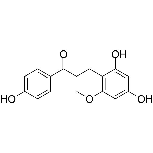
Loureirin D is a selective P2Y12 receptor antagonist that penetrates the blood-brain barrier. Loureirin D inhibits the MAPK/JNK/ERK/p38 signaling pathway in microglia by acting on CD38, and inhibits ADP (HY-W010918)-induced platelet aggregation, MMP2/9-mediated tight junction disruption, and ischemia-related blood-brain barrier damage. Loureirin D protects endothelial cells from OGD-induced apoptosis. Loureirin D is used in studies of ischemic stroke and thrombosis.
For research use only. We do not sell to patients.
- Purity : 99.30%
- CAS No.: 119425-91-1
- Formula: C16H16O5
- Molecular Weight:288.30
-
Storage:
4°C, protect from light
* In solvent : -80°C, 6 months; -20°C, 1 month (protect from light)

To place orders, for customer services and technical support, please contact: MedChemExpress USA
Tel: 609-228-6898 E-mail: [email protected] [email protected]


-
 Biological Activity
-
Chemical Information
-
Solvent & Solubility
- Protocols
- Purity & Documentation
- References
-
 Help & FAQs


All P2Y Receptor Isoforms
More

Biological Activity
Description
In Vitro
Loureirin D (10-20 μM) inhibits microglial activation and suppresses the MAPK/JNK/ERK signaling cascade in OGD-exposed BV2 microglial cells[1].
Loureirin D inhibits the MAPK signaling pathway in BV2 microglia and mediates endothelial barrier protection in a CD38-dependent manner, and CD38 is an essential upstream mediator of its inhibition of microglial activation[1].
Loureirin D (25-60 ℃) directly binds to CD38 in BV2 microglial cells, as demonstrated by an increase in the thermal stability of this protein[1].
Loureirin D (10-20 μM) attenuates the secretion of MMP2 and MMP9 from OGD-activated BV2 microglia[1].
Loureirin D maintains endothelial tight junction integrity by inhibiting pro-inflammatory microglial activation in the BV2-bEnd.3 co-culture system[1].
Loureirin D (compound 17) (100 ns) forms a stable complex with the P2Y12 receptor and maintains stable intermolecular interactions throughout the 100 ns molecular dynamics simulation[2].
Loureirin D (5-20 μM) protects bEnd.3 endothelial cells from OGD-induced apoptosis[1].
Loureirin D (5-20 μM) attenuates the upregulation of pro-apoptotic proteins in OGD-exposed bEnd.3 endothelial cells[1].
Loureirin D (5-20 μM) alleviates oxidative stress in OGD-exposed bEnd.3 endothelial cells[1].
MedChemExpress (MCE) has not independently confirmed the accuracy of these methods. They are for reference only. Further protocols information, click here.
Parmacokinetics
| Species | Dose | Route | AUC0-t | AUC0-∞ | MRT0-t | MRT0-∞ | T1/2 (Elimination) | CLz/F | Vz/F | Cmax | Bioavailability |
|---|---|---|---|---|---|---|---|---|---|---|---|
| Rat[3] | 1 mg/kg | i.v. | 2074.7 ng·h/mL | 2096.8 ng·h/mL | 3.5 h | 3.7 h | 3.3 h | 0.5 L/h/kg | 2.3 L/kg | 924.4 ng/mL | / |
| Rat[3] | 5 mg/kg | p.o. | 563.4 ng·h/mL | 572.6 ng·h/mL | 4.2 h | 4.8 h | 4.2 h | 8.8 L/h/kg | 52.7 L/kg | 222.2 ng/mL | 5.4 % |
In Vivo
Compound 17 (Loureirin D) (6-24 mg/kg; i.g.; once daily; 3 days) alleviates ischemic stroke injury in PT-stroke mice by attenuating BBB disruption and inhibiting platelet aggregation via P2Y12 downregulation[2].
MedChemExpress (MCE) has not independently confirmed the accuracy of these methods. They are for reference only.
-
Animal Model:Sprague-Dawley (SD) (Male; 260-280 g)[1]
-
Dosage:12 mg/kg/day; 24 mg/kg/day
-
Administration:i.g.; once daily; 3 consecutive days
-
Result:Ameliorated pMCAO-induced behavioral deficits, including reduced average locomotor velocity and total distance moved in the OFT, and markedly prolonged latency to fall in the rotarod test at 24 mg/kg.
Attenuated elevated Bederson neurological deficit scores at 24 mg/kg.
Mitigated histopathological changes and preserved neuronal density and morphological integrity in the hippocampal CA2 region.
Attenuated Evans blue leakage and brain water content in the ipsilateral ischemic hemisphere.
Markedly attenuated IgG extravasation.
Restored the expression of ZO-1 and Occludin and significantly attenuated the upregulation of MMP2 and MMP9.
Identified 99 DEGs commonly dysregulated in both "Model vs. Sham" and "This compound vs. Model" comparisons.
Significantly inhibited the mRNA expression of CD38 and Mapk11 in brain tissues.
Significantly attenuated CD38 protein expression.
Markedly attenuated CD38 signal intensity specifically within IBA-1-positive microglial cells.
-
Animal Model:Mice (male, 20-25 g)[2]
-
Dosage:6, 12, 24 mg/kg
-
Administration:i.g.; once daily; 3 days
-
Result:Ameliorated neurological deficits and improved motor function at 24 mg/kg.
Dose-dependently preserved BBB integrity with significant reduction in dye leakage.
Significantly inhibited platelet aggregation.
Significantly reduced P2Y12 protein levels in brain tissue and downregulated P2Y12 mRNA in cortical tissue.


Chemical Information
-
CAS No. 119425-91-1
-
Appearance Solid
-
Molecular Weight 288.30
-
Formula C16H16O5
-
Color White to pink
-
SMILES
O=C(C1=CC=C(O)C=C1)CCC2=C(OC)C=C(O)C=C2O
-
Structure Classification
-
Initial Source
-
Shipping
Room temperature in continental US; may vary elsewhere.
-
Storage
4°C, protect from light
* In solvent : -80°C, 6 months; -20°C, 1 month (protect from light)


Solvent & Solubility
In Vitro:
DMSO : ≥ 50 mg/mL (173.43 mM; Hygroscopic DMSO has a significant impact on the solubility of product, please use newly opened DMSO)
* "≥" means soluble, but saturation unknown.
Please refer to the solubility information to select the appropriate solvent. Once prepared, please aliquot and store the solution to prevent product inactivation from repeated freeze-thaw cycles.
Storage method and period of stock solution: -80°C, 6 months; -20°C, 1 month (protect from light). When stored at -80°C, please use it within 6 months. When stored at -20°C, please use it within 1 month.
Please refer to the solubility information to select the appropriate solvent. Once prepared, please aliquot and store the solution to prevent product inactivation from repeated freeze-thaw cycles.
Storage method and period of stock solution: -80°C, 6 months; -20°C, 1 month (protect from light). When stored at -80°C, please use it within 6 months. When stored at -20°C, please use it within 1 month.
Concentration (start) × Volume (start) = Concentration (final) × Volume (final)
In Vivo:
Select the appropriate dissolution method based on your experimental animal and administration route.
- For the following dissolution methods, please ensure to first prepare a clear stock solution using an In Vitro approach and then sequentially add co-solvents:
- To ensure reliable experimental results, the clarified stock solution can be appropriately stored based on storage conditions. As for the working solution for In Vivo experiments, it is recommended to prepare freshly and use it on the same day.
- The percentages shown for the solvents indicate their volumetric ratio in the final prepared solution. If precipitation or phase separation occurs during preparation, heat and/or sonication can be used to aid dissolution.
Add each solvent one by one: 10% DMSO 40% PEG300 5% Tween-80 45% Saline
Solubility: ≥ 2.5 mg/mL (8.67 mM); Clear solution
This protocol yields a clear solution of ≥ 2.5 mg/mL (saturation unknown).
Taking 1 mL working solution as an example, add 100 μL DMSO stock solution (25.0 mg/mL) to 400 μL PEG300, and mix evenly; then add 50 μL Tween-80 and mix evenly; then add 450 μL Saline to adjust the volume to 1 mL.
Preparation of Saline: Dissolve 0.9 g sodium chloride in ddH₂O and dilute to 100 mL to obtain a clear Saline solution.
Add each solvent one by one: 10% DMSO 90% (20% SBE-β-CD in Saline)
Solubility: ≥ 2.5 mg/mL (8.67 mM); Clear solution
This protocol yields a clear solution of ≥ 2.5 mg/mL (saturation unknown).
Taking 1 mL working solution as an example, add 100 μL DMSO stock solution (25.0 mg/mL) to 900 μL 20% SBE-β-CD in Saline, and mix evenly.
Preparation of 20% SBE-β-CD in Saline (4°C, storage for one week): 2 g SBE-β-CD powder is dissolved in 10 mL Saline, completely dissolve until clear.
In Vivo Dissolution Calculator
Please enter the basic information of animal experiments:
-
-
-
-
Recommended: Prepare an additional quantity of animals to account for potential losses during experiments.
Please enter your animal formula composition:
-
%DMSO +
Recommended: Keep the proportion of DMSO in working solution below 2% if your animal is weak.
-
%+
-
+%Tween-80 + +
-
%Saline +
The co-solvents required include: DMSO, . All of co-solvents are available by MedChemExpress (MCE). , Tween 80. All of co-solvents are available by MedChemExpress (MCE).
Working solution concentration: 0.22 mg/mL
Method for preparing stock solution: mg drug dissolved in μL DMSO. Stock solution concentration: mg/mL. * In solvent : -80°C, 6 months; -20°C, 1 month (protect from light)
1. Take μL DMSO stock solution;
2. Add μL .
μL , mix evenly;
3. Then add μL Tween 80, mix evenly;
4. Then add μL
Please ensure that the stock solution in the first step is dissolved to a clear state, and add co-solvents in sequence. You can use ultrasonic heating (ultrasonic cleaner, recommended frequency 20-40 kHz), vortexing, etc. to assist dissolution.

Protocols
-
Apoptosis
Apoptosis, also called programmed cell death, is generally characterized by distinct morphological characteristics.
-
TUNEL staining for apoptotic DNA fragmentation
TUNEL staining detects DNA strand breaks by using terminal deoxynucleotidyl transferase to add labeled nucleotides to exposed 3′-OH DNA termini, generating either microscopic staining in fixed cells or tissue sections, or fluorescence/cytometric signal in cell suspensions. TUNEL positivity reflects DNA fragmentation but should not be interpreted alone as definitive apoptosis, because TUNEL can also label necrotic, autolytic, mechanically damaged, or DNA-repair-associated DNA breaks.
-
Annexin V plus membrane-impermeant dye apoptosis staining
Annexin V-based apoptosis assays rely on the detection of phosphatidylserine (PS) externalization from the inner leaflet of the plasma membrane to the outer leaflet, an early biochemical hallmark of apoptosis. Fluorescently labeled Annexin V binds PS in a calcium-dependent manner, enabling identification of early apoptotic cells by flow cytometry or fluorescence microscopy. When combined with a membrane-impermeant DNA-binding dye (e. g. , propidium iodide), this approach allows discrimination between viable (Annexin V−/dye−), early apoptotic (Annexin V+/dye−), and late apoptotic or necrotic (Annexin V+/dye+) cell populations by assessing membrane integrity and PS exposure.
-
Endothelial Tube Formation Assay
Endothelial tube formation assay evaluates the ability of endothelial cells to attach, migrate, align, and organize into capillary-like networks when cultured on gelled basement membrane extract or Matrigel; the readout is the morphology and quantity of tube-like networks, which reflects an in vitro endothelial morphogenesis step related to angiogenesis. Basement membrane extract/Matrigel provides laminin-rich extracellular matrix cues that support endothelial differentiation into capillary-like structures, but it can contain biologically active growth factors, so growth-factor-reduced matrix is preferred when testing defined angiogenic stimulators or inhibitors.
-
Apoptosis Solutions
Apoptosis is a regulated, generally non-lytic cell-death pathway that removes unwanted, damaged, infected, or abnormal cells through coordinated morphological changes, caspase activation, DNA fragmentation, and membrane remodeling. The intrinsic apoptosis pathway is controlled mainly by mitochondrial outer membrane permeabilization, BCL-2 family proteins, cytochrome c release, apoptosome formation, caspase-9 activation, and downstream executioner caspase-3/7 activation. The extrinsic apoptosis pathway is initiated by death receptors such as Fas, TNFR, and TRAIL receptors, which recruit adaptor proteins and activate caspase-8 before engaging executioner caspases or mitochondrial amplification through BID cleavage. Apoptosis is linked to many phenotypes, including cancer cell killing, tissue homeostasis, immune regulation, neurodegeneration, infection response, and treatment-induced cytotoxicity; unresolved questions include how apoptosis interacts with necroptosis, pyroptosis, ferroptos


Purity & Documentation
-
Data Sheet (285 KB)
-
SDS (393 KB)
- English - EN (393 KB)
- Français - FR (393 KB)
- Deutsch - DE (393 KB)
- Norwegian - NO (393 KB)
- Español - ES (393 KB)
- Swedish - SV (393 KB)
- Italian - IT (393 KB)
- Korean - KR (393 KB)
- Portuguese - PT (393 KB)
-
Handling Instructions (2659 KB)
References


Complete Stock Solution Preparation Table
Please refer to the solubility information to select the appropriate solvent. Once prepared, please aliquot and store the solution to prevent product inactivation from repeated freeze-thaw cycles.
Storage method and period of stock solution: -80°C, 6 months; -20°C, 1 month (protect from light). When stored at -80°C, please use it within 6 months. When stored at -20°C, please use it within 1 month.
| Optional Solvent | Concentration Solvent Mass | 1 mg | 5 mg | 10 mg | 25 mg |
|---|---|---|---|---|---|
| DMSO | 1 mM | 3.4686 mL | 17.3430 mL | 34.6861 mL | 86.7152 mL |
| 5 mM | 0.6937 mL | 3.4686 mL | 6.9372 mL | 17.3430 mL | |
| 10 mM | 0.3469 mL | 1.7343 mL | 3.4686 mL | 8.6715 mL | |
| 15 mM | 0.2312 mL | 1.1562 mL | 2.3124 mL | 5.7810 mL | |
| 20 mM | 0.1734 mL | 0.8672 mL | 1.7343 mL | 4.3358 mL | |
| 25 mM | 0.1387 mL | 0.6937 mL | 1.3874 mL | 3.4686 mL | |
| 30 mM | 0.1156 mL | 0.5781 mL | 1.1562 mL | 2.8905 mL | |
| 40 mM | 0.0867 mL | 0.4336 mL | 0.8672 mL | 2.1679 mL | |
| 50 mM | 0.0694 mL | 0.3469 mL | 0.6937 mL | 1.7343 mL | |
| 60 mM | 0.0578 mL | 0.2891 mL | 0.5781 mL | 1.4453 mL | |
| 80 mM | 0.0434 mL | 0.2168 mL | 0.4336 mL | 1.0839 mL | |
| 100 mM | 0.0347 mL | 0.1734 mL | 0.3469 mL | 0.8672 mL |


Loureirin D Related Classifications
HY-N8189 Related Classifications
 Powered by Bioz