



Plantamajoside
Based on 2 publication(s) in Google Scholar
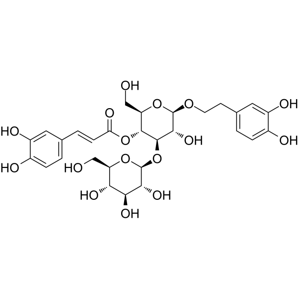
Plantamajoside is an orally active phenylpropanoid glycoside. Plantamajoside can be isolated from Plantago asiatica L.(Plantaginaceae). Plantamajoside inactivates NF-κB, PI3K/akt, induces Apoptosis, and improves Autophagy. Plantamajoside regulates MAPK, integrin-linked kinase/c-Src. Plantamajoside inhibits multiple cancers, improves lung and kidney damage. Plantamajoside has neuroprotective and anti-inflammatory effects.
For research use only. We do not sell to patients.
- Purity : 99.80%
- CAS No.: 104777-68-6
- Formula: C29H36O16
- Molecular Weight:640.59
-
Storage:
4°C, protect from light
* In solvent : -80°C, 6 months; -20°C, 1 month (protect from light)

To place orders, for customer services and technical support, please contact: MedChemExpress USA
Tel: 609-228-6898 E-mail: [email protected] [email protected]


-
 Biological Activity
-
Chemical Information
-
Solvent & Solubility
- Protocols
- Purity & Documentation
- References
-
 Help & FAQs


-
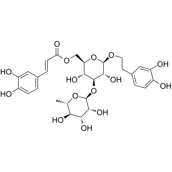
HY-N0022
IsoacteosideNatural Product -
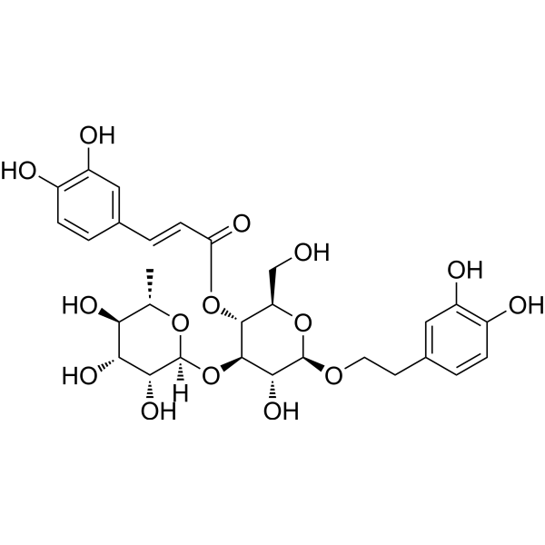
HY-N0021
VerbascosideBacterial Inhibitor -
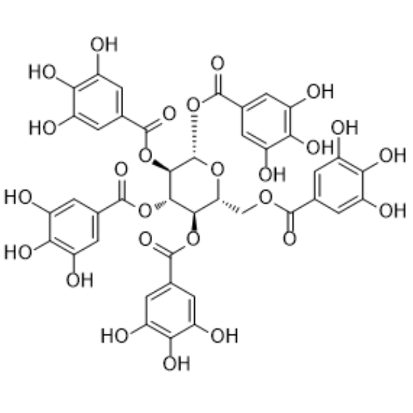
HY-N0527
PentagalloylglucoseAntioxidant -
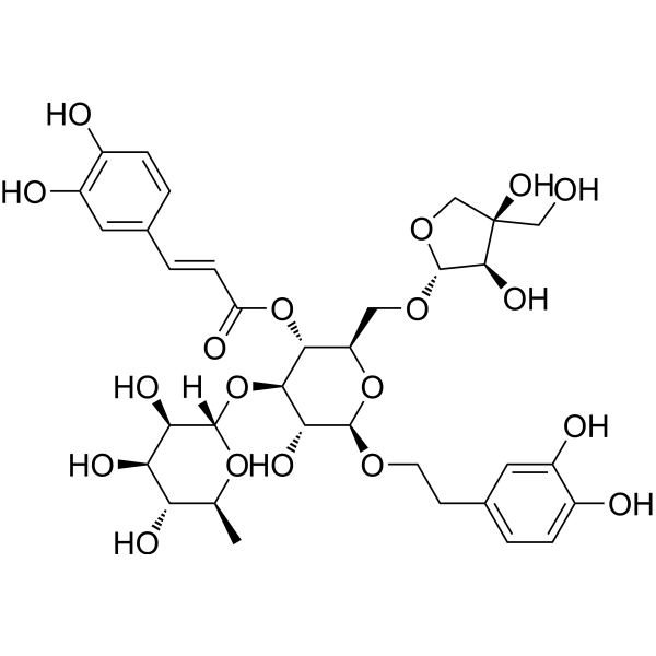
HY-N0029
Forsythoside BTNF Inhibitor -
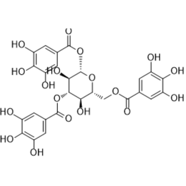
HY-N6006
1,3,6-Tri-O-galloyl-beta-D-glucosePhenolic Compound -
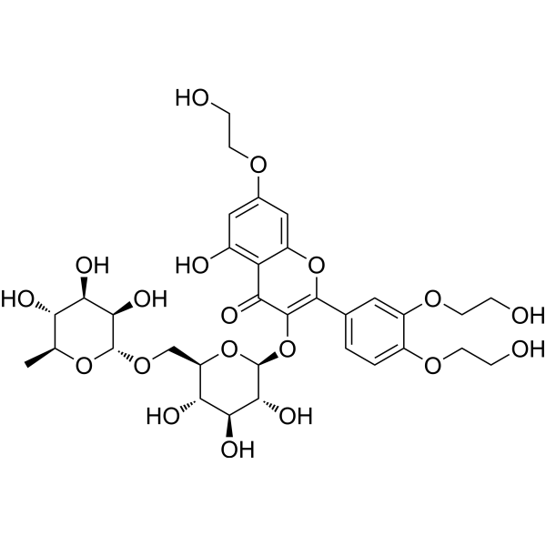
HY-N0139
TroxerutinNOD-like Receptor (NLR) Inhibitor -
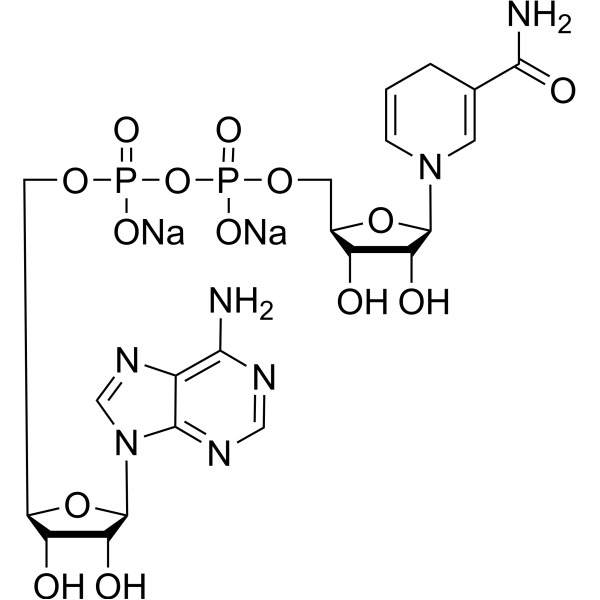
HY-F0001
NADH disodium saltReduced Coenzyme -
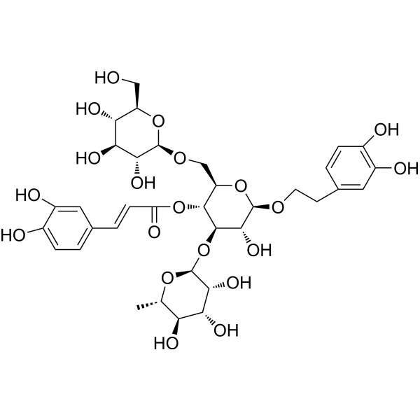
HY-N0020
EchinacosideWnt/β-catenin Signaling Inhibitor -
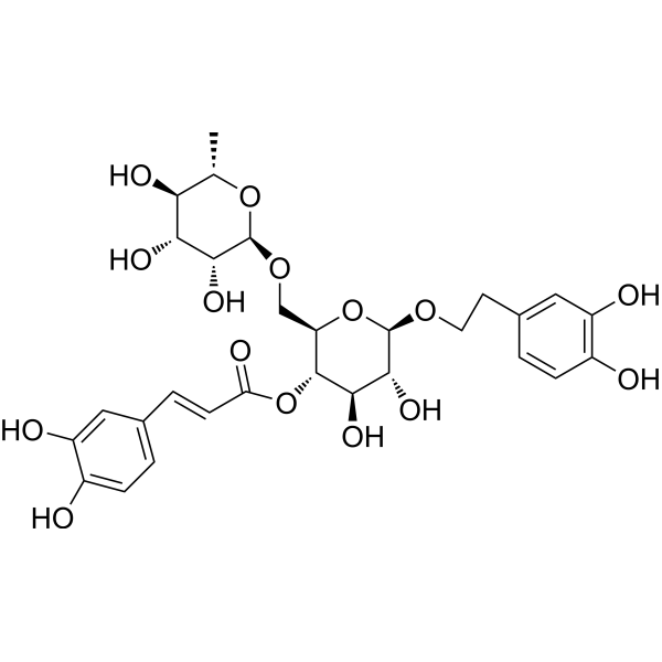
HY-N0028
Forsythiaside AAnti-Inflammatory Agent -

HY-13259
MG-132Proteasome Inhibitor -
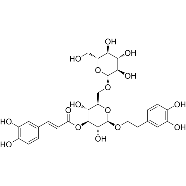
HY-N12084
IsolugrandosideNature Product -
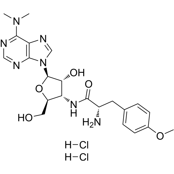
HY-B1743A
Puromycin dihydrochlorideProtein Synthesis Inhibitor
Publications Citing Use of MedChemExpress (MCE) Plantamajoside
More Customer Validation & Images

-
Histological Imaging/Staining

-
IF

-
RT-PCR

-
WB

-
In Vivo Efficacy Study


Biological Activity
Description
In Vitro
Plantamajoside (1-800 μg/mL, 24 h-2 weeks) inhibits proliferation, stemness, and migration, and initiates apoptosis in 95D cells and dose-dependently suppresses the survival of 95D cells[3].
Plantamajoside (10 μM, 1-24 h) from Plantago asiatica modulates human umbilical vein endothelial cell dysfunction by glyceraldehyde-induced AGEs via MAPK/NF-κB[4].
Plantamajoside (10-40 μM, 24 h) inhibits high glucose-induced oxidative stress, inflammation, and extracellular matrix accumulation in rat glomerular mesangial cells HBZY-1 through the inactivation of Akt/NF-κB pathway[5].
Plantamajoside (5-120 μM 48 h) alleviates hypoxia-reoxygenation injury through integrin-linked kinase/c-Src/Akt and the mitochondrial apoptosis signaling pathways in H9c2 myocardial cells[6].
Plantamajoside (10-20 μM, 12-48 h) inhibits hypoxia-induced migration and invasion of human cervical cancer cells (SiHa and CaSki) through blocking the NF-κB and PI3K/akt pathways[7].
Plantamajoside (20-320 μg/mL, 24 h) ameliorates TGFβ2-induced autophagy, epithelial-mesenchymal transition and inflammatory processes in human lens epithelial cells (hLECs)[8].
Plantamajoside (20-160 μg/mL, 48 h) inhibits proliferation, migration, invasion and induces apoptosis in activated HSC-T6 cell[9].
Plantamajoside (20-160 μg/mL, 24-72 h) inhibits the invasion, migration and viability of malignant melanoma cells A2058[10].
Plantamajoside (2.5-40 μg/mL, 7 days) promotes metformin-induced apoptosis, autophagy and proliferation arrest of liver cancer cells via suppressing Akt/GSK3β signaling[11].
Plantamajoside (31.25-500 μg/mL, 12-48 h) inhibits breast cancer cells (MDA-MB-231) growth by decreasing the activity of matrix metalloproteinase-9 and -2[12].
Plantamajoside (10-80 μM, 24 h) protects H9c2 cells against hypoxia/reoxygenation-induced injury through regulating the akt/Nrf2/HO-1 and NF-κB signaling pathways[13].
Plantamajoside (20-160 g/mL) inhibits HCC cells progression through regulating cell viability, apoptosis, migration, invasion and PI3K/AKT pathway[14].
MedChemExpress (MCE) has not independently confirmed the accuracy of these methods. They are for reference only. Further protocols information, click here.
-
Cell Line:95D
-
Concentration:50, 100, 200 µg/mL
-
Incubation Time:24 h
-
Result:Dose-dependently induced 95D cell apoptosis.
Induced effective apoptosis at 100 µg/mL and was more significant at 200 µg/mL.
-
Cell Line:95D
-
Concentration:1, 2.5, 5, 10, 25, 50, 100, 200, 300, 400, 500, 600, and 800 µg/mL
-
Incubation Time:24 h
-
Result:Exhibited significantly lower viability than the control group (100 µg/mL).
In Vivo
Plantamajoside (25-100 mg/kg, i.p., 24 h) alleviates acute sepsis-induced organ dysfunction through inhibiting the TRAF6/NF-κB axis in mice[16].
Plantamajoside (10-40 mg/kg, p.o., 4 weeks) has protective activities against Cd-induced renal injury in rats[17].
Plantamajoside (25-100 mg/kg, i.p., three times at 6, 12, 18 h) ameliorates lipopolysaccharide-induced acute lung injury via suppressing NF-κB and MAPK activation in mice[18].
Plantamajoside (20-80 mg/kg, i.p., once a day for 4 weeks) modulates immune dysregulation and hepatic lipid metabolism in rats with nonalcoholic fatty liver disease via AMPK/Nrf2 elevation[19].
MedChemExpress (MCE) has not independently confirmed the accuracy of these methods. They are for reference only.
-
Animal Model:Acute sepsis C57BL/6 male mice model by caecal ligation and perforation (CLP)[16]
-
Dosage:25, 50, 100 mg/kg
-
Administration:Intraperitoneal injection (i.p.), 24 h
-
Result:Extended survival in CLP model.
Improved acute sepsis-triggered organ damage.
Alleviated acute sepsis-triggered apoptosis.
Relieved acute sepsis-triggered inflammatory response.
Improved acute sepsis-triggered organ damage via mediating the TRAF6/NF-κB pathway.
Improved acute sepsis-triggered apoptosis and inflammation via regulating the TRAF6/NF-κB pathway.


Chemical Information
-
CAS No. 104777-68-6
-
Appearance Solid
-
Molecular Weight 640.59
-
Formula C29H36O16
-
Color White to yellow
-
SMILES
O[C@H]([C@H]([C@H](OC(/C=C/C1=CC=C(O)C(O)=C1)=O)[C@@H](CO)O2)O[C@H]3[C@@H]([C@H]([C@@H]([C@@H](CO)O3)O)O)O)[C@@H]2OCCC4=CC=C(O)C(O)=C4
-
Structure Classification
-
Initial Source
-
Shipping
Room temperature in continental US; may vary elsewhere.
-
Storage
4°C, protect from light
* In solvent : -80°C, 6 months; -20°C, 1 month (protect from light)


Publications (2)
-
Journal Impact Factor
-
Most Recent
-
Phytomedicine
Plantamajoside alleviates DSS-induced ulcerative colitis by modulating gut microbiota, upregulating CBS, and inhibiting NF-κB. [Abstract]2025 Jul 25:143:156827. PMID: 40381501
Plantamajoside purchased from MedChemExpress. Usage Cited in: Phytomedicine. 2025 Jul 25:143:156827. [Abstract]
Representative hematoxylin and eosin (H-E)-stained histological images of colon tissues in each group treated with Plantamajoside (PMS) (12.5, 25, 50 mg/kg, i.g.).
Plantamajoside purchased from MedChemExpress. Usage Cited in: Phytomedicine. 2025 Jul 25:143:156827. [Abstract]
Representative immunofluorescence images of claudin-3 in colon tissues treated with Plantamajoside (PMS) (12.5, 25, 50 mg/kg, i.g.).
Plantamajoside purchased from MedChemExpress. Usage Cited in: Phytomedicine. 2025 Jul 25:143:156827. [Abstract]
Reverse transcription-quantitative polymerase chain reaction (RT-qPCR) analysis of claudin-3 expression levels in colon tissues treated with Plantamajoside (PMS) (12.5, 25, 50 mg/kg, i.g.).
Plantamajoside purchased from MedChemExpress. Usage Cited in: Phytomedicine. 2025 Jul 25:143:156827. [Abstract]
Representative bands of western blotting analysis of claudin-3, occludin, and ZO-1 in colon tissues treated with Plantamajoside (PMS) (12.5, 25, 50 mg/kg, i.g.).
Plantamajoside purchased from MedChemExpress. Usage Cited in: Phytomedicine. 2025 Jul 25:143:156827. [Abstract]
Representative images of colons treated with Plantamajoside (PMS) (12.5, 25, 50 mg/kg, i.g.).
-
Int J Mol Sci
Plantago major and Plantago lanceolata Exhibit Antioxidant and Borrelia burgdorferi Inhibiting Activities. [Abstract]2024 Jun 28;25(13):7112. PMID: 39000214


Solvent & Solubility
In Vitro:
DMSO : 100 mg/mL (156.11 mM; Need ultrasonic; Hygroscopic DMSO has a significant impact on the solubility of product, please use newly opened DMSO)
Please refer to the solubility information to select the appropriate solvent. Once prepared, please aliquot and store the solution to prevent product inactivation from repeated freeze-thaw cycles.
Storage method and period of stock solution: -80°C, 6 months; -20°C, 1 month (protect from light). When stored at -80°C, please use it within 6 months. When stored at -20°C, please use it within 1 month.
Please refer to the solubility information to select the appropriate solvent. Once prepared, please aliquot and store the solution to prevent product inactivation from repeated freeze-thaw cycles.
Storage method and period of stock solution: -80°C, 6 months; -20°C, 1 month (protect from light). When stored at -80°C, please use it within 6 months. When stored at -20°C, please use it within 1 month.
Concentration (start) × Volume (start) = Concentration (final) × Volume (final)
In Vivo:
Select the appropriate dissolution method based on your experimental animal and administration route.
- For the following dissolution methods, please ensure to first prepare a clear stock solution using an In Vitro approach and then sequentially add co-solvents:
- To ensure reliable experimental results, the clarified stock solution can be appropriately stored based on storage conditions. As for the working solution for In Vivo experiments, it is recommended to prepare freshly and use it on the same day.
- The percentages shown for the solvents indicate their volumetric ratio in the final prepared solution. If precipitation or phase separation occurs during preparation, heat and/or sonication can be used to aid dissolution.
Add each solvent one by one: 10% DMSO 40% PEG300 5% Tween-80 45% Saline
Solubility: ≥ 0.83 mg/mL (1.30 mM); Clear solution
This protocol yields a clear solution of ≥ 0.83 mg/mL (saturation unknown).
Taking 1 mL working solution as an example, add 100 μL DMSO stock solution (8.3 mg/mL) to 400 μL PEG300, and mix evenly; then add 50 μL Tween-80 and mix evenly; then add 450 μL Saline to adjust the volume to 1 mL.
Preparation of Saline: Dissolve 0.9 g sodium chloride in ddH₂O and dilute to 100 mL to obtain a clear Saline solution.
Add each solvent one by one: 10% DMSO 90% (20% SBE-β-CD in Saline)
Solubility: ≥ 0.83 mg/mL (1.30 mM); Clear solution
This protocol yields a clear solution of ≥ 0.83 mg/mL (saturation unknown).
Taking 1 mL working solution as an example, add 100 μL DMSO stock solution (8.3 mg/mL) to 900 μL 20% SBE-β-CD in Saline, and mix evenly.
Preparation of 20% SBE-β-CD in Saline (4°C, storage for one week): 2 g SBE-β-CD powder is dissolved in 10 mL Saline, completely dissolve until clear.
In Vivo Dissolution Calculator
Please enter the basic information of animal experiments:
-
-
-
-
Recommended: Prepare an additional quantity of animals to account for potential losses during experiments.
Please enter your animal formula composition:
-
%DMSO +
Recommended: Keep the proportion of DMSO in working solution below 2% if your animal is weak.
-
%+
-
+%Tween-80 + +
-
%Saline +
The co-solvents required include: DMSO, . All of co-solvents are available by MedChemExpress (MCE). , Tween 80. All of co-solvents are available by MedChemExpress (MCE).
Working solution concentration: 0.22 mg/mL
Method for preparing stock solution: mg drug dissolved in μL DMSO. Stock solution concentration: mg/mL. * In solvent : -80°C, 6 months; -20°C, 1 month (protect from light)
1. Take μL DMSO stock solution;
2. Add μL .
μL , mix evenly;
3. Then add μL Tween 80, mix evenly;
4. Then add μL
Please ensure that the stock solution in the first step is dissolved to a clear state, and add co-solvents in sequence. You can use ultrasonic heating (ultrasonic cleaner, recommended frequency 20-40 kHz), vortexing, etc. to assist dissolution.

Protocols
-
Apoptosis
Apoptosis, also called programmed cell death, is generally characterized by distinct morphological characteristics.
-
TUNEL staining for apoptotic DNA fragmentation
TUNEL staining detects DNA strand breaks by using terminal deoxynucleotidyl transferase to add labeled nucleotides to exposed 3′-OH DNA termini, generating either microscopic staining in fixed cells or tissue sections, or fluorescence/cytometric signal in cell suspensions. TUNEL positivity reflects DNA fragmentation but should not be interpreted alone as definitive apoptosis, because TUNEL can also label necrotic, autolytic, mechanically damaged, or DNA-repair-associated DNA breaks.
-
Annexin V plus membrane-impermeant dye apoptosis staining
Annexin V-based apoptosis assays rely on the detection of phosphatidylserine (PS) externalization from the inner leaflet of the plasma membrane to the outer leaflet, an early biochemical hallmark of apoptosis. Fluorescently labeled Annexin V binds PS in a calcium-dependent manner, enabling identification of early apoptotic cells by flow cytometry or fluorescence microscopy. When combined with a membrane-impermeant DNA-binding dye (e. g. , propidium iodide), this approach allows discrimination between viable (Annexin V−/dye−), early apoptotic (Annexin V+/dye−), and late apoptotic or necrotic (Annexin V+/dye+) cell populations by assessing membrane integrity and PS exposure.
-
Autophagy
Autophagy is a process in which eukaryotic cells use lysosomes to degrade their own cytoplasmic proteins and damaged organelles under the regulation of autophagy related gene (Atg). Microtubule-associated proteins light chain 3 (LC3) is recognized as autophagy marker, which transfers from cytoplasmic LC3 (LC3-I) to membrane type (LC3-II). LC3-II/I ratio could be detected by Western Blot and fluorescence microscopy.
-
Apoptosis Solutions
Apoptosis is a regulated, generally non-lytic cell-death pathway that removes unwanted, damaged, infected, or abnormal cells through coordinated morphological changes, caspase activation, DNA fragmentation, and membrane remodeling. The intrinsic apoptosis pathway is controlled mainly by mitochondrial outer membrane permeabilization, BCL-2 family proteins, cytochrome c release, apoptosome formation, caspase-9 activation, and downstream executioner caspase-3/7 activation. The extrinsic apoptosis pathway is initiated by death receptors such as Fas, TNFR, and TRAIL receptors, which recruit adaptor proteins and activate caspase-8 before engaging executioner caspases or mitochondrial amplification through BID cleavage. Apoptosis is linked to many phenotypes, including cancer cell killing, tissue homeostasis, immune regulation, neurodegeneration, infection response, and treatment-induced cytotoxicity; unresolved questions include how apoptosis interacts with necroptosis, pyroptosis, ferroptos
-
Research Protocol for Inflammation-related Diseases
The NLRP3 inflammasome is a cytosolic innate immune signaling platform that integrates priming signals and danger-signal activation to promote caspase-1 activation, maturation of IL-1β and IL-18, and gasdermin D-mediated pyroptotic cell death. The core experimental logic is to determine whether inflammatory disease phenotypes are driven by increased NLRP3 expression, ASC-containing inflammasome assembly, caspase-1 cleavage, GSDMD cleavage, and extracellular release of IL-1β/IL-18 rather than by nonspecific cell injury alone. The pathway is strongly linked to inflammation-related disease phenotypes because monosodium urate crystals activate NALP3/NLRP3 inflammasome signaling in gout-like crystal inflammation, cholesterol crystals activate NLRP3 inflammasomes in atherogenesis models, and DSS-induced intestinal inflammation has been reported to involve NLRP3 inflammasome activity. However, experimental colitis studies also show context-dependent protective effects of NLRP3 inflammasome co
-
Lysosome and acidic-vesicle live-cell staining
Lysosome and acidic-vesicle live-cell staining detects acidic intracellular compartments by using membrane-permeant acidotropic probes that accumulate in low-pH vesicles, including lysosomes, late endosomes, autolysosomes, and acidic phagosomes. LysoTracker staining is commonly used as an intensity-based readout of acidic lysosomal compartment abundance or enlargement, while acridine orange produces green fluorescence in less concentrated compartments and red fluorescence after concentration-dependent accumulation in acidic vesicular organelles. Loss or reduction of acridine-orange red signal can be used as a readout of lysosomal membrane permeabilization or reduced acidic-vesicle integrity. This protocol is designed for live cultured cells and can be adapted for fluorescence microscopy, high-content imaging, plate-reader readout, or flow cytometry when the selected literature supports the readout. Because these dyes report acidotropic accumulation rather than lysosome identity alone,
-
Macroautophagy Solutions
Macroautophagy is a conserved lysosome-dependent degradation pathway in which cytoplasmic material is sequestered into double-membrane autophagosomes and delivered to lysosomes for degradation and recycling. The pathway supports cellular homeostasis during nutrient limitation, organelle stress, protein-aggregate accumulation, infection, differentiation, and tissue remodeling by coupling cargo sequestration, autophagosome maturation, lysosomal fusion, and degradation of cargo-derived macromolecules. The core molecular sequence includes initiation by nutrient- and stress-regulated autophagy machinery, autophagosome nucleation, LC3/ATG8-family conjugation to autophagosomal membranes, cargo selection through receptors such as SQSTM1/p62, autophagosome-lysosome fusion, and lysosomal degradation. LC3 was identified as a mammalian homolog of yeast Atg8 that localizes to autophagosomal membranes after processing, and p62/SQSTM1 was shown to connect ubiquitinated cargo with autophagic degradati


Purity & Documentation
-
Data Sheet (288 KB)
-
SDS (394 KB)
- English - EN (394 KB)
- Français - FR (394 KB)
- Deutsch - DE (394 KB)
- Norwegian - NO (394 KB)
- Español - ES (394 KB)
- Swedish - SV (394 KB)
- Italian - IT (394 KB)
- Korean - KR (394 KB)
- Portuguese - PT (394 KB)
-
Handling Instructions (2659 KB)
References
[1]. Wu H, et al. Plantamajoside ameliorates lipopolysaccharide-induced acute lung injury via suppressing NF-κB and MAPK activation.Int Immunopharmacol. 2016 Jun;35:315-322. [Content Brief]
[2]. Zuo X, Li L, Sun L. Plantamajoside inhibits hypoxia-induced migration and invasion of human cervical cancer cells through the NF-κB and PI3K/akt pathways. J Recept Signal Transduct Res. 2021;41(4):339-348. [Content Brief]
[3]. Li Y, et al. Plantamajoside modulates the proliferation, stemness, and apoptosis of lung carcinoma via restraining p38MAPK and AKT phosphorylation. Transl Cancer Res. 2020 Jun;9(6):3828-3841. [Content Brief]
[4]. Son WR, et al. Plantamajoside from Plantago asiatica modulates human umbilical vein endothelial cell dysfunction by glyceraldehyde-induced AGEs via MAPK/NF-κB. BMC Complement Altern Med. 2017 Jan 21;17(1):66. [Content Brief]
[5]. Xiao D, et al. Plantamajoside inhibits high glucose-induced oxidative stress, inflammation, and extracellular matrix accumulation in rat glomerular mesangial cells through the inactivation of Akt/NF-κB pathway. J Recept Signal Transduct Res. 2021 Feb;41(1):45-52. [Content Brief]
[6]. Du Y, et al. Plantamajoside alleviates hypoxia-reoxygenation injury through integrin-linked kinase/c-Src/Akt and the mitochondrial apoptosis signaling pathways in H9c2 myocardial cells. BMC Complement Med Ther. 2023 Feb 24;23(1):64. [Content Brief]
[7]. Zuo X, et al. Plantamajoside inhibits hypoxia-induced migration and invasion of human cervical cancer cells through the NF-κB and PI3K/akt pathways. J Recept Signal Transduct Res. 2021 Aug;41(4):339-348. [Content Brief]
[9]. Wang Y, et al. Plantamajoside exerts antifibrosis effects in the liver by inhibiting hepatic stellate cell activation. Exp Ther Med. 2019 Oct;18(4):2421-2428. [Content Brief]
[10]. Wang Y, et al. Plantamajoside represses the growth and metastasis of malignant melanoma. Exp Ther Med. 2020 Mar;19(3):2296-2302. [Content Brief]
[11]. Wang Z, et al. Plantamajoside promotes metformin-induced apoptosis, autophagy and proliferation arrest of liver cancer cells via suppressing Akt/GSK3β signaling. Hum Exp Toxicol. 2022 Jan-Dec;41:9603271221078868. [Content Brief]
[12]. Pei S, et al. Plantamajoside, a potential anti-tumor herbal medicine inhibits breast cancer growth and pulmonary metastasis by decreasing the activity of matrix metalloproteinase-9 and -2. BMC Cancer. 2015 Dec 16;15:965. [Content Brief]
[13]. Zeng G, et al. Plantamajoside protects H9c2 cells against hypoxia/reoxygenation-induced injury through regulating the akt/Nrf2/HO-1 and NF-κB signaling pathways. J Recept Signal Transduct Res. 2022 Apr;42(2):125-132. [Content Brief]
[15]. Hu H, et al. The protective mechanism of action of plantamajoside on a rat model of acute spinal cord injury. Exp Ther Med. 2021 Apr;21(4):378. [Content Brief]
[16]. Feng D, et al. Plantamajoside alleviates acute sepsis-induced organ dysfunction through inhibiting the TRAF6/NF-κB axis. Pharm Biol. 2023 Dec;61(1):897-906. [Content Brief]
[17]. Jung HY, et al. Nephroprotection of plantamajoside in rats treated with cadmium. Environ Toxicol Pharmacol. 2015 Jan;39(1):125-36. [Content Brief]
[18]. Wu H, et al. Plantamajoside ameliorates lipopolysaccharide-induced acute lung injury via suppressing NF-κB and MAPK activation. Int Immunopharmacol. 2016 Jun;35:315-322. [Content Brief]
[19]. Wu JM, et al. Plantamajoside modulates immune dysregulation and hepatic lipid metabolism in rats with nonalcoholic fatty liver disease via AMPK/Nrf2 elevation. Kaohsiung J Med Sci. 2023 Aug;39(8):801-810. [Content Brief]


Complete Stock Solution Preparation Table
Please refer to the solubility information to select the appropriate solvent. Once prepared, please aliquot and store the solution to prevent product inactivation from repeated freeze-thaw cycles.
Storage method and period of stock solution: -80°C, 6 months; -20°C, 1 month (protect from light). When stored at -80°C, please use it within 6 months. When stored at -20°C, please use it within 1 month.
| Optional Solvent | Concentration Solvent Mass | 1 mg | 5 mg | 10 mg | 25 mg |
|---|---|---|---|---|---|
| DMSO | 1 mM | 1.5611 mL | 7.8053 mL | 15.6106 mL | 39.0265 mL |
| 5 mM | 0.3122 mL | 1.5611 mL | 3.1221 mL | 7.8053 mL | |
| 10 mM | 0.1561 mL | 0.7805 mL | 1.5611 mL | 3.9027 mL | |
| 15 mM | 0.1041 mL | 0.5204 mL | 1.0407 mL | 2.6018 mL | |
| 20 mM | 0.0781 mL | 0.3903 mL | 0.7805 mL | 1.9513 mL | |
| 25 mM | 0.0624 mL | 0.3122 mL | 0.6244 mL | 1.5611 mL | |
| 30 mM | 0.0520 mL | 0.2602 mL | 0.5204 mL | 1.3009 mL | |
| 40 mM | 0.0390 mL | 0.1951 mL | 0.3903 mL | 0.9757 mL | |
| 50 mM | 0.0312 mL | 0.1561 mL | 0.3122 mL | 0.7805 mL | |
| 60 mM | 0.0260 mL | 0.1301 mL | 0.2602 mL | 0.6504 mL | |
| 80 mM | 0.0195 mL | 0.0976 mL | 0.1951 mL | 0.4878 mL | |
| 100 mM | 0.0156 mL | 0.0781 mL | 0.1561 mL | 0.3903 mL |


Plantamajoside Related Classifications
HY-N0031 Related Classifications
 Powered by Bioz