



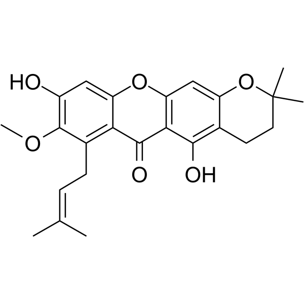
3-Isomangostin
Based on 1 publication(s) in Google Scholar
3-Isomangostin is a flavonoid compound that acts as an inhibitor of MTH1 and acetylcholinesterase, with an IC50 of 52 nM against MTH1 and IC50 values of 5.75 μM and 12.96 μM against AChE and BChE, respectively. 3-Isomangostin also inhibits human aldose reductase and CDK4/Cyclin D1 with IC50 values of 3.48 μM and 15.6 μM, respectively. 3-Isomangostin inhibits polyol pathway flux, competes with 8-oxo-dGTP for binding to the MTH1 active site through hydrogen bonding and hydrophobic xanthone interactions, blocks cell cycle progression, reduces cancer cell viability, and exhibits growth inhibitory effects against both normal and penicillin-resistant Staphylococcus aureus. 3-Isomangostin is used in research on Alzheimer's disease, diabetic complications, Staphylococcus aureus infections, and colon cancer.
For research use only. We do not sell to patients.
- Purity : 99.70%
- CAS No.: 19275-46-8
- Formula: C24H26O6
- Molecular Weight:410.46
-
Storage:
4°C, protect from light
* In solvent : -80°C, 6 months; -20°C, 1 month (protect from light)

To place orders, for customer services and technical support, please contact: MedChemExpress USA
Tel: 609-228-6898 E-mail: [email protected] [email protected]


-
 Biological Activity
-
Chemical Information
-
Solvent & Solubility
- Protocols
- Purity & Documentation
- References
-
 Help & FAQs


Publications Citing Use of MedChemExpress (MCE) 3-Isomangostin
MoreAll DNA/RNA Synthesis Isoforms
More

Biological Activity
Description
|
MTH1 52 nM (IC50) |
Aldose reductase 3.48 μM (IC50) |
AChE 5.75 μM (IC50) |
BChE 12.96 μM (IC50) |
Cdk4/cyclin D1 15.6 μM (IC50) |
Cellular Effect
|
Cell Line

|
Type | Value | Description | References |
|---|---|---|---|---|
| HCC1937 | IC50 |
59.9 μM
|
Reduction of viability of human HCC1937 breast cancer cells incubated for 48 h assessed by MTT assay with absorbance read at 570 nm relative to vehicle control.
Reduction of viability of human HCC1937 breast cancer cells incubated for 48 h assessed by MTT assay with absorbance read at 570 nm relative to vehicle control.
|
31363494 |
| HCT-116 | EC50 |
47.4 μM
|
Reduction of viability of human HCT116 colon cancer cells incubated for 48 h assessed by MTT assay with absorbance read at 570 nm relative to vehicle control.
Reduction of viability of human HCT116 colon cancer cells incubated for 48 h assessed by MTT assay with absorbance read at 570 nm relative to vehicle control.
|
31363494 |
| HT-29 | ED50 |
4.9 μM
|
Cytotoxic activity against human HT-29 colon cancer cells determined from dose-response testing.
Cytotoxic activity against human HT-29 colon cancer cells determined from dose-response testing.
|
19839614 |
In Vitro
3-Isomangostin inhibits electric eel acetylcholinesterase with an IC50 of 5.75 μM, and also inhibits equine serum butyrylcholinesterase with an IC50 of 12.96 μM[1].
3-Isomangostin potently inhibits human recombinant aldose reductase in vitro with an IC50 of 3.48 μM[2].
3-Isomangostin (3.3 nM-100 μM; 30 min pre-incubation, 40 min reaction) potently inhibits purified human MTH1 enzymatic activity with an IC50 of 0.052 μM[3].
3-Isomangostin inhibits the growth of normal Staphylococcus aureus ATCC 25923 with a minimal inhibitory concentration of 125 μg/mL[4].
3-Isomangostin inhibits the growth of 41 penicillin-resistant Staphylococcus aureus strains with a minimal inhibitory concentration of 250 μg/mL[4].
3-Isomangostin (10-40 μM) inhibits purified CDK4/Cyclin D1 kinase activity with an IC50 of 15.6 μM in a cell-free biochemical assay[5].
3-Isomangostin (1-100 μM; 48 h) decreases viability of HCC1937 human breast cancer cells with an IC50 of 59.9 μM after 48 h of treatment[5].
3-Isomangostin (0-100 μM; 48 h) decreases viability of HCT116 human colon cancer cells with an EC50 of 47.4 μM after 48 h of treatment[5].
3-Isomangostin (4.9 μM) is cytotoxic to HT-29 human colon cancer cells with an ED50 of 4.9 μM[6].
MedChemExpress (MCE) has not independently confirmed the accuracy of these methods. They are for reference only. Further protocols information, click here.


Chemical Information
-
CAS No. 19275-46-8
-
Appearance Solid
-
Molecular Weight 410.46
-
Formula C24H26O6
-
Color Off-white to light yellow
-
SMILES
C/C(C)=C\CC1=C2C(OC3=C(C(O)=C4C(OC(C)(C)CC4)=C3)C2=O)=CC(O)=C1OC
-
Structure Classification
-
Initial Source
-
Shipping
Room temperature in continental US; may vary elsewhere.
-
Storage
4°C, protect from light
* In solvent : -80°C, 6 months; -20°C, 1 month (protect from light)


Publications (1)
-
Journal Impact Factor
-
Most Recent


Solvent & Solubility
In Vitro:
DMSO : 100 mg/mL (243.63 mM; Need ultrasonic; Hygroscopic DMSO has a significant impact on the solubility of product, please use newly opened DMSO)
Please refer to the solubility information to select the appropriate solvent. Once prepared, please aliquot and store the solution to prevent product inactivation from repeated freeze-thaw cycles.
Storage method and period of stock solution: -80°C, 6 months; -20°C, 1 month (protect from light). When stored at -80°C, please use it within 6 months. When stored at -20°C, please use it within 1 month.
Please refer to the solubility information to select the appropriate solvent. Once prepared, please aliquot and store the solution to prevent product inactivation from repeated freeze-thaw cycles.
Storage method and period of stock solution: -80°C, 6 months; -20°C, 1 month (protect from light). When stored at -80°C, please use it within 6 months. When stored at -20°C, please use it within 1 month.
Concentration (start) × Volume (start) = Concentration (final) × Volume (final)
In Vivo:
Select the appropriate dissolution method based on your experimental animal and administration route.
- For the following dissolution methods, please ensure to first prepare a clear stock solution using an In Vitro approach and then sequentially add co-solvents:
- To ensure reliable experimental results, the clarified stock solution can be appropriately stored based on storage conditions. As for the working solution for In Vivo experiments, it is recommended to prepare freshly and use it on the same day.
- The percentages shown for the solvents indicate their volumetric ratio in the final prepared solution. If precipitation or phase separation occurs during preparation, heat and/or sonication can be used to aid dissolution.
Add each solvent one by one: 10% DMSO 90% (20% SBE-β-CD in Saline)
Solubility: ≥ 2.5 mg/mL (6.09 mM); Clear solution
This protocol yields a clear solution of ≥ 2.5 mg/mL (saturation unknown).
Taking 1 mL working solution as an example, add 100 μL DMSO stock solution (25.0 mg/mL) to 900 μL 20% SBE-β-CD in Saline, and mix evenly.
Preparation of 20% SBE-β-CD in Saline (4°C, storage for one week): 2 g SBE-β-CD powder is dissolved in 10 mL Saline, completely dissolve until clear.
In Vivo Dissolution Calculator
Please enter the basic information of animal experiments:
-
-
-
-
Recommended: Prepare an additional quantity of animals to account for potential losses during experiments.
Please enter your animal formula composition:
-
%DMSO +
Recommended: Keep the proportion of DMSO in working solution below 2% if your animal is weak.
-
%+
-
+%Tween-80 + +
-
%Saline +
The co-solvents required include: DMSO, . All of co-solvents are available by MedChemExpress (MCE). , Tween 80. All of co-solvents are available by MedChemExpress (MCE).
Working solution concentration: 0.22 mg/mL
Method for preparing stock solution: mg drug dissolved in μL DMSO. Stock solution concentration: mg/mL. * In solvent : -80°C, 6 months; -20°C, 1 month (protect from light)
1. Take μL DMSO stock solution;
2. Add μL .
μL , mix evenly;
3. Then add μL Tween 80, mix evenly;
4. Then add μL
Please ensure that the stock solution in the first step is dissolved to a clear state, and add co-solvents in sequence. You can use ultrasonic heating (ultrasonic cleaner, recommended frequency 20-40 kHz), vortexing, etc. to assist dissolution.

Protocols
-
Cell Cytotoxicity Assay
Cytotoxicity assays are usually based on the assessment of cell membrane damage, which can also be indirectly detected by measuring cell viability. Detection methods include MTT assay, CKK-8 assay, LDH assay and ATP assay, etc.
-
Flow cytometric DNA-content cell-cycle staining
Flow cytometric DNA-content cell-cycle staining measures the fluorescence intensity of DNA-bound fluorochromes in single cells or nuclei to estimate DNA content distributions, allowing assignment of populations to G0/G1, S, and G2/M phases by DNA histogram deconvolution. Propidium iodide (PI) intercalates into DNA, and PI fluorescence is proportional to cellular DNA content when staining is performed under conditions that make DNA accessible and minimize non-DNA signal. Cells with G2/M DNA content are expected to show approximately twice the fluorescence intensity of G0/G1 cells, while S-phase cells occupy intermediate fluorescence values. PI-based DNA-content analysis can also detect cells with fractional DNA content, often reported as sub-G1, when DNA fragmentation and extraction during staining reduce retained DNA signal in apoptotic cells. DAPI is an alternative DNA fluorochrome for univariate DNA-content analysis, while bivariate approaches combining DNA content with proliferation
-
Mammalian live/dead viability and cytotoxicity staining
Live/dead viability and cytotoxicity staining assays are based on the simultaneous detection of intracellular esterase activity in metabolically active (viable) cells and membrane integrity loss in non-viable cells. In commonly used dual-staining approaches, membrane-permeant fluorogenic substrates are converted by intracellular esterases into fluorescent products in live cells, while impermeant DNA-binding dyes selectively enter cells with compromised plasma membranes and label nucleic acids in dead or dying cells, enabling discrimination between viable and non-viable populations by fluorescence microscopy or flow cytometry.
-
BrdU Incorporation Assay
Bromodeoxyuridine (BrdU) incorporation assay is based on the principle that BrdU, a thymidine analog, is incorporated into newly synthesized DNA during the S phase of the cell cycle, thereby serving as a marker of DNA replication and cellular proliferation. Incorporated BrdU can be detected using anti-BrdU antibodies following DNA denaturation, enabling visualization or quantification of proliferating cells through immunochemical detection methods such as immunofluorescence or immunohistochemistry.
-
Protocol for Cell Cycle
Cell-cycle analysis by flow cytometry measures DNA content in single cells to estimate the fraction of cells in G0/G1, S, and G2/M phases. Propidium iodide intercalates into DNA, and after RNA removal with RNase, fluorescence intensity reflects cellular DNA content: 2N cells are assigned to G0/G1, cells between 2N and 4N to S phase, and 4N cells to G2/M. DNA-content analysis alone cannot reliably separate G0 from G1 or G2 from M. Ki-67 can distinguish quiescent G0 cells from cycling cells, EdU or BrdU incorporation marks active DNA synthesis in S phase, and phospho-histone H3 staining identifies mitotic cells within the 4N population.
-
CCK-8/WST-8 Cell Proliferation Assay
The CCK-8/WST-8 assay is based on the reduction of the water-soluble tetrazolium salt WST-8 to a water-soluble formazan product by cellular dehydrogenases in metabolically active cells, where the generated formazan amount is proportional to the number of living cells and is quantified by measuring absorbance in the visible range, providing a colorimetric readout for cell viability and proliferation assessment. This class of tetrazolium-based assays improves upon earlier MTT-based systems by producing a water-soluble formazan, eliminating the need for organic solubilization steps and enabling direct spectrophotometric measurement in culture medium.
-
MTT Cell Proliferation Assay
The MTT assay is a colorimetric endpoint assay for estimating viable cell number, cell growth, cytotoxicity, or cell activation in cultured mammalian cells. Living cells reduce the yellow tetrazolium salt MTT into purple/blue formazan, while dead cells do not generate the same signal; the resulting color can be quantified with a multiwell spectrophotometer. MTT reduction is commonly interpreted as a readout of metabolic activity that often correlates with viable cell number, but it should not be treated as a direct cell-counting method unless the assay is optimized for the cell type and experimental condition. Studies show that MTT reduction can involve mitochondrial and non-mitochondrial reducing systems, and formazan may accumulate in intracellular lipid droplets rather than simply marking mitochondria.
-
Cell Viability Determination by MTT Colorimetric Assay
The following protocol uses the MTT colorimetric assay as a classic literature-established method for assessing cell viability/metabolic activity in cultured mammalian cells. MTT[3-(4,5-dimethylthiazol-2-yl)-2,5-diphenyltetrazolium bromide] is reduced by metabolically active cells to a colored formazan product; the amount of formazan is quantified spectrophotometrically and provides an indirect measure of metabolically active viable cells. Importantly, MTT reduction reflects cellular oxidoreductase/metabolic activity rather than an absolute direct count of living cells, so changes in cellular metabolism can alter the signal independently of cell number.
-
Alzheimer’s Disease Modeling
Alzheimer’s Disease (AD) is a neurodegenerative disorder characterized by a progressive decline in cognitive functions and loss of specific types of neurons and synapses. Alzheimer's symptoms can be simulated in mice by injecting drugs (such as Aβ) or genetically modified.


Purity & Documentation
-
Data Sheet (289 KB)
-
SDS (393 KB)
- English - EN (393 KB)
- Français - FR (393 KB)
- Deutsch - DE (393 KB)
- Norwegian - NO (393 KB)
- Español - ES (393 KB)
- Swedish - SV (393 KB)
- Italian - IT (393 KB)
- Korean - KR (393 KB)
- Portuguese - PT (393 KB)
-
Handling Instructions (2659 KB)
References
[1]. Khaw KY, et al. Prenylated xanthones from mangosteen as promising cholinesterase inhibitors and their molecular docking studies. Phytomedicine : international journal of phytotherapy and phytopharmacology. 2014 Sep 25;21(11):1303-9. [Content Brief]
[3]. Yokoyama T, et al. Discovery of a new class of MTH1 inhibitor by X-ray crystallographic screening. European journal of medicinal chemistry. 2019 Apr 01;167:153-160. [Content Brief]
[6]. Han AR, et al. Cytotoxic xanthone constituents of the stem bark of Garcinia mangostana (mangosteen). Journal of natural products. 2009 Nov;72(11):2028-31. [Content Brief]


Complete Stock Solution Preparation Table
Please refer to the solubility information to select the appropriate solvent. Once prepared, please aliquot and store the solution to prevent product inactivation from repeated freeze-thaw cycles.
Storage method and period of stock solution: -80°C, 6 months; -20°C, 1 month (protect from light). When stored at -80°C, please use it within 6 months. When stored at -20°C, please use it within 1 month.
| Optional Solvent | Concentration Solvent Mass | 1 mg | 5 mg | 10 mg | 25 mg |
|---|---|---|---|---|---|
| DMSO | 1 mM | 2.4363 mL | 12.1815 mL | 24.3629 mL | 60.9073 mL |
| 5 mM | 0.4873 mL | 2.4363 mL | 4.8726 mL | 12.1815 mL | |
| 10 mM | 0.2436 mL | 1.2181 mL | 2.4363 mL | 6.0907 mL | |
| 15 mM | 0.1624 mL | 0.8121 mL | 1.6242 mL | 4.0605 mL | |
| 20 mM | 0.1218 mL | 0.6091 mL | 1.2181 mL | 3.0454 mL | |
| 25 mM | 0.0975 mL | 0.4873 mL | 0.9745 mL | 2.4363 mL | |
| 30 mM | 0.0812 mL | 0.4060 mL | 0.8121 mL | 2.0302 mL | |
| 40 mM | 0.0609 mL | 0.3045 mL | 0.6091 mL | 1.5227 mL | |
| 50 mM | 0.0487 mL | 0.2436 mL | 0.4873 mL | 1.2181 mL | |
| 60 mM | 0.0406 mL | 0.2030 mL | 0.4060 mL | 1.0151 mL | |
| 80 mM | 0.0305 mL | 0.1523 mL | 0.3045 mL | 0.7613 mL | |
| 100 mM | 0.0244 mL | 0.1218 mL | 0.2436 mL | 0.6091 mL |


3-Isomangostin Related Classifications
HY-N6845 Related Classifications
 Powered by Bioz