



JG26
Based on 1 publication(s) in Google Scholar
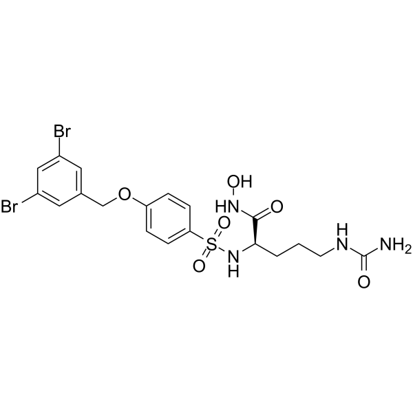
JG26 is an ADAM inhibitor with IC50 values of 12 nM, 1.9 nM, and 150 nM for ADAM8, ADAM17, and ADAM10, respectively. JG26 inhibits MMP-12 with an IC50 value of 9.4 nM. JG26 inhibits AngII (HY-13948)-induced EGFR transactivation and ERK activation. JG26 increases the expression of ACE2, inhibits the cleavage of CD23, reduces the infection of SARS-CoV-2. JG26 inhibits colorectal cancer metastasis. JG26 can be used for research on Hodgkin lymphoma and vascular diseases.
Nur für Forschungszwecke. Wir verkaufen nicht an Patienten.
- Reinheit : 98.14%
- CAS. Nr.: 1464910-32-4
- Formel: C19H22Br2N4O6S
- Molecular Weight:594.27
-
Speicherung:
4°C, protect from light
* In solvent : -80°C, 6 months; -20°C, 1 month (protect from light)

To place orders, for customer services and technical support, please contact: MedChemExpress USA
Tel: 609-228-6898 E-mail: [email protected] [email protected]


-
 Biologische Aktivität
-
Chemical Information
-
Lösungsmittel & Löslichkeit
- Protokoll
- Reinheit & Dokumentation
- Verweise
-
 Help & FAQs


Publications Citing Use of MedChemExpress (MCE) JG26
MoreAlle EGFR Isoform-spezifische Produkte anzeigen
More

Biologische Aktivität
Beschreibung
IC50 & Target
[1]|
ADAM17 1.9 nM (IC50) |
MMP-12 9.4 nM (IC50) |
ADAM8 12 nM (IC50) |
ADAM10 150 nM (IC50) |
MMP-8 240 nM (IC50) |
MMP-9 1630 nM (IC50) |
MMP14 19500 nM (IC50) |
MMP-9 >500000 nM (IC50) |
Cellular Effect
|
Cell Line

|
Type | Value | Description | References |
|---|---|---|---|---|
| A2780 | IC50 |
<10 nM
Compound: 21
|
Inhibition of ADAM-17 in human A2780 cells assessed as inhibition of pervanadate-induced sALCAM release incubated for 30 mins prior to pervanadate induction measured after 90 mins by ELISA
Inhibition of ADAM-17 in human A2780 cells assessed as inhibition of pervanadate-induced sALCAM release incubated for 30 mins prior to pervanadate induction measured after 90 mins by ELISA
|
[PMID: 24044434] |
| A2780 | IC50 |
845 nM
Compound: 21
|
Inhibition of ADAM-17 in human A2780 cells assessed as inhibition of EGF-induced sALCAM release incubated for 30 mins prior to EGF induction measured after 18 hrs by ELISA
Inhibition of ADAM-17 in human A2780 cells assessed as inhibition of EGF-induced sALCAM release incubated for 30 mins prior to EGF induction measured after 18 hrs by ELISA
|
[PMID: 24044434] |
| SK-OV-3 | IC50 |
4272 nM
Compound: 21
|
Inhibition of ADAM-17 in human SKOV3 cells transfected with luciferase gene assessed as inhibition of EGF-induced sALCAM release incubated for 30 mins prior to EGF induction measured after 18 hrs by ELISA
Inhibition of ADAM-17 in human SKOV3 cells transfected with luciferase gene assessed as inhibition of EGF-induced sALCAM release incubated for 30 mins prior to EGF induction measured after 18 hrs by ELISA
|
[PMID: 24044434] |
| SK-OV-3 | IC50 |
58 nM
Compound: 21
|
Inhibition of ADAM-17 in human SKOV3 cells transfected with luciferase gene assessed as inhibition of pervanadate-induced sALCAM release incubated for 30 mins prior to pervanadate induction measured after 90 mins by ELISA
Inhibition of ADAM-17 in human SKOV3 cells transfected with luciferase gene assessed as inhibition of pervanadate-induced sALCAM release incubated for 30 mins prior to pervanadate induction measured after 90 mins by ELISA
|
[PMID: 24044434] |
In Vitro
JG26 (5-25 µM; 24-48 h) has no significant effect on the viability of Calu-3 cells (CC50: >25 µM) and increases the expression of ACE2 on the surface of Calu-3 cells[1].
JG26 (25 µM) can significantly reduce the infection of SARS-CoV-2 in Calu-3 cells, with LR values of 1.44 and 2.37, respectively[1].
JG26 (10 µM; 24 h) can reduce the shedding of NKG2D ligands in Hodgkin lymphoma cell lines[2].
JG26 (125 nM-10 µM; 24 h) can inhibit the cleavage of CD23 and the release of its soluble form in HEK293 cells[3].
JG26 (25 nM; 2 h) can effectively reduce the permeability of HUVECs and tumor cell adhesion induced by EMT-HCT116 exosomes[4].
JG26 (1 μM; 30 min) inhibits AngII-induced extracellular collagen accumulation in vascular smooth muscle cells (VSMCs)[5].
MedChemExpress (MCE) has not independently confirmed the accuracy of these methods. They are for reference only. Further protocols information, click here.
In Vivo
MedChemExpress (MCE) has not independently confirmed the accuracy of these methods. They are for reference only.
-
Animal Model:Six-week-old male athymic BALB/c-nu/nu mice, orthotropic metastasis model of colorectal cancer[4]
-
Dosage:3 mg/kg
-
Administration:Intravenous injection
-
Result:Significantly reduced the decrease in VE-cadherin expression at the invasive front of CRC tumors.
Decreased the number of CTCs in mouse blood and the proportion of mice with both lung and liver metastasis.
Reduced the number and size of metastatic nodules in the lungs and liver.


Chemical Information
-
CAS. Nr. 1464910-32-4
-
Appearance Solid
-
Molecular Weight 594.27
-
Formel C19H22Br2N4O6S
-
Color White to off-white
-
SMILES
O=C([C@@H](CCCNC(N)=O)NS(=O)(C1=CC=C(C=C1)OCC2=CC(Br)=CC(Br)=C2)=O)NO
-
Versand
Room temperature in continental US; may vary elsewhere.
-
Speicherung
4°C, protect from light
* In solvent : -80°C, 6 months; -20°C, 1 month (protect from light)


Publications (1)
-
Journal Impact Factor
-
Most Recent
-
Cell Discov
2024 Dec 9;10(1):121. PMID: 39648224


Lösungsmittel & Löslichkeit
In Vitro:
DMSO : 100 mg/mL (168.27 mM; Need ultrasonic; Hygroscopic DMSO has a significant impact on the solubility of product, please use newly opened DMSO)
Please refer to the solubility information to select the appropriate solvent. Once prepared, please aliquot and store the solution to prevent product inactivation from repeated freeze-thaw cycles.
Storage method and period of stock solution: -80°C, 6 months; -20°C, 1 month (protect from light). When stored at -80°C, please use it within 6 months. When stored at -20°C, please use it within 1 month.
Please refer to the solubility information to select the appropriate solvent. Once prepared, please aliquot and store the solution to prevent product inactivation from repeated freeze-thaw cycles.
Storage method and period of stock solution: -80°C, 6 months; -20°C, 1 month (protect from light). When stored at -80°C, please use it within 6 months. When stored at -20°C, please use it within 1 month.
Konzentration (Stammlösung) × Volumen (Stammlösung) = Konzentration (Ziellösung) × Volumen (Ziellösung)
In Vivo:
Select the appropriate dissolution method based on your experimental animal and administration route.
- For the following dissolution methods, please ensure to first prepare a clear stock solution using an In Vitro approach and then sequentially add co-solvents:
- To ensure reliable experimental results, the clarified stock solution can be appropriately stored based on storage conditions. As for the working solution for In Vivo experiments, it is recommended to prepare freshly and use it on the same day.
- The percentages shown for the solvents indicate their volumetric ratio in the final prepared solution. If precipitation or phase separation occurs during preparation, heat and/or sonication can be used to aid dissolution.
Add each solvent one by one: 10% DMSO 40% PEG300 5% Tween-80 45% Saline
Solubility: ≥ 2.5 mg/mL (4.21 mM); Clear solution
This protocol yields a clear solution of ≥ 2.5 mg/mL (saturation unknown).
Taking 1 mL working solution as an example, add 100 μL DMSO stock solution (25.0 mg/mL) to 400 μL PEG300, and mix evenly; then add 50 μL Tween-80 and mix evenly; then add 450 μL Saline to adjust the volume to 1 mL.
Preparation of Saline: Dissolve 0.9 g sodium chloride in ddH₂O and dilute to 100 mL to obtain a clear Saline solution.
Add each solvent one by one: 10% DMSO 90% (20% SBE-β-CD in Saline)
Solubility: ≥ 2.5 mg/mL (4.21 mM); Clear solution
This protocol yields a clear solution of ≥ 2.5 mg/mL (saturation unknown).
Taking 1 mL working solution as an example, add 100 μL DMSO stock solution (25.0 mg/mL) to 900 μL 20% SBE-β-CD in Saline, and mix evenly.
Preparation of 20% SBE-β-CD in Saline (4°C, storage for one week): 2 g SBE-β-CD powder is dissolved in 10 mL Saline, completely dissolve until clear.
In Vivo Dissolution Calculator
Please enter the basic information of animal experiments:
-
-
-
-
Recommended: Prepare an additional quantity of animals to account for potential losses during experiments.
Please enter your animal formula composition:
-
%DMSO +
Recommended: Keep the proportion of DMSO in working solution below 2% if your animal is weak.
-
%+
-
+%Tween-80 + +
-
%Saline +
The co-solvents required include: DMSO, . All of co-solvents are available by MedChemExpress (MCE). , Tween 80. All of co-solvents are available by MedChemExpress (MCE).
Working solution concentration: 0.22 mg/mL
Method for preparing stock solution: mg drug dissolved in μL DMSO. Stock solution concentration: mg/mL. * In solvent : -80°C, 6 months; -20°C, 1 month (protect from light)
1. Take μL DMSO stock solution;
2. Add μL .
μL , mix evenly;
3. Then add μL Tween 80, mix evenly;
4. Then add μL
Please ensure that the stock solution in the first step is dissolved to a clear state, and add co-solvents in sequence. You can use ultrasonic heating (ultrasonic cleaner, recommended frequency 20-40 kHz), vortexing, etc. to assist dissolution.

Protokoll
-
RNA extraction experimental
By lysing cells, releasing RNA, and removing impurities such as proteins and DNA, high-purity RNA products are finally obtained. The commonly used traditional method is the guanidine isothiocyanate/phenol/chloroform method (Trizol), which is suitable for a variety of animal materials including animal tissues, microorganisms, cultured cells, etc., and most plant materials.
-
Research Protocol for Infectious Diseases
Infectious-disease experiments test how pathogens interact with host barriers, innate immune receptors, inflammatory signaling, pathogen replication, and tissue injury; pattern-recognition receptors such as TLRs, RIG-I-like receptors, NOD-like receptors, and inflammasomes detect microbial molecules and activate NF-κB, interferon, and cytokine responses. The central hypothesis is that infection severity reflects the balance between pathogen burden and host response: protective inflammation restricts pathogen growth, whereas excessive or mislocalized inflammation contributes to tissue damage and disease phenotype. Unresolved questions include which host pathways are protective versus pathogenic, why some infection models fail to translate to human disease, and which combined readouts best predict clinically relevant infection outcomes.
-
Mitochondrial membrane-potential fluorescent assay
Mitochondrial membrane potential fluorescent assays estimate ΔΨm in living cells using lipophilic cationic dyes such as TMRM, TMRE, rhodamine 123, and JC-1, which accumulate in mitochondria according to membrane polarization; loss of signal after FCCP or CCCP treatment is interpreted as mitochondrial depolarization. TMRM/TMRE and rhodamine 123 are commonly used for semi-quantitative live-cell microscopy or flow cytometry, while JC-1 can report a shift from red aggregate fluorescence to green monomer fluorescence during depolarization; interpretation requires controls because dye concentration, quenching mode, cell type, dye efflux, and mitochondrial mass can affect fluorescence independently of ΔΨm.
-
Mitochondrial membrane-potential and mitochondrial mass staining
Mitochondrial membrane potential staining measures the electrochemical polarization across the mitochondrial inner membrane in live cells using lipophilic cationic fluorescent probes; early rhodamine-based work showed that selective mitochondrial dye accumulation is lost when the mitochondrial transmembrane potential is dissipated. JC-1 reports mitochondrial polarization by shifting from green monomer fluorescence to red J-aggregate fluorescence as dye concentration increases within energized mitochondria; therefore, the red/green fluorescence ratio is used as a relative readout of mitochondrial membrane potential. TMRE or TMRM staining provides a single-channel relative readout because these cationic rhodamine esters accumulate in polarized mitochondria, and lower fluorescence indicates reduced mitochondrial polarization when acquisition and dye-loading conditions are controlled. Mitochondrial mass staining is commonly performed with MitoTracker Green FM or related MitoTracker dyes as
-
Orthotopic Cell-Line Xenograft
Orthotopic cell-line xenograft models involve implantation of human cancer cell lines into the anatomically corresponding organ of immunodeficient mice to reproduce tumor growth within a native microenvironment, enabling more clinically relevant tumor behavior compared with subcutaneous models. These models are widely used because orthotopic placement better recapitulates tumor progression, including invasion and metastatic spread, which are often underrepresented in heterotopic implantation systems. Compared with conventional xenografts, orthotopic implantation is described as more technically complex but provides improved simulation of tumor-microenvironment interactions and metastatic behavior, making it particularly valuable for translational oncology research. Surgical orthotopic implantation approaches have been emphasized as enabling faithful reproduction of clinical cancer features, including metastasis and disease progression patterns that align with the tumor’s organ of origi


Reinheit & Dokumentation
-
Data Sheet (275 KB)
-
SDS (252 KB)
- English - EN (252 KB)
- Français - FR (252 KB)
- Deutsch - DE (252 KB)
- Norwegian - NO (252 KB)
- Español - ES (252 KB)
- Swedish - SV (252 KB)
- Italian - IT (252 KB)
- Korean - KR (252 KB)
- Portuguese - PT (252 KB)
-
Handling Instructions (2659 KB)
Verweise
[1]. Gentili V, et al. JG26 attenuates ADAM17 metalloproteinase-mediated ACE2 receptor processing and SARS-CoV-2 infection in vitro. Pharmacol Rep. 2025 Feb;77(1):260-273. [Content Brief]
[2]. Zocchi MR, et al. ADAM10 new selective inhibitors reduce NKG2D ligand release sensitizing Hodgkin lymphoma cells to NKG2D-mediated killing. Oncoimmunology. 2015 Dec 29;5(5):e1123367. [Content Brief]
[3]. Cuffaro D, et al. Discovery of Dimeric Arylsulfonamides as Potent ADAM8 Inhibitors. ACS Med Chem Lett. 2021 Oct 8;12(11):1787-1793. [Content Brief]
[4]. Li K, et al. Tumor-derived exosomal ADAM17 promotes pre-metastatic niche formation by enhancing vascular permeability in colorectal cancer. J Exp Clin Cancer Res. 2024 Feb 27;43(1):59. [Content Brief]
[6]. Doretta Cuffaro, et al. Discovery of Dimeric Arylsulfonamides as Potent ADAM8 Inhibitors. ACS Med Chem Lett. 2021 Oct 8;12(11):1787-1793. [Content Brief]


Complete Stock Solution Preparation Table
Please refer to the solubility information to select the appropriate solvent. Once prepared, please aliquot and store the solution to prevent product inactivation from repeated freeze-thaw cycles.
Storage method and period of stock solution: -80°C, 6 months; -20°C, 1 month (protect from light). When stored at -80°C, please use it within 6 months. When stored at -20°C, please use it within 1 month.
| Optional Solvent | Concentration Solvent Mass | 1 mg | 5 mg | 10 mg | 25 mg |
|---|---|---|---|---|---|
| DMSO | 1 mM | 1.6827 mL | 8.4137 mL | 16.8274 mL | 42.0684 mL |
| 5 mM | 0.3365 mL | 1.6827 mL | 3.3655 mL | 8.4137 mL | |
| 10 mM | 0.1683 mL | 0.8414 mL | 1.6827 mL | 4.2068 mL | |
| 15 mM | 0.1122 mL | 0.5609 mL | 1.1218 mL | 2.8046 mL | |
| 20 mM | 0.0841 mL | 0.4207 mL | 0.8414 mL | 2.1034 mL | |
| 25 mM | 0.0673 mL | 0.3365 mL | 0.6731 mL | 1.6827 mL | |
| 30 mM | 0.0561 mL | 0.2805 mL | 0.5609 mL | 1.4023 mL | |
| 40 mM | 0.0421 mL | 0.2103 mL | 0.4207 mL | 1.0517 mL | |
| 50 mM | 0.0337 mL | 0.1683 mL | 0.3365 mL | 0.8414 mL | |
| 60 mM | 0.0280 mL | 0.1402 mL | 0.2805 mL | 0.7011 mL | |
| 80 mM | 0.0210 mL | 0.1052 mL | 0.2103 mL | 0.5259 mL | |
| 100 mM | 0.0168 mL | 0.0841 mL | 0.1683 mL | 0.4207 mL |


JG26 Related Classifications
HY-120852 Related Classifications
 Powered by Bioz