



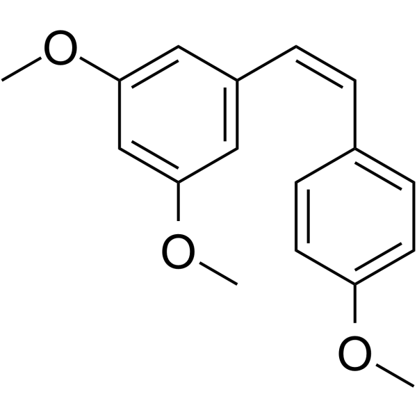
cis-Trismethoxy resveratrol

Based on 1 Customer Validation
cis-Trismethoxy resveratrol ((Z)-3,5,4'-Trimethoxystilbene) is an anti-HCV agent and Tubulin inhibitor, with an IC50 of 4 μM against Tubulin. cis-Trismethoxy resveratrol induces G2/M phase cell cycle arrest, reduces DCLK1, decreases CDK1 levels, blocks phosphorylation of Akt Ser473, and induces the expression of p21Cip1/Waf1. cis-Trismethoxy resveratrol exhibits anti-tumor and hepatoprotective activities. cis-Trismethoxy resveratrol can be used in studies related to colon adenocarcinoma, hepatocellular carcinoma, hepatitis C, and liver injury.
For research use only. We do not sell to patients.
- Purity : 99.87%
- CAS No.: 94608-23-8
- Formula: C17H18O3
- Molecular Weight:270.32
-
Storage:
Solution, -20°C, protect from light, 2 years

To place orders, for customer services and technical support, please contact: MedChemExpress USA
Tel: 609-228-6898 E-mail: [email protected] [email protected]


-
 Biological Activity
-
Chemical Information
- Protocols
- Purity & Documentation
- References
-
 Help & FAQs




Biological Activity
Description
IC50 & Target
[1]|
CDK1 |
Cellular Effect
|
Cell Line

|
Type | Value | Description | References |
|---|---|---|---|---|
| A549 | ED50 |
1.3 μM
Compound: 6i
|
Compound was evaluated for cytotoxicity against human A-549 non-small cell lung cancer cell line.
Compound was evaluated for cytotoxicity against human A-549 non-small cell lung cancer cell line.
|
[PMID: 1613753] |
| B16-F10 | IC50 |
1 μM
Compound: 72
|
Anti cancer activity against mouse B16-F10 cells assessed as cell growth inhibition incubated for 40 hrs by Almar Blue stained fluorescence-based assay
Anti cancer activity against mouse B16-F10 cells assessed as cell growth inhibition incubated for 40 hrs by Almar Blue stained fluorescence-based assay
|
[PMID: 32485531] |
| BXPC-3 | GI50 |
0.0034 μg/mL
Compound: 1d
|
Growth inhibition of human BxPC3 cells after 48 hrs by sulforhodamine B assay
Growth inhibition of human BxPC3 cells after 48 hrs by sulforhodamine B assay
|
[PMID: 19719153] |
| BXPC-3 | GI50 |
0.0034 μg/mL
Compound: 4a
|
Cell growth inhibition against pancreatic BXPC-3 cells, 50% reduction in the net protein increase
Cell growth inhibition against pancreatic BXPC-3 cells, 50% reduction in the net protein increase
|
[PMID: 12036362] |
| Caco-2 | IC50 |
0.08 μM
Compound: 10
|
Cytotoxicity against human Caco-2 cells after 3 days by [3H]thymidine incorporation assay
Cytotoxicity against human Caco-2 cells after 3 days by [3H]thymidine incorporation assay
|
[PMID: 20627379] |
| Caco-2 | IC50 |
0.145 μM
Compound: 72
|
Anti cancer activity against human Caco-2 cell assessed as cell growth inhibition incubated for 48 hrs by Brdu assay
Anti cancer activity against human Caco-2 cell assessed as cell growth inhibition incubated for 48 hrs by Brdu assay
|
[PMID: 32485531] |
| DU-145 | GI50 |
0.0054 μg/mL
Compound: 1d
|
Growth inhibition of human DU145 cells after 48 hrs by sulforhodamine B assay
Growth inhibition of human DU145 cells after 48 hrs by sulforhodamine B assay
|
[PMID: 19719153] |
| DU-145 | GI50 |
0.0054 μg/mL
Compound: 4a
|
Cell growth inhibition of prostate DU-145 cells, expressed as 50% reduction in the net protein increase
Cell growth inhibition of prostate DU-145 cells, expressed as 50% reduction in the net protein increase
|
[PMID: 12036362] |
| HCT-116 | IC50 |
0.22 μM
Compound: 8Z
|
Cytotoxicity against human HCT116 cells after 24 hrs by MTT assay
Cytotoxicity against human HCT116 cells after 24 hrs by MTT assay
|
[PMID: 21215623] |
| HepG2 | IC50 |
0.473 μM
Compound: 72
|
Anti cancer activity against human HepG2 cell assessed as cell growth inhibition incubated for 48 hrs by Brdu assay
Anti cancer activity against human HepG2 cell assessed as cell growth inhibition incubated for 48 hrs by Brdu assay
|
[PMID: 32485531] |
| HL-60 | IC50 |
0.15 μM
Compound: 6b
|
Antiproliferative activity against human HL60 cells
Antiproliferative activity against human HL60 cells
|
[PMID: 16580204] |
| HT-29 | ED50 |
3.4 μM
Compound: 6i
|
Cytotoxicity against human HT-29 colon cell line.
Cytotoxicity against human HT-29 colon cell line.
|
[PMID: 1613753] |
| HT-29 | IC50 |
0.04 μM
Compound: 10
|
Cytotoxicity against human HT-29 cells after 3 days by [3H]thymidine incorporation assay
Cytotoxicity against human HT-29 cells after 3 days by [3H]thymidine incorporation assay
|
[PMID: 20627379] |
| HT-29 | IC50 |
0.115 μM
Compound: 72
|
Anti cancer activity against human HT-29 cell assessed as cell growth inhibition incubated for 48 hrs by Brdu assay
Anti cancer activity against human HT-29 cell assessed as cell growth inhibition incubated for 48 hrs by Brdu assay
|
[PMID: 32485531] |
| HT-29 | IC50 |
54 nM
Compound: 17
|
Antiproliferative activity against human HT29 cell line by methylene blue dye assay
Antiproliferative activity against human HT29 cell line by methylene blue dye assay
|
[PMID: 17034147] |
| IMR-90 | IC50 |
0.23 μM
Compound: 8Z
|
Cytotoxicity against human IMR90 cells after 24 hrs by MTT assay
Cytotoxicity against human IMR90 cells after 24 hrs by MTT assay
|
[PMID: 21215623] |
| KB | IC50 |
280 nM
Compound: 17
|
Antiproliferative activity against human KB cell line by methylene blue dye assay
Antiproliferative activity against human KB cell line by methylene blue dye assay
|
[PMID: 17034147] |
| MCF7 | ED50 |
1.6 μM
Compound: 6i
|
Cytotoxicity against human MCF-7 breast cancer cell line.
Cytotoxicity against human MCF-7 breast cancer cell line.
|
[PMID: 1613753] |
| MCF7 | IC50 |
0.12 μM
Compound: 8Z
|
Cytotoxicity against human MCF7 cells after 24 hrs by MTT assay
Cytotoxicity against human MCF7 cells after 24 hrs by MTT assay
|
[PMID: 21215623] |
| MKN-45 | IC50 |
218 nM
Compound: 17
|
Antiproliferative activity against human MKN45 cell line by methylene blue dye assay
Antiproliferative activity against human MKN45 cell line by methylene blue dye assay
|
[PMID: 17034147] |
| MLM | ED50 |
9.8 μM
Compound: 6i
|
Cytotoxicity against human MLM melanoma cell line.
Cytotoxicity against human MLM melanoma cell line.
|
[PMID: 1613753] |
| NCI-H460 | GI50 |
0.0028 μg/mL
Compound: 1d
|
Growth inhibition of human NCI-H460 cells after 48 hrs by sulforhodamine B assay
Growth inhibition of human NCI-H460 cells after 48 hrs by sulforhodamine B assay
|
[PMID: 19719153] |
| NCI-H460 | GI50 |
0.0028 μg/mL
Compound: 4a
|
Cell growth inhibition of lung-NSC NCI-H460 cells, expressed as 50% reduction in the net protein increase
Cell growth inhibition of lung-NSC NCI-H460 cells, expressed as 50% reduction in the net protein increase
|
[PMID: 12036362] |
| NCI-H460 | IC50 |
253 nM
Compound: 17
|
Antiproliferative activity against human H460 cell line by methylene blue dye assay
Antiproliferative activity against human H460 cell line by methylene blue dye assay
|
[PMID: 17034147] |
| P388 | ED50 |
0.0262 μg/mL
Compound: 4a
|
Evaluated for cell growth inhibition of the mouse leukemia P388 cells in 10% horse serum/Fisher media
Evaluated for cell growth inhibition of the mouse leukemia P388 cells in 10% horse serum/Fisher media
|
[PMID: 12036362] |
| SF-268 | GI50 |
0.0044 μg/mL
Compound: 1d
|
Growth inhibition of human SF268 cells after 48 hrs by sulforhodamine B assay
Growth inhibition of human SF268 cells after 48 hrs by sulforhodamine B assay
|
[PMID: 19719153] |
| SF-268 | GI50 |
0.0044 μg/mL
Compound: 4a
|
Cell growth inhibition of central nervous system (CNS) SF-268 cells, expressed as 50% reduction in the net protein increase
Cell growth inhibition of central nervous system (CNS) SF-268 cells, expressed as 50% reduction in the net protein increase
|
[PMID: 12036362] |
| SK-MEL-5 | ED50 |
4.2 μM
Compound: 6i
|
Compound was evaluated for cytotoxicity against human SKMEL-5 melanoma cell line.
Compound was evaluated for cytotoxicity against human SKMEL-5 melanoma cell line.
|
[PMID: 1613753] |
| SW480 | IC50 |
0.3 μM
Compound: 4
|
Antiproliferative activity against human SW480 cells assessed as cell viability using propidium iodide staining after 48 hrs
Antiproliferative activity against human SW480 cells assessed as cell viability using propidium iodide staining after 48 hrs
|
[PMID: 20395019] |
| SW480 | IC50 |
0.3 μM
Compound: 4
|
Antiproliferative activity against human SW480 cells assessed as cell viability using propidium iodide staining after 72 hrs
Antiproliferative activity against human SW480 cells assessed as cell viability using propidium iodide staining after 72 hrs
|
[PMID: 20395019] |
| TSGH 9201 | IC50 |
324 nM
Compound: 17
|
Antiproliferative activity against human TSGH stomach carcinoma cell line by methylene blue dye assay
Antiproliferative activity against human TSGH stomach carcinoma cell line by methylene blue dye assay
|
[PMID: 17034147] |
In Vitro
cis-Trismethoxy resveratrol potently inhibits the growth of various cancer cell lines, with IC50 values ranging from 0.08 μM (KB cells) to 0.25 μM (Caco-2 cells), and the activity of its cis conformation is 100-fold higher than that of the trans isomer[1].
cis-Trismethoxy resveratrol (0.3 μM; 16-48 hr, second treatment after 48 h) induces G2/M phase arrest in Caco-2 cells, and a second treatment administered after 48 h potentiates this effect[1].
cis-Trismethoxy resveratrol (0.1-30 μM; up to 30 min) inhibits tubulin polymerization in vitro, with an IC50 of 4 μM, and completely blocks the polymerization process at a concentration of 30 μM[1].
cis-Trismethoxy resveratrol (0.3 μM; 1-3 days) reduces ODC activity by 52% after 1 day, AdoMetDC activity by 40% after 2 days, and AdoMetDC activity by 50% after 3 days in Caco-2 cells[1].
cis-Trismethoxy resveratrol (0.05-2.5 μM; 48 h) dose-dependently inhibits the expression of HCV NS5B polymerase in FCA4 hepatocellular carcinoma cells; when applied at concentrations ranging from 0.1 to 1.0 μM for 48 h, it shows no cytotoxicity to either FCA4 cells or normal hepatocytes[2].
cis-Trismethoxy resveratrol (1 μM; 48 h) inhibits HCV replication by approximately 10-fold in JFH1 HCVcc-infected Huh7.5 hepatocellular carcinoma cells and reduces NS3 protein levels after 48 h of treatment[2].
Treatment with cis-Trismethoxy resveratrol (1 μM; 48 h) induces G2/M phase arrest in over 90% of GS5, Huh7.5, Huh7 and FCA4 hepatocellular carcinoma cells[2].
MedChemExpress (MCE) has not independently confirmed the accuracy of these methods. They are for reference only. Further protocols information, click here.
-
Cell Line:human colon adenocarcinoma Caco-2 cells
-
Concentration:0.3 μM
-
Incubation Time:16 hr, 48 hr; second treatment initiated after 48 hr
-
Result:Caused accumulation of Caco-2 cells at the G2/M phase, reaching 54% of cells at 16 hours and 46% at 48 hours.
Increased the proportion of cells blocked at G2/M to 77% after a second 0.3 μM treatment at 48 hours.
Decreased the proportion of cells in the G1 phase, while the S phase proportion remained unchanged.
-
Cell Line:GS5, Huh7.5, Huh7, and FCA4 hepatoma cells
-
Concentration:1 μM
-
Incubation Time:48 h
-
Result:Caused >90% of cells to arrest in the G2/M phase, with concomitant reduction in G1 and S phase populations.
In Vivo
MedChemExpress (MCE) has not independently confirmed the accuracy of these methods. They are for reference only.
-
Animal Model:C57BL/6 (8 weeks old, in-house bred, 5 per group, DEN/CCl4-induced liver injury)[2]
-
Dosage:20 mg/kg BW; 40 mg/kg BW
-
Administration:injection; twice weekly; 7 weeks
-
Result:Reduced serum total bilirubin to 0.6 and alanine aminotransferase to 60.0 at 20 mg/kg BW compared to DEN/CCl4-only controls (ALT 78.6).
Reduced serum total bilirubin to 0.7 at 40 mg/kg BW compared to DEN/CCl4-only controls, but resulted in an alanine aminotransferase level of 109.0, which is higher than the DEN/CCl4-only control's 78.6.
Significantly reduced the number of DCLK1+ cells in liver injury sites and improved liver histologic appearance at both doses.
Showed serum liver biomarker levels (ALB 2.6, ALT 50.7, AST 128.7, ALKP 52.3, TBIL 0.9) within or near normal ranges in mice treated with 40 mg/kg BW alone, indicating no hepatotoxicity.
Caused gastrointestinal emptying complications in 20% of treated mice.


Chemical Information
-
CAS No. 94608-23-8
-
Appearance Liquid
-
Molecular Weight 270.32
-
Formula C17H18O3
-
Color Colorless to light yellow
-
SMILES
COC1=CC(/C=C\C2=CC=C(C=C2)OC)=CC(OC)=C1
-
Synonyms
(Z)-3,5,4'-Trimethoxystilbene
-
Shipping
Room temperature in continental US; may vary elsewhere.
-
Storage
Solution, -20°C, protect from light, 2 years

Protocols
-
Kinase activity and phosphorylation assays
Kinase activity assays measure the ability of kinases to transfer phosphate groups from ATP to specific substrates, while phosphorylation assays detect the presence and levels of phosphorylated proteins. Common methods include radiolabeled ATP incorporation (e. g. ,), ADP release detection via bioluminescence (e. g. ,[3]), enzyme-linked immunosorbent assays (ELISA) for phospho-specific epitopes (e. g. ,[6]), and microtiter-based formats for high-throughput screening (e. g. ,[8]). The ADP-Glo assay quantifies kinase activity by measuring ADP produced during phosphorylation using a luciferase-based system. Radiometric assays involve autoradiography or scintillation counting after incorporation of 32P-labeled ATP into substrate proteins. ELISA-based approaches rely on phospho-specific antibodies to detect activated kinases in cell lysates or purified samples.
-
Western Blot
Western blotting (WB) is a commonly used experimental method in molecular biology, biochemistry, and immunogenetics for identifying and quantifying target proteins. It combines gel electrophoresis with immunoassay, enabling researchers to analyze protein expression, post-translational modifications, and molecular weight.
-
RNA extraction experimental
By lysing cells, releasing RNA, and removing impurities such as proteins and DNA, high-purity RNA products are finally obtained. The commonly used traditional method is the guanidine isothiocyanate/phenol/chloroform method (Trizol), which is suitable for a variety of animal materials including animal tissues, microorganisms, cultured cells, etc., and most plant materials.
-
Cell Cytotoxicity Assay
Cytotoxicity assays are usually based on the assessment of cell membrane damage, which can also be indirectly detected by measuring cell viability. Detection methods include MTT assay, CKK-8 assay, LDH assay and ATP assay, etc.
-
Flow cytometric DNA-content cell-cycle staining
Flow cytometric DNA-content cell-cycle staining measures the fluorescence intensity of DNA-bound fluorochromes in single cells or nuclei to estimate DNA content distributions, allowing assignment of populations to G0/G1, S, and G2/M phases by DNA histogram deconvolution. Propidium iodide (PI) intercalates into DNA, and PI fluorescence is proportional to cellular DNA content when staining is performed under conditions that make DNA accessible and minimize non-DNA signal. Cells with G2/M DNA content are expected to show approximately twice the fluorescence intensity of G0/G1 cells, while S-phase cells occupy intermediate fluorescence values. PI-based DNA-content analysis can also detect cells with fractional DNA content, often reported as sub-G1, when DNA fragmentation and extraction during staining reduce retained DNA signal in apoptotic cells. DAPI is an alternative DNA fluorochrome for univariate DNA-content analysis, while bivariate approaches combining DNA content with proliferation
-
BrdU Incorporation Assay
Bromodeoxyuridine (BrdU) incorporation assay is based on the principle that BrdU, a thymidine analog, is incorporated into newly synthesized DNA during the S phase of the cell cycle, thereby serving as a marker of DNA replication and cellular proliferation. Incorporated BrdU can be detected using anti-BrdU antibodies following DNA denaturation, enabling visualization or quantification of proliferating cells through immunochemical detection methods such as immunofluorescence or immunohistochemistry.
-
Protocol for Cell Cycle
Cell-cycle analysis by flow cytometry measures DNA content in single cells to estimate the fraction of cells in G0/G1, S, and G2/M phases. Propidium iodide intercalates into DNA, and after RNA removal with RNase, fluorescence intensity reflects cellular DNA content: 2N cells are assigned to G0/G1, cells between 2N and 4N to S phase, and 4N cells to G2/M. DNA-content analysis alone cannot reliably separate G0 from G1 or G2 from M. Ki-67 can distinguish quiescent G0 cells from cycling cells, EdU or BrdU incorporation marks active DNA synthesis in S phase, and phospho-histone H3 staining identifies mitotic cells within the 4N population.
-
Protocol for Kinase activity and phosphorylation assays
Kinase activity assays measure transfer of phosphate from ATP to a protein or peptide substrate, generating phosphorylated substrate, ADP, or incorporated radiolabeled phosphate as the readout; phosphorylation assays measure site-specific phosphorylation in cells or tissues as a proxy for kinase-pathway activation, inhibition, or substrate regulation. Phosphorylation can be detected by phospho-specific Western blot, immunoprecipitation kinase assay, phospho-immunofluorescence, phospho-flow cytometry, luminescent ADP detection, radiolabeled ATP incorporation, or reporter-based pathway assays, and these readouts can be applied to cancer cells, primary neurons, mouse tumors, organoids, inflammatory macrophages, ferroptosis studies, and mitophagy studies when the kinase target is biologically relevant.
-
Liver Cancer Modeling
Liver cancer can be classified into primary liver cancer and secondary liver cancer. Secondary liver cancer is the metastatic liver cancer. Primary liver cancer includes hepatocellular carcinoma (HCC), intrahepatic cholangiocarcinoma (ICC) and fibrolamellar HCC, of which HCC is the most common form, accounting for approximately 90% of primary liver cancers[1]. HCC mouse models include chemical agent-induced models, transplanted tumor models, and genetic engineered models.


Purity & Documentation
-
Data Sheet (278 KB)
-
SDS (393 KB)
- English - EN (393 KB)
- Français - FR (393 KB)
- Deutsch - DE (393 KB)
- Norwegian - NO (393 KB)
- Español - ES (393 KB)
- Swedish - SV (393 KB)
- Italian - IT (393 KB)
- Korean - KR (393 KB)
- Portuguese - PT (393 KB)
-
Handling Instructions (2659 KB)
References
[1]. Schneider Y, et al. Resveratrol analog (Z)-3,5,4'-trimethoxystilbene is a potent anti-mitotic drug inhibiting tubulin polymerization. Int J Cancer. 2003;107(2):189-196. [Content Brief]
[2]. Nguyen CB, et al. (Z)-3,5,4'-Trimethoxystilbene Limits Hepatitis C and Cancer Pathophysiology by Blocking Microtubule Dynamics and Cell-Cycle Progression. Cancer Res. 2016;76(16):4887-4896. [Content Brief]

Calculators
Concentration (start) × Volume (start) = Concentration (final) × Volume (final)


cis-Trismethoxy resveratrol Related Classifications
HY-121989 Related Classifications
 Powered by Bioz