



Pemrametostat succinate
Based on 35 publication(s) in Google Scholar
GSK3326595 succinate (EPZ015938 succinate) is a protein arginine methyltransferase 5 (PRMT5) inhibitor. GSK3326595 succinate decreases SARS-CoV-2 infection, inhibits cancer cell proliferation and induces pro-inflammatory macrophage polarization and increases hepatic triglyceride levels without affecting atherosclerosis. GSK3326595 succinate can be used for research of relapsed/refractory mantle cell lymphoma.
For research use only. We do not sell to patients.
- CAS No.: 1848944-46-6
- Formula: C28H38N6O7
- Molecular Weight:570.64
-
Storage:
Please store the product under the recommended conditions in the Certificate of Analysis.


To place orders, for customer services and technical support, please contact: MedChemExpress USA
Tel: 609-228-6898 E-mail: [email protected] [email protected]


-
 Biological Activity
-
Chemical Information
- Protocols
- Purity & Documentation
- References
-
 Help & FAQs


Publications Citing Use of MedChemExpress (MCE) Pemrametostat succinate
More- Gut. 2024 Sep 11:gutjnl-2024-332998. [Abstract]
- Nat Commun. 2025 Dec 3;16(1):10129. [Abstract]
- Nat Commun. 2021 Jun 8;12(1):3444. [Abstract]
- J Adv Res. 2025 Nov 5:S2090-1232(25)00874-4. [Abstract]
- J Exp Clin Cancer Res. 2022 Oct 5;41(1):293. [Abstract]
- Adv Sci (Weinh). 2025 Sep;12(33):e08280. [Abstract]
- MedComm (2020). 2024 Sep 20;5(10):e705. [Abstract]
- Cell Death Dis. 2026 Feb 2;17(1):195. [Abstract]
- Cancer Lett. 2026 May 1:645:218395. [Abstract]
- Clin Cancer Res. 2025 Jun 24. [Abstract]
- Oncogene. 2021 Apr;40(15):2711-2724. [Abstract]
- EMBO Mol Med. 2023 Aug 7;15(8):e17248. [Abstract]
- Blood Adv. 2022 Apr 28;bloodadvances.2021006306. [Abstract]
- Cell Mol Life Sci. 2023 Jan 17;80(2):43. [Abstract]
- Biomed J. 2026 Jun 10:101002. [Abstract]
- EMBO Rep. 2025 Oct 6. [Abstract]
- Invest Ophthalmol Vis Sci. 2025 Jun 2;66(6):8. [Abstract]
- Transl Oncol. 2025 Feb:52:102264. [Abstract]
- Sci Rep. 2023 Jul 3;13(1):10752. [Abstract]
- Cancers (Basel). 2023 Apr 27;15(9):2501. [Abstract]
- J Cell Mol Med. 2024 Mar;28(5):e17856. [Abstract]
- J Biol Chem. 2022 Oct;298(10):102434. [Abstract]
- Cancer Res Commun. 2026 Sep 1;6(9):2039-2055.
- Breast Cancer (Dove Med Press). 2023 Nov 6:15:785-799. [Abstract]
- Mol Carcinog. 2023 Aug;62(8):1119-1135. [Abstract]
- Glycobiology. 2024 Dec 10;34(12):cwae081. [Abstract]
- Biomed Pharmacother. 2026 Sep:202:119803.
- bioRxiv. 2026 Apr 13.
- bioRxiv. 2026 Feb 18:2026.02.16.706212. [Abstract]
- SSRN. 2025 Dec 1.
- Res Sq. 2025 Jul 14.
- bioRxiv. 2024 Aug 16:2024.08.15.608201. [Abstract]
- Medical University of South Carolina. 2023 Aug 12.
- Research Square Print. November 11th, 2022.
- Springer. 2021 May 12.
 Customer Validation & Images

-
WB
-
Cell Proliferation/Viability Assay
-
WB
-
IHC
-
Cell Proliferation/Viability Assay
All Histone Methyltransferase Isoforms
More

Biological Activity
Description
IC50 & Target
|
PRMT5 |
CDK4 |
CDK6 |
In Vitro
GSK3326595 (10-100 nM, 24-72 h) succinate (EPZ015938 succinate) inhibits SARS-CoV-2spike pseudovirus infection HEK-293 cells and A549 cells by attenuating ACE2-RBD interaction[1].
GSK3326595 (100 nM, 12 h) succinate primes peritoneal macrophages to IFN-gamma-induced M1 polarization[3].
GSK3326595 (0.15-10 μM, 72 h) succinate induces cell death in MCL cell[4].
MedChemExpress (MCE) has not independently confirmed the accuracy of these methods. They are for reference only. Further protocols information, click here.
-
Cell Line:HEK-293T cells, A549 cells
-
Concentration:10 nM, 25 nM, 50 nM, 100 nM
-
Incubation Time:48 h
-
Result:Strongly inhibited ACE2-RBD interaction at low concentration. Inhibited SARS-CoV-2 Omicron and other variants Spike1 binding with ACE2. Inhibits SARS-CoV-2 spike pseudovirus infection host cells.
-
Cell Line:MCL cells
-
Concentration:0.15 μM, 0.3 μM, 0.6 μM, 1.25 μM, 2.5 μM, 5 μM, 10 μM
-
Incubation Time:72 h
-
Result:Resulted in modest growth inhibition in MCL cells.
In Vivo
MedChemExpress (MCE) has not independently confirmed the accuracy of these methods. They are for reference only.
-
Animal Model:LDL receptor knockout mice[3]
-
Dosage:5 mg/kg
-
Administration:Intraperitoneal injection (i.p.)
-
Result:Did not alter atherosclerosis susceptibility. Increased hepatic triglyceride levels without changing the hyperlipidemia extent. Activated genes involved in fatty acid acquisition.
-
Animal Model:myelocytomatosis transgene turned on mice[5]
-
Dosage:25 mg/kg, 50 mg/kg
-
Administration:Oral
-
Result:Significantly suppressed tumor growth at 50 mg/kg. Showed better therapeutic efficacy at 25 mg/kg.


Chemical Information
-
CAS No. 1848944-46-6
-
Molecular Weight 570.64
-
Formula C28H38N6O7
-
SMILES
OC(CCC(O)=O)=O.O=C(C1=NC=NC(NC2CCN(CC2)C(C)=O)=C1)NC[C@H](O)CN3CC4=CC=CC=C4CC3
-
Synonyms
GSK3326595 succinate; EPZ015938 succinate
-
Shipping
Room temperature in continental US; may vary elsewhere.
-
Storage
Please store the product under the recommended conditions in the Certificate of Analysis.


Publications (35)
-
Journal Impact Factor
-
Most Recent
-
Gut
2024 Sep 11:gutjnl-2024-332998. PMID: 39266051
Pemrametostat succinate purchased from MedChemExpress. Usage Cited in: Gut. 2024 Sep 11:gutjnl-2024-332998. [Abstract]
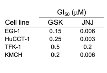
GI50 values for the PRMT5 inhibitors GSK3326595 and JNJ64619178 (Onametostat) in the indicated CCA cell lines.
Pemrametostat succinate purchased from MedChemExpress. Usage Cited in: Gut. 2024 Sep 11:gutjnl-2024-332998. [Abstract]
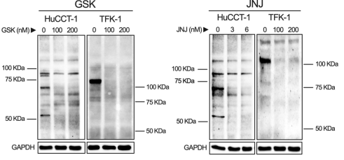
Immunoblot analysis of PRMT5-dependent symmetric dimethylarginine (SMDA) protein marks in control and GSK3326595 or JNJ64619178 (Onametostat) treated HuCCT-1 and TFK-1 cells at the indicated doses for 5 days.
Pemrametostat succinate purchased from MedChemExpress. Usage Cited in: Gut. 2024 Sep 11:gutjnl-2024-332998. [Abstract]
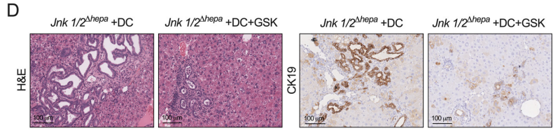
Immunohistochemical analysis of CK19 and H&E stainings showing the extent of CCA lesions in control JnkΔhepa+DEN+CCl4 and GSK3326595-treated mice. Representative images are shown.
-
Nat Commun
2025 Dec 3;16(1):10129. PMID: 41339334 -
Nat Commun
2021 Jun 8;12(1):3444. PMID: 34103528
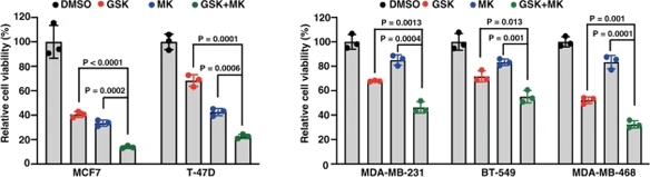
Pemrametostat succinate purchased from MedChemExpress. Usage Cited in: Nat Commun. 2021 Jun 8;12(1):3444. [Abstract]
Breast cancer cells are treated with DMSO, 0.5 μM GSK3326595 (GSK) for 4 days before examining cell viability.
-
J Adv Res
The aryl hydrocarbon receptor inhibits antigen presentation to promote progression of pancreatic ductal adenocarcinoma. [Abstract]2025 Nov 5:S2090-1232(25)00874-4. PMID: 41203070 -
J Exp Clin Cancer Res
2022 Oct 5;41(1):293. PMID: 36199122 -
Adv Sci (Weinh)
2025 Sep;12(33):e08280. PMID: 40642900
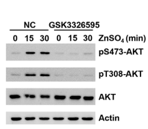
Pemrametostat succinate purchased from MedChemExpress. Usage Cited in: Adv Sci (Weinh). 2025 Sep;12(33):e08280. [Abstract]
HCT116 cells were treated with GSK3326595 (10 μM) for 6 h,HCT116 cells were starved in amino acid-free medium for 1 h, and then supplemented with ZnSO4 for the indicated time. The level of pS473-AKT,binding
-
MedComm (2020)
SCR-7952, a highly selective MAT2A inhibitor, demonstrates synergistic antitumor activities in combination with the S-adenosylmethionine-competitive or the methylthioadenosine-cooperative protein arginine methyltransferase 5 inhibitors in methylthioadenosine phosphorylase-deleted tumors. [Abstract]2024 Sep 20;5(10):e705. PMID: 39309689 -
Cell Death Dis
Reversible arginine methylation regulates mitochondrial IDH2 activity: coordinated control by CARM1 and KDM3A/4A. [Abstract]2026 Feb 2;17(1):195. PMID: 41629283 -
Cancer Lett
DysUFMylation reprograms immunosuppressive neutrophils to potentiate anti-PD-1 therapy in hepatocellular carcinoma. [Abstract]2026 May 1:645:218395. PMID: 41780840 -
Clin Cancer Res
MTA-Cooperative PRMT5 Inhibitors are Efficacious in MTAP-Deleted Malignant Peripheral Nerve Sheath Tumor Models. [Abstract]2025 Jun 24. PMID: 40553452 -
Oncogene
2021 Apr;40(15):2711-2724. PMID: 33712705 -
EMBO Mol Med
2023 Aug 7;15(8):e17248. PMID: 37458145 -
Blood Adv
Loss of METTL3 attenuates blastic plasmacytoid dendritic cell neoplasm response to PRMT5 inhibition via IFN signaling. [Abstract]2022 Apr 28;bloodadvances.2021006306. PMID: 35482445 -
Cell Mol Life Sci
Opposing USP19 splice variants in TGF-β signaling and TGF-β-induced epithelial-mesenchymal transition of breast cancer cells. [Abstract]2023 Jan 17;80(2):43. PMID: 36646950 -
Biomed J
2026 Jun 10:101002. PMID: 42270077 -
EMBO Rep
2025 Oct 6. PMID: 41053315 -
Invest Ophthalmol Vis Sci
PRMT5 Regulates Senescence in Retinal Ganglion Cells by Targeting the Wnt/β-Catenin Signaling Cascade. [Abstract]2025 Jun 2;66(6):8. PMID: 40459496 -
Transl Oncol
TNG908 is a brain-penetrant, MTA-cooperative PRMT5 inhibitor developed for the treatment of MTAP-deleted cancers. [Abstract]2025 Feb:52:102264. PMID: 39756156 -
Sci Rep
2023 Jul 3;13(1):10752. PMID: 37400460 -
Cancers (Basel)
2023 Apr 27;15(9):2501. PMID: 37173967 -
J Cell Mol Med
2024 Mar;28(5):e17856. PMID: 37461162 -
J Biol Chem
Protein arginine methyltransferase 5 is essential for oncogene product EWSR1-ATF1-mediated gene transcription in clear cell sarcoma. [Abstract]2022 Oct;298(10):102434. PMID: 36041632 -
-
Breast Cancer (Dove Med Press)
Therapeutic Advantage of Targeting PRMT5 in Combination with Chemotherapies or EGFR/HER2 Inhibitors in Triple-Negative Breast Cancers. [Abstract]2023 Nov 6:15:785-799. PMID: 37954171 -
Mol Carcinog
PRMT1 inhibition promotes ferroptosis sensitivity via ACSL1 upregulation in acute myeloid leukemia. [Abstract]2023 Aug;62(8):1119-1135. PMID: 37144835 -
Glycobiology
Compromised CDK12 activity causes dependency on the high activity of O-GlcNAc transferase. [Abstract]2024 Dec 10;34(12):cwae081. PMID: 39361894 -
-
-
bioRxiv
Radiation synergizes with BET inhibition to stimulate durable, systemic anti-tumor immunity in murine cancer models. [Abstract]2026 Feb 18:2026.02.16.706212. PMID: 41757027 -
-
-
bioRxiv
2024 Aug 16:2024.08.15.608201. PMID: 39211199 -
-
-

Protocols
-
Research Protocol for Infectious Diseases
Infectious-disease experiments test how pathogens interact with host barriers, innate immune receptors, inflammatory signaling, pathogen replication, and tissue injury; pattern-recognition receptors such as TLRs, RIG-I-like receptors, NOD-like receptors, and inflammasomes detect microbial molecules and activate NF-κB, interferon, and cytokine responses. The central hypothesis is that infection severity reflects the balance between pathogen burden and host response: protective inflammation restricts pathogen growth, whereas excessive or mislocalized inflammation contributes to tissue damage and disease phenotype. Unresolved questions include which host pathways are protective versus pathogenic, why some infection models fail to translate to human disease, and which combined readouts best predict clinically relevant infection outcomes.
-
BrdU Incorporation Assay
Bromodeoxyuridine (BrdU) incorporation assay is based on the principle that BrdU, a thymidine analog, is incorporated into newly synthesized DNA during the S phase of the cell cycle, thereby serving as a marker of DNA replication and cellular proliferation. Incorporated BrdU can be detected using anti-BrdU antibodies following DNA denaturation, enabling visualization or quantification of proliferating cells through immunochemical detection methods such as immunofluorescence or immunohistochemistry.
-
Somatic Cell Culture
A method of simulating the in vivo environment in vitro to maintain the cell growth, differentation and main functions.
-
CFSE Dye Dilution Proliferation Assay
The CFSE (carboxyfluorescein diacetate succinimidyl ester) dye dilution proliferation assay is based on the covalent labeling of intracellular proteins by a cell-permeant fluorescent dye that becomes fluorescent upon intracellular ester cleavage and then is stably retained within cells. As labeled cells divide, the dye is partitioned equally between daughter cells, resulting in a stepwise halving of fluorescence intensity that can be quantified by flow cytometry to determine the number of cell divisions undergone by each cell population. This fluorescence dilution approach enables quantitative tracking of lymphocyte proliferation at the single-cell level over multiple rounds of division. CFSE-based proliferation analysis has been widely applied to measure antigen-driven lymphocyte expansion in vitro, where discrete fluorescence peaks correspond to successive cell divisions and allow reconstruction of proliferative history within heterogeneous populations.
-
Dye-dilution cell tracking and proliferation staining
Dye-dilution cell tracking assays quantify cell proliferation by covalently labeling intracellular proteins with a stable fluorescent dye that is equally partitioned between daughter cells during mitosis, resulting in stepwise halving of fluorescence intensity with each cell division as measured by flow cytometry histograms. Carboxyfluorescein diacetate succinimidyl ester (CFSE) is a prototypical dye that diffuses into cells, is enzymatically converted into a fluorescent compound, and then covalently binds intracellular amine groups, producing long-lived fluorescence suitable for tracking multiple rounds of division in vitro and in vivo. Successive generations of dividing cells form discrete peaks of decreasing fluorescence intensity, enabling estimation of proliferation history, precursor frequency, and division index within heterogeneous populations. Alternative dyes such as CellTrace Violet (CTV) and far-red membrane dyes (e. g. , PKH26) follow the same dilution principle but differ
-
CCK-8/WST-8 Cell Proliferation Assay
The CCK-8/WST-8 assay is based on the reduction of the water-soluble tetrazolium salt WST-8 to a water-soluble formazan product by cellular dehydrogenases in metabolically active cells, where the generated formazan amount is proportional to the number of living cells and is quantified by measuring absorbance in the visible range, providing a colorimetric readout for cell viability and proliferation assessment. This class of tetrazolium-based assays improves upon earlier MTT-based systems by producing a water-soluble formazan, eliminating the need for organic solubilization steps and enabling direct spectrophotometric measurement in culture medium.
-
Protocol for Cell Cycle
Cell-cycle analysis by flow cytometry measures DNA content in single cells to estimate the fraction of cells in G0/G1, S, and G2/M phases. Propidium iodide intercalates into DNA, and after RNA removal with RNase, fluorescence intensity reflects cellular DNA content: 2N cells are assigned to G0/G1, cells between 2N and 4N to S phase, and 4N cells to G2/M. DNA-content analysis alone cannot reliably separate G0 from G1 or G2 from M. Ki-67 can distinguish quiescent G0 cells from cycling cells, EdU or BrdU incorporation marks active DNA synthesis in S phase, and phospho-histone H3 staining identifies mitotic cells within the 4N population.
-
Research Protocol for Cardiovascular Diseases
Cardiovascular disease can be modeled as maladaptive cardiac remodeling, where ischemic injury or pressure overload activates inflammatory signaling, fibroblast activation, extracellular-matrix deposition, cardiomyocyte hypertrophy, vascular remodeling, and progressive ventricular dysfunction. The TGF-β/SMAD axis is a central profibrotic pathway after myocardial injury and pressure overload, while innate immune and cytokine pathways regulate leukocyte recruitment, scar formation, and adverse remodeling. Key unresolved questions include which inflammatory signals are reparative versus harmful, when fibrosis is protective versus maladaptive, and whether pathway inhibition improves function without weakening necessary infarct healing or compensatory remodeling.
-
Cell Counting-Based Growth Curve Assay
Cell counting-based growth curve assays quantify cell proliferation by directly measuring changes in viable cell number over time using manual or automated counting methods such as hemocytometer-based counting or instrument-assisted cell enumeration, enabling construction of growth curves that reflect population expansion dynamics in response to culture conditions. A widely used approach is trypan blue exclusion with hemocytometer counting, where membrane-compromised (non-viable) cells take up the dye, allowing discrimination between viable and non-viable cells while simultaneously enabling total cell number quantification. Repeated sampling across time points allows estimation of proliferation rate, growth phases, and comparative growth kinetics between experimental conditions.
-
Research Protocol for Inflammation-related Diseases
The NLRP3 inflammasome is a cytosolic innate immune signaling platform that integrates priming signals and danger-signal activation to promote caspase-1 activation, maturation of IL-1β and IL-18, and gasdermin D-mediated pyroptotic cell death. The core experimental logic is to determine whether inflammatory disease phenotypes are driven by increased NLRP3 expression, ASC-containing inflammasome assembly, caspase-1 cleavage, GSDMD cleavage, and extracellular release of IL-1β/IL-18 rather than by nonspecific cell injury alone. The pathway is strongly linked to inflammation-related disease phenotypes because monosodium urate crystals activate NALP3/NLRP3 inflammasome signaling in gout-like crystal inflammation, cholesterol crystals activate NLRP3 inflammasomes in atherogenesis models, and DSS-induced intestinal inflammation has been reported to involve NLRP3 inflammasome activity. However, experimental colitis studies also show context-dependent protective effects of NLRP3 inflammasome co
-
Colony Formation (Clonogenic) Assay
The clonogenic (colony formation) assay measures the ability of a single cell to retain reproductive viability and form a macroscopic colony, typically defined as a cluster derived from one progenitor cell after a defined growth period. This assay is widely used to evaluate cell survival after exposure to ionizing radiation or cytotoxic treatments and is considered a standard method in radiation biology for generating dose-response relationships of reproductive cell death. Colony formation reflects long-term proliferative capacity rather than short-term metabolic activity, and survival is quantified by comparing treated versus untreated conditions based on colony number and derived survival fractions.
-
Cell Viability Determination by MTT Colorimetric Assay
The following protocol uses the MTT colorimetric assay as a classic literature-established method for assessing cell viability/metabolic activity in cultured mammalian cells. MTT[3-(4,5-dimethylthiazol-2-yl)-2,5-diphenyltetrazolium bromide] is reduced by metabolically active cells to a colored formazan product; the amount of formazan is quantified spectrophotometrically and provides an indirect measure of metabolically active viable cells. Importantly, MTT reduction reflects cellular oxidoreductase/metabolic activity rather than an absolute direct count of living cells, so changes in cellular metabolism can alter the signal independently of cell number.
-
Phagocytosis Functional Assay
A phagocytosis functional assay measures the ability of phagocytic cells, such as neutrophils, macrophages, monocytes, or microglia/macrophages, to bind and internalize particulate targets including bacteria, yeast particles, beads, or myelin particles. Fluorescent flow-cytometry assays detect target uptake as fluorescence associated with gated phagocytes, while pH-sensitive dyes such as pHrodo increase signal in acidic phagosomal compartments and therefore preferentially report internalized particles rather than particles remaining outside the cell. Microscopy or high-content imaging can be used to confirm intracellular localization and, in some protocols, to follow uptake kinetics.
-
EdU Incorporation Assay (Click Chemistry-Based DNA Synthesis Measurement)
The EdU incorporation assay measures DNA synthesis by adding the thymidine analog 5-ethynyl-2′-deoxyuridine to cells or tissues, where it is incorporated into newly synthesized DNA during S phase. Incorporated EdU is detected by copper-catalyzed azide-alkyne cycloaddition, in which a fluorescent azide covalently reacts with the ethynyl group on EdU, allowing S-phase cells to be detected by fluorescence microscopy, flow cytometry, or high-content imaging. EdU detection does not require DNA denaturation or anti-BrdU antibody access, which preserves sample structure and improves compatibility with immunostaining and multiparameter cytometry compared with BrdU-based detection. EdU can be cytotoxic in a cell-type- and exposure-dependent manner, so pulse duration, concentration, and continuous-labeling designs should be validated for each cell type.
-
Ki-67 Immunostaining Proliferation Assay
Ki-67 immunostaining measures the growth fraction of a cell population by detecting Ki-67, a nuclear antigen present in proliferating cells and absent in quiescent G0 cells. The readout is the percentage of Ki-67-positive nuclei among total counted cells, commonly called the Ki-67 labeling index or proliferation index.
-
PCNA Immunodetection Proliferation Assay
PCNA immunodetection measures proliferative activity by detecting proliferating cell nuclear antigen, a nuclear protein associated with DNA polymerase δ function and DNA replication. The assay readout is the proportion of PCNA-positive nuclei among total counted cells, but PCNA labeling is not identical to BrdU labeling because PCNA can mark late G1/early S-associated replication competence and may persist beyond active DNA synthesis depending on fixation and extraction conditions.
-
Protocol for Cell Counting and Cell Density Analysis
Cell counting and cell-density analysis estimate the number of cells in a known volume or field area. Manual hemocytometer counting uses a chamber of defined geometry to convert counted cells into cells/mL, while automated counters and image-analysis workflows detect cell objects from optical, brightfield, fluorescence, impedance, or digital-image features. Trypan blue viability counting is based on dye exclusion: viable cells with intact membranes exclude dye, while non-viable cells with compromised membranes stain blue. The readout is total cell density, viable-cell density, dead-cell density, and percent viability. Cell density can also be estimated from microscopy images by counting objects per image area, from flow cytometry using calibrated volume or reference particles, or from in situ microscopy in bioreactors after calibration against reference methods such as hemocytometer or flow cytometry.
-
Primary monocyte-to-macrophage differentiation
Primary human monocytes can be differentiated ex vivo into monocyte-derived macrophages by culturing purified blood monocytes for approximately 5-7 days in macrophage-supporting cytokine conditions; M-CSF commonly yields CD14^high/CD163^high macrophages, while GM-CSF yields a phenotypically distinct macrophage population, so the cytokine condition should be chosen according to the downstream model. The readout of successful differentiation is a combined change in morphology, adherence, surface phenotype, and function: differentiated macrophages become adherent, enlarge, acquire macrophage-associated markers such as CD14, CD68, CD163, CD206, or HLA-DR depending on culture condition, and show increased phagocytic capacity compared with starting monocytes.
-
MTT Cell Proliferation Assay
The MTT assay is a colorimetric endpoint assay for estimating viable cell number, cell growth, cytotoxicity, or cell activation in cultured mammalian cells. Living cells reduce the yellow tetrazolium salt MTT into purple/blue formazan, while dead cells do not generate the same signal; the resulting color can be quantified with a multiwell spectrophotometer. MTT reduction is commonly interpreted as a readout of metabolic activity that often correlates with viable cell number, but it should not be treated as a direct cell-counting method unless the assay is optimized for the cell type and experimental condition. Studies show that MTT reduction can involve mitochondrial and non-mitochondrial reducing systems, and formazan may accumulate in intracellular lipid droplets rather than simply marking mitochondria.


Purity & Documentation
References
[1]. Che Y, et al. Exploiting PRMT5 as a target for combination therapy in mantle cell lymphoma characterized by frequent ATM and TP53 mutations [J]. Blood cancer journal, 2023, 13(1): 27. [Content Brief]
[5]. Luo Y, et al. Myelocytomatosis-protein arginine N-methyltransferase 5 Axis defines the tumorigenesis and immune response in hepatocellular carcinoma [J]. Hepatology, 2021, 74(4): 1932-1951. [Content Brief]

Calculators
Concentration (start) × Volume (start) = Concentration (final) × Volume (final)


Pemrametostat succinate Related Classifications
HY-101563A Related Classifications
 Powered by Bioz