



HNHA

Based on 1 Customer Validation
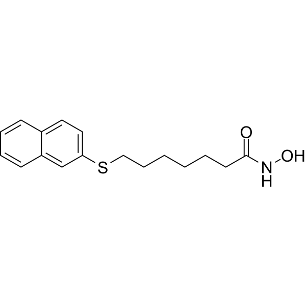
HNHA is a potent HDAC inhibitor with an IC50 of 100 nM. HNHA arrests the cell cycle at the G1/S phase via p21 induction. HNHA inhibits tumor growth and tumor neovascularization. HNHA may be a potent anti-cancer agent against breast cancer.
For research use only. We do not sell to patients.
- Purity : 99.71%
- CAS No.: 926908-04-5
- Formula: C17H21NO2S
- Molecular Weight:303.42
-
Storage:Powder -20°C, 3 years , 4°C, 2 years ; In solvent -80°C, 6 months , -20°C, 1 month

To place orders, for customer services and technical support, please contact: MedChemExpress USA
Tel: 609-228-6898 E-mail: [email protected] [email protected]


-
 Biological Activity
-
Chemical Information
-
Solvent & Solubility
- Protocols
- Purity & Documentation
- References
-
 Help & FAQs


-
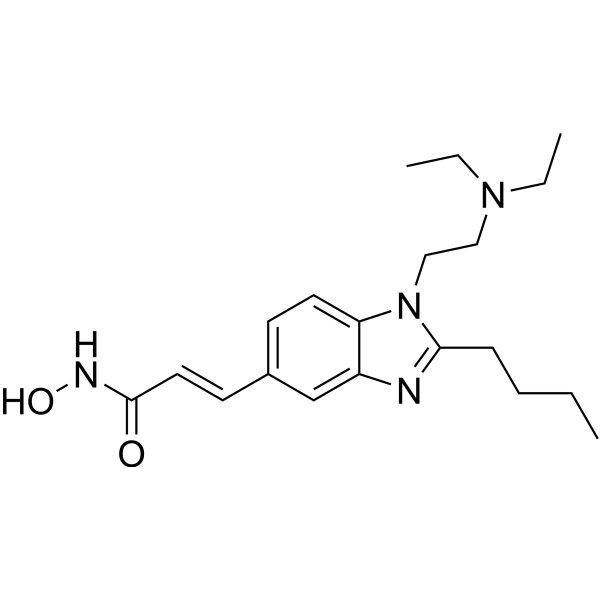
HY-13322
PracinostatHDAC/MBLAC2 Inhibitor -
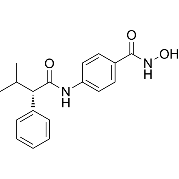
HY-13265
AR-42Pan HDAC Inhibitor -
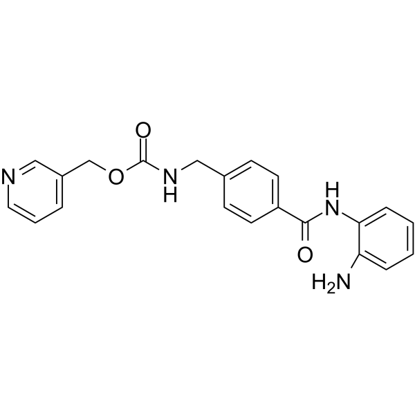
HY-12163
EntinostatHDAC Class I Inhibitor -
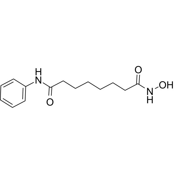
HY-10221
VorinostatHDAC Inhibitor -
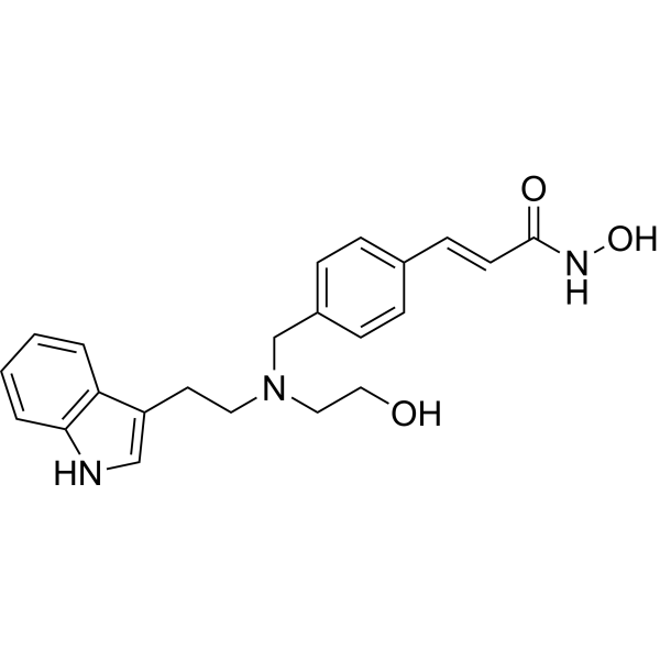
HY-13606
DacinostatAutophagy Inducer -
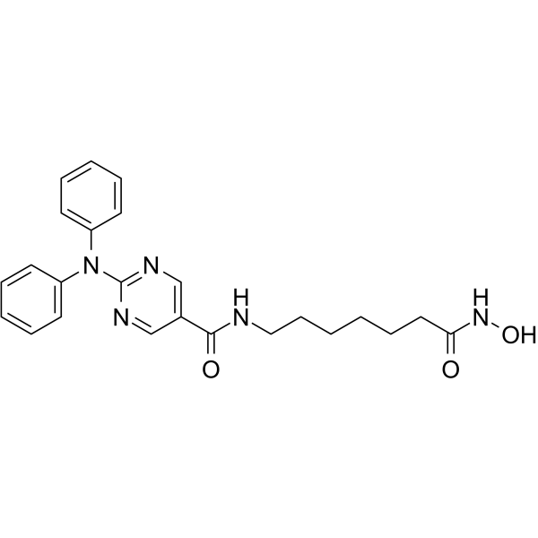
HY-16026
RicolinostatHDAC6 Inhibitor -
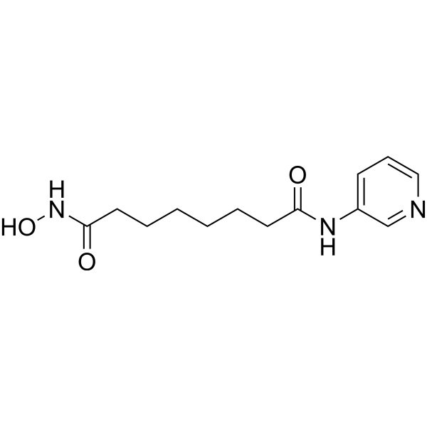
HY-13216
PyroxamideHDAC1 Inhibitor -
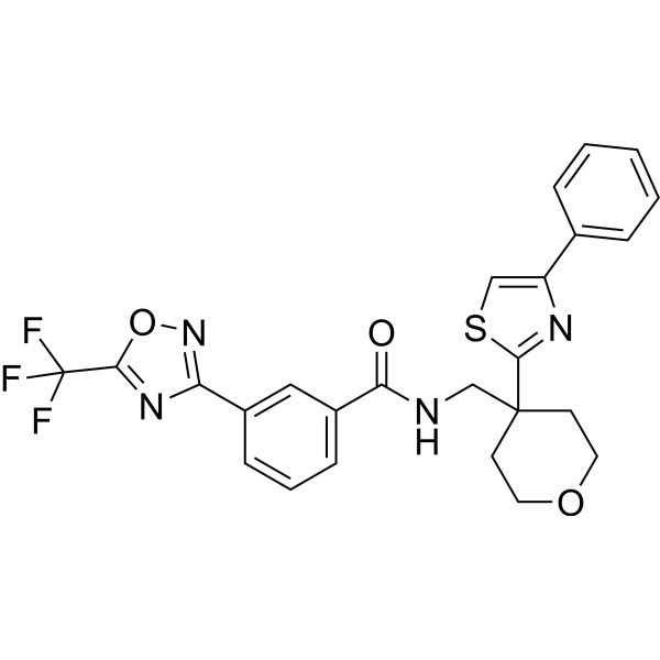
HY-18360
TMP269HDACIIa Inhibitor -
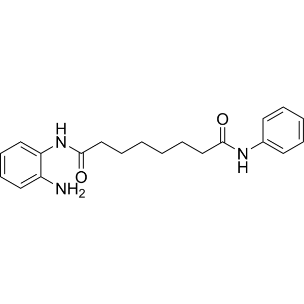
HY-19350
BML-210HDAC Inhibitor -
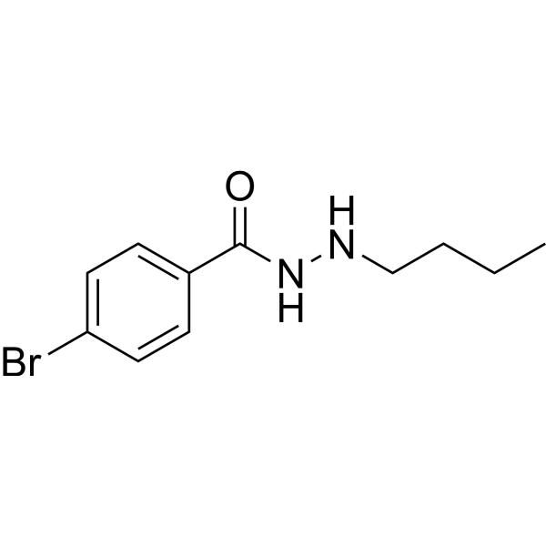
HY-18976
UF010HDAC Inhibitor -

HY-10221R
Vorinostat (Standard)HDAC Inhibitor -
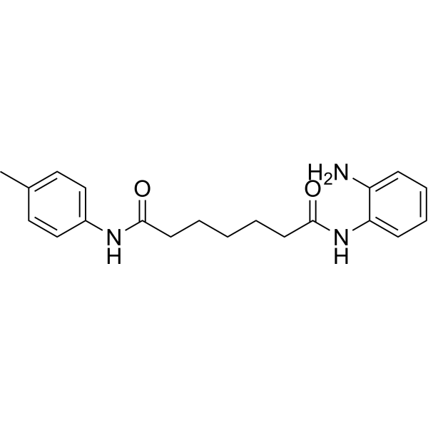
HY-19348
Pimelic Diphenylamide 106HDAC Inhibitor


Biological Activity
Description
IC50 & Target
|
MMP-2 |
MMP-9 |
Cellular Effect
|
Cell Line

|
Type | Value | Description | References |
|---|---|---|---|---|
| HT-1080 | IC50 |
3 μM
Compound: HNHA
|
Cytotoxicity against human HT1080 cells
Cytotoxicity against human HT1080 cells
|
[PMID: 23953690] |
In Vitro
HNHA (0-100 μM, 96 h) shows strong inhibition at lower concentrations on cancer cell lines, especially on breast cancer cells, mouse FM3A and human MCF-7[1].
HNHA (15 μM, 24 h) arrests cancer cells at the G1/S phase of the cell cycle, activates p21and rescues strongly protein acetylation[1].
HNHA (15 μM, 12 h) inhibits angiogenic proteins in breast cancer cells, effectively inactivates MMP-2, MMP-9, VEGF and HIF-1α[1].
MedChemExpress (MCE) has not independently confirmed the accuracy of these methods. They are for reference only. Further protocols information, click here.
-
Cell Line:FM3A, C1300, LA-N-1, LA-N-2, LA-N-5, NB16, NB19, NB69, SK-N-SH, MCF-7 and HT-29[1]
-
Concentration:0-100 μM
-
Incubation Time:96 h
-
Result:Showed strong inhibition at lower concentrations on all cancer cell lines (FM3A, C1300, LA-N-1, LA-N-2, LA-N-5, NB16, NB19, NB69, SK-N-SH, MCF-7 and HT-29), with IC50 values of 15.70, 55.63, 22.78, 23.18, 26.70, 19.64, 21.26, 22.31, 65.09, 14.33, and 16.98 μM, respectively.
-
Cell Line:FM3A and MCF-7[1]
-
Concentration:0, 0.1, 1, 5, 10, 15, 20, 25,30 μM
-
Incubation Time:48 h
-
Result:Showed dose-dependent inhibition of viability in mouse and human breast cancer cells.
-
Cell Line:FM3A and MCF-7 cells[1]
-
Concentration:15 μM
-
Incubation Time:24 h
-
Result:Arrested FM3A and MCF-7 cells in the G1/S phase.
-
Cell Line:FM3A and MCF-7 cells[1]
-
Concentration:0, 0.1, 1, 10, and 20 μM (24 h)
-
Incubation Time:1, 6, 24, 48, and 72 h (15 μM)
-
Result:Activated a cell proliferation arrestor p21, increased histone and non-histone protein acetylation and inhibited FM3A and MCF-7 proliferation in vitro, and was very effective in increasing the acetylation level of histone H3 protein in FM3A and MCF-7. The most effective dose point for acetylation of histone H3 was 10-20 μM. Histone H3 acetylation peaked after 1 h of exposure to the drugs and remained stable for 1-6 h.
-
Cell Line:FM3A and MCF-7 cells[1]
-
Concentration:15 μM
-
Incubation Time:12 h
-
Result:Showed a strong induction of TIMP-1 and TIMP-2, and effectively inactivated MMP-2, MMP-9, VEGF and HIF-1α.
In Vivo
MedChemExpress (MCE) has not independently confirmed the accuracy of these methods. They are for reference only.
-
Animal Model:C3H/HeJ-FasL mice (FM3A breast cancer cell tumor xenograft, 6 weeks, n = 25/group)[1]
-
Dosage:20 μM/mouse
-
Administration:IP, once every 2 days for a total of six injections
-
Result:Reduced tumor burden and extended the survival rate. Effectively inhibited cancer development and angiogenesis in vivo. Increased TIMP-1, TIMP-2 and p21, decreased MMP-2, MMP-9, HIF-1α and VEGF protein expression, and reduced the distribution of CD34, HIF-1α and VEGF.


Chemical Information
-
CAS No. 926908-04-5
-
Appearance Solid
-
Molecular Weight 303.42
-
Formula C17H21NO2S
-
Color White to off-white
-
SMILES
O=C(NO)CCCCCCSC1=CC=C2C=CC=CC2=C1
-
Shipping
Room temperature in continental US; may vary elsewhere.
-
Storage
Powder -20°C 3 years 4°C 2 years In solvent -80°C 6 months -20°C 1 month


Solvent & Solubility
In Vitro:
DMSO : 100 mg/mL (329.58 mM; Need ultrasonic; Hygroscopic DMSO has a significant impact on the solubility of product, please use newly opened DMSO)
Please refer to the solubility information to select the appropriate solvent. Once prepared, please aliquot and store the solution to prevent product inactivation from repeated freeze-thaw cycles.
Storage method and period of stock solution: -80°C, 6 months; -20°C, 1 month. When stored at -80°C, please use it within 6 months. When stored at -20°C, please use it within 1 month.
Please refer to the solubility information to select the appropriate solvent. Once prepared, please aliquot and store the solution to prevent product inactivation from repeated freeze-thaw cycles.
Storage method and period of stock solution: -80°C, 6 months; -20°C, 1 month. When stored at -80°C, please use it within 6 months. When stored at -20°C, please use it within 1 month.
Concentration (start) × Volume (start) = Concentration (final) × Volume (final)

Protocols
-
EdU Incorporation Assay (Click Chemistry-Based DNA Synthesis Measurement)
The EdU incorporation assay measures DNA synthesis by adding the thymidine analog 5-ethynyl-2′-deoxyuridine to cells or tissues, where it is incorporated into newly synthesized DNA during S phase. Incorporated EdU is detected by copper-catalyzed azide-alkyne cycloaddition, in which a fluorescent azide covalently reacts with the ethynyl group on EdU, allowing S-phase cells to be detected by fluorescence microscopy, flow cytometry, or high-content imaging. EdU detection does not require DNA denaturation or anti-BrdU antibody access, which preserves sample structure and improves compatibility with immunostaining and multiparameter cytometry compared with BrdU-based detection. EdU can be cytotoxic in a cell-type- and exposure-dependent manner, so pulse duration, concentration, and continuous-labeling designs should be validated for each cell type.
-
Cell Cytotoxicity Assay
Cytotoxicity assays are usually based on the assessment of cell membrane damage, which can also be indirectly detected by measuring cell viability. Detection methods include MTT assay, CKK-8 assay, LDH assay and ATP assay, etc.
-
Flow cytometric DNA-content cell-cycle staining
Flow cytometric DNA-content cell-cycle staining measures the fluorescence intensity of DNA-bound fluorochromes in single cells or nuclei to estimate DNA content distributions, allowing assignment of populations to G0/G1, S, and G2/M phases by DNA histogram deconvolution. Propidium iodide (PI) intercalates into DNA, and PI fluorescence is proportional to cellular DNA content when staining is performed under conditions that make DNA accessible and minimize non-DNA signal. Cells with G2/M DNA content are expected to show approximately twice the fluorescence intensity of G0/G1 cells, while S-phase cells occupy intermediate fluorescence values. PI-based DNA-content analysis can also detect cells with fractional DNA content, often reported as sub-G1, when DNA fragmentation and extraction during staining reduce retained DNA signal in apoptotic cells. DAPI is an alternative DNA fluorochrome for univariate DNA-content analysis, while bivariate approaches combining DNA content with proliferation
-
BrdU Incorporation Assay
Bromodeoxyuridine (BrdU) incorporation assay is based on the principle that BrdU, a thymidine analog, is incorporated into newly synthesized DNA during the S phase of the cell cycle, thereby serving as a marker of DNA replication and cellular proliferation. Incorporated BrdU can be detected using anti-BrdU antibodies following DNA denaturation, enabling visualization or quantification of proliferating cells through immunochemical detection methods such as immunofluorescence or immunohistochemistry.
-
Patient-Derived Xenograft (PDX)
Patient-derived xenograft (PDX) models are generated by engrafting primary human tumor tissue directly into immunodeficient mice, allowing in vivo propagation of patient tumor biology without initial in vitro adaptation. These models are used to preserve key histopathological and molecular characteristics of the original tumor and enable assessment of tumor growth dynamics and therapeutic response in a living organism. The biological readout is tumor engraftment and subsequent growth in the murine host, which reflects the ability of human tumor cells to survive, vascularize, and expand in an immunocompromised microenvironment.
-
Patient-Derived Orthotopic Xenograft (PDOX)
Patient-derived orthotopic xenograft (PDOX) modeling implants fresh patient tumor tissue or patient-derived tumor cells into the anatomically corresponding organ or tissue site of immunodeficient mice, usually by surgical orthotopic implantation, to preserve patient tumor histology, local microenvironmental context, invasion, metastatic behavior, and treatment-response features better than subcutaneous implantation. PDOX readouts include tumor engraftment, orthotopic tumor growth, local invasion, metastasis, recurrence after resection, histologic similarity to the donor tumor, biomarker retention, molecular concordance, survival, and response or resistance to therapy. PDOX models are used for preclinical drug testing and individualized therapy evaluation, but engraftment success varies by tumor type and specimen quality.
-
Protocol for Cell Cycle
Cell-cycle analysis by flow cytometry measures DNA content in single cells to estimate the fraction of cells in G0/G1, S, and G2/M phases. Propidium iodide intercalates into DNA, and after RNA removal with RNase, fluorescence intensity reflects cellular DNA content: 2N cells are assigned to G0/G1, cells between 2N and 4N to S phase, and 4N cells to G2/M. DNA-content analysis alone cannot reliably separate G0 from G1 or G2 from M. Ki-67 can distinguish quiescent G0 cells from cycling cells, EdU or BrdU incorporation marks active DNA synthesis in S phase, and phospho-histone H3 staining identifies mitotic cells within the 4N population.
-
Subcutaneous Cell-Line-Derived Xenograft
Subcutaneous cell-line-derived xenograft (CDX) models are established by implanting cultured human cancer cell lines into immunodeficient mice, where the injected cells form localized tumors that can be monitored in vivo as a measure of tumorigenic potential, growth kinetics, and treatment response. These models are widely used in oncology research because they allow reproducible tumor formation and enable comparative assessment of tumor growth between different cell lines or genetic manipulations in a controlled in vivo microenvironment. Subcutaneous implantation of cancer cells in immunodeficient mice is a standard approach for evaluating tumor growth behavior and therapeutic response across multiple cancer types, including prostate, esophageal, pancreatic, and colon cancer models.
-
Breast Cancer Modeling
Breast cancer is a heterogeneous cancer, and it has been distinguished into four subtypes: luminal A, luminal B, HER2-positive and basal-like. Molecular mutations, epigenetic alterations, hormone exposure and immune microenvironment are related to the progression of breast cancer.
-
Orthotopic Cell-Line Xenograft
Orthotopic cell-line xenograft models involve implantation of human cancer cell lines into the anatomically corresponding organ of immunodeficient mice to reproduce tumor growth within a native microenvironment, enabling more clinically relevant tumor behavior compared with subcutaneous models. These models are widely used because orthotopic placement better recapitulates tumor progression, including invasion and metastatic spread, which are often underrepresented in heterotopic implantation systems. Compared with conventional xenografts, orthotopic implantation is described as more technically complex but provides improved simulation of tumor-microenvironment interactions and metastatic behavior, making it particularly valuable for translational oncology research. Surgical orthotopic implantation approaches have been emphasized as enabling faithful reproduction of clinical cancer features, including metastasis and disease progression patterns that align with the tumor’s organ of origi


Purity & Documentation
-
Data Sheet (273 KB)
-
SDS (254 KB)
- English - EN (254 KB)
- Français - FR (254 KB)
- Deutsch - DE (254 KB)
- Norwegian - NO (254 KB)
- Español - ES (254 KB)
- Swedish - SV (254 KB)
- Italian - IT (254 KB)
- Korean - KR (254 KB)
- Portuguese - PT (254 KB)
-
Handling Instructions (2659 KB)
References


Complete Stock Solution Preparation Table
Please refer to the solubility information to select the appropriate solvent. Once prepared, please aliquot and store the solution to prevent product inactivation from repeated freeze-thaw cycles.
Storage method and period of stock solution: -80°C, 6 months; -20°C, 1 month. When stored at -80°C, please use it within 6 months. When stored at -20°C, please use it within 1 month.
| Optional Solvent | Concentration Solvent Mass | 1 mg | 5 mg | 10 mg | 25 mg |
|---|---|---|---|---|---|
| DMSO | 1 mM | 3.2958 mL | 16.4788 mL | 32.9576 mL | 82.3940 mL |
| 5 mM | 0.6592 mL | 3.2958 mL | 6.5915 mL | 16.4788 mL | |
| 10 mM | 0.3296 mL | 1.6479 mL | 3.2958 mL | 8.2394 mL | |
| 15 mM | 0.2197 mL | 1.0986 mL | 2.1972 mL | 5.4929 mL | |
| 20 mM | 0.1648 mL | 0.8239 mL | 1.6479 mL | 4.1197 mL | |
| 25 mM | 0.1318 mL | 0.6592 mL | 1.3183 mL | 3.2958 mL | |
| 30 mM | 0.1099 mL | 0.5493 mL | 1.0986 mL | 2.7465 mL | |
| 40 mM | 0.0824 mL | 0.4120 mL | 0.8239 mL | 2.0599 mL | |
| 50 mM | 0.0659 mL | 0.3296 mL | 0.6592 mL | 1.6479 mL | |
| 60 mM | 0.0549 mL | 0.2746 mL | 0.5493 mL | 1.3732 mL | |
| 80 mM | 0.0412 mL | 0.2060 mL | 0.4120 mL | 1.0299 mL | |
| 100 mM | 0.0330 mL | 0.1648 mL | 0.3296 mL | 0.8239 mL |


HNHA Related Classifications
HY-118672 Related Classifications
 Powered by Bioz