



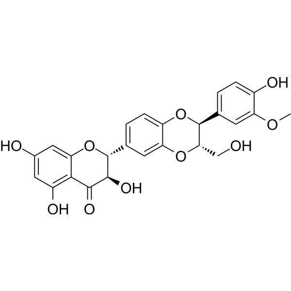
Isosilybin B
Based on 2 publication(s) in Google Scholar
Isosilybin B is a flavonolignan. Isosilybin B can be isolated from Silybum marianum. Isosilybin B can regulate cell cycle-related proteins (e.g., reduce cyclins (D3, D1, A, E), Cdk4, Cdk2, Cdc25A), and activate Caspases (Caspase-9 and Caspase-3). Isosilybin B can promote Apoptosis, reduce androgen receptor (AR) and PSA. Isosilybin B has anticancer activity against prostate cancer.
For research use only. We do not sell to patients.
- Purity : 98.94%
- CAS No.: 142796-22-3
- Formula: C25H22O10
- Molecular Weight:482.44
-
Storage:Powder -20°C, 3 years , 4°C, 2 years ; In solvent -80°C, 6 months , -20°C, 1 month

To place orders, for customer services and technical support, please contact: MedChemExpress USA
Tel: 609-228-6898 E-mail: [email protected] [email protected]


-
 Biological Activity
-
Chemical Information
-
Solvent & Solubility
- Protocols
- Purity & Documentation
- References
-
 Help & FAQs



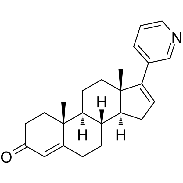
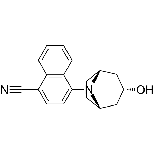
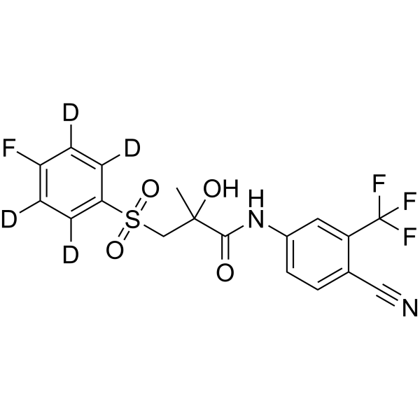
Publications Citing Use of MedChemExpress (MCE) Isosilybin B
MoreAll Caspase Isoforms
More

Biological Activity
Description
|
CDK2 |
CDK4 |
Caspase-9 |
Caspase-3 |
Cellular Effect
|
Cell Line

|
Type | Value | Description | References |
|---|---|---|---|---|
| B16-4A5 | IC50 |
6.1 μM
Compound: 8
|
Inhibition of theophylline-stimulated melanogenesis in mouse B16-4A5 cells after 72 hrs
Inhibition of theophylline-stimulated melanogenesis in mouse B16-4A5 cells after 72 hrs
|
[PMID: 20189399] |
| Huh-7 | IC50 |
27 μM
Compound: 7
|
Cytotoxicity against human Huh7.5.1 cells after 72 hrs
Cytotoxicity against human Huh7.5.1 cells after 72 hrs
|
[PMID: 23673225] |
In Vitro
Isosilybin B (10-90 μM; 24-48 h) causes a decrease in cell number, increased cell death, G1 arrest, and increased apoptosis in human prostate cancer LNCaP and 22Rv1 cells[1].
Isosilybin B (10-90 μM; 24-72 h) decreases the levels of androgen receptor (AR) and prostate-specific antigen (PSA) in human prostate cancer LNCaP, 22Rv1 and LAPC4 cells[2].
MedChemExpress (MCE) has not independently confirmed the accuracy of these methods. They are for reference only.
-
Cell Line:LNCaP cells, 22Rv1 cells
-
Concentration:10 μM, 30 μM, 60 μM, 90 μM
-
Incubation Time:24 h, 48 h
-
Result:Decreased the levels of cyclins (D3, D1, A, E), Cdk4, Cdk2 and Cdc25A (LNCaP, 22Rv1).
Resulted in a dose-dependent increase in PARP cleavage, a slight increase in the levels of cleaved caspase-9 and cleaved caspase-3, and a decrease in survivin levels (LNCaP).


Chemical Information
-
CAS No. 142796-22-3
-
Appearance Solid
-
Molecular Weight 482.44
-
Formula C25H22O10
-
Color White to off-white
-
SMILES
O=C1[C@H](O)[C@@H](C2=CC=C(O[C@@H](C3=CC=C(O)C(OC)=C3)[C@H](CO)O4)C4=C2)OC5=CC(O)=CC(O)=C15
-
Structure Classification
-
Shipping
Room temperature in continental US; may vary elsewhere.
-
Storage
Powder -20°C 3 years 4°C 2 years In solvent -80°C 6 months -20°C 1 month


Publications (2)
-
Journal Impact Factor
-
Most Recent
-
Chem Biol Interact
Isosilybin regulates lipogenesis and fatty acid oxidation via the AMPK/SREBP-1c/PPARα pathway. [Abstract]2022 Dec 1:368:110250. PMID: 36347319 -
Discov Oncol
Isosilybin B: a potential novel therapeutic agent with hepatoprotective, anticancer and antifibrotic properties. [Abstract]2025 Aug 8;16(1):1502. PMID: 40779034


Solvent & Solubility
In Vitro:
Ethanol : 1 mg/mL (2.07 mM; Need ultrasonic)
Please refer to the solubility information to select the appropriate solvent. Once prepared, please aliquot and store the solution to prevent product inactivation from repeated freeze-thaw cycles.
Storage method and period of stock solution: -80°C, 6 months; -20°C, 1 month. When stored at -80°C, please use it within 6 months. When stored at -20°C, please use it within 1 month.
Please refer to the solubility information to select the appropriate solvent. Once prepared, please aliquot and store the solution to prevent product inactivation from repeated freeze-thaw cycles.
Storage method and period of stock solution: -80°C, 6 months; -20°C, 1 month. When stored at -80°C, please use it within 6 months. When stored at -20°C, please use it within 1 month.
Concentration (start) × Volume (start) = Concentration (final) × Volume (final)


Purity & Documentation
-
Data Sheet (274 KB)
-
SDS (393 KB)
- English - EN (393 KB)
- Français - FR (393 KB)
- Deutsch - DE (393 KB)
- Norwegian - NO (393 KB)
- Español - ES (393 KB)
- Swedish - SV (393 KB)
- Italian - IT (393 KB)
- Korean - KR (393 KB)
- Portuguese - PT (393 KB)
-
Handling Instructions (2659 KB)
References
[1]. Deep G, et al. Isosilybin B and isosilybin A inhibit growth, induce G1 arrest and cause apoptosis in human prostatecancer LNCaP and 22Rv1 cells. Carcinogenesis. 2007 Jul;28(7):1533-42. [Content Brief]
[2]. Deep G, et al. Isosilybin B causes androgen receptor degradation in human prostate carcinoma cells via PI3K-Akt-Mdm2-mediated pathway. Oncogene. 2008 Jun 26;27(28):3986-98. [Content Brief]


Complete Stock Solution Preparation Table
Please refer to the solubility information to select the appropriate solvent. Once prepared, please aliquot and store the solution to prevent product inactivation from repeated freeze-thaw cycles.
Storage method and period of stock solution: -80°C, 6 months; -20°C, 1 month. When stored at -80°C, please use it within 6 months. When stored at -20°C, please use it within 1 month.
| Optional Solvent | Concentration Solvent Mass | 1 mg | 5 mg | 10 mg | 25 mg |
|---|---|---|---|---|---|
| Ethanol | 1 mM | 2.0728 mL | 10.3640 mL | 20.7280 mL | 51.8199 mL |


Isosilybin B Related Classifications
HY-N7045 Related Classifications
 Powered by Bioz