



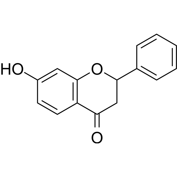
7-Hydroxyflavanone

Based on 1 Customer Validation
7-Hydroxyflavanone is an aromatase (CYP19) activity inhibitor found in plants with an IC50 of 65 μM, and it also possesses 20S proteasome inhibitory activity. 7-Hydroxyflavanone significantly inhibits primary humoral immunity to SRBC in mice and weakly suppresses DTH. 7-Hydroxyflavanone 1 attenuates Doxorubicin (HY-15142A)-induced oxidative stress, IL-6 inflammation, mitochondrial dysfunction, caspase 3/7 activation, apoptosis, and necrosis, and upregulates the PGC-1α/pAMPK/Beclin-1 autophagy-related pathway. 7-Hydroxyflavanone is extensively metabolized by various fungi, undergoing multiple structural modifications. 7-Hydroxyflavanone can be used in research related to Doxorubicin-induced cardiomyopathy and immune system diseases.
商品は「研究用試薬」です。人や動物の医療用・臨床診断用・食品用の製品ではありません。
研究用途以外に使用した場合、当社は一切の責任を負いかねます。
- 純度 : 99.59%
- CAS 番号: 6515-36-2
- 分子式: C15H12O3
- 分子量:240.25
-
保管条件:
4°C, stored under nitrogen
* In solvent : -80°C, 6 months; -20°C, 1 month (stored under nitrogen)

To place orders, for customer services and technical support, please contact: MedChemExpress USA
Tel: 609-228-6898 E-mail: [email protected] [email protected]


-
 生物活性
生物活性
-
化学情報
-
溶剤 & 溶解度
- プロトコル
- 純度とドキュメンテーション
- 参考文献
-
 Help & FAQs


-
HY-W028263
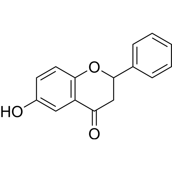
6-HydroxyflavanoneNatural Product -
HY-N7128
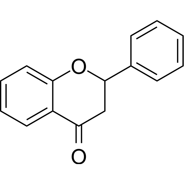
FlavanoneFlavones -
HY-N7108
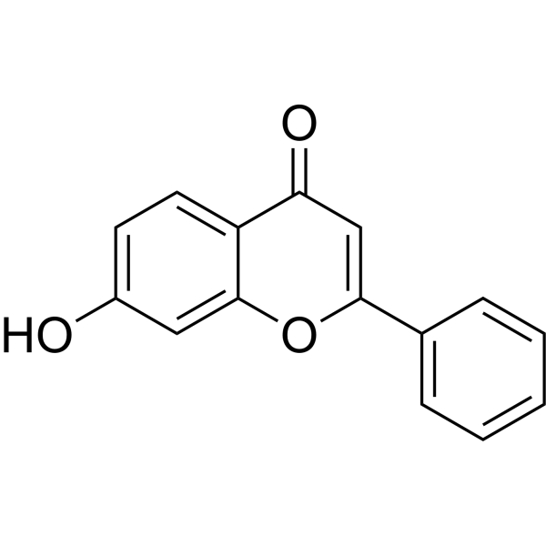
7-HydroxyflavoneAnti-inflammatory Agent -
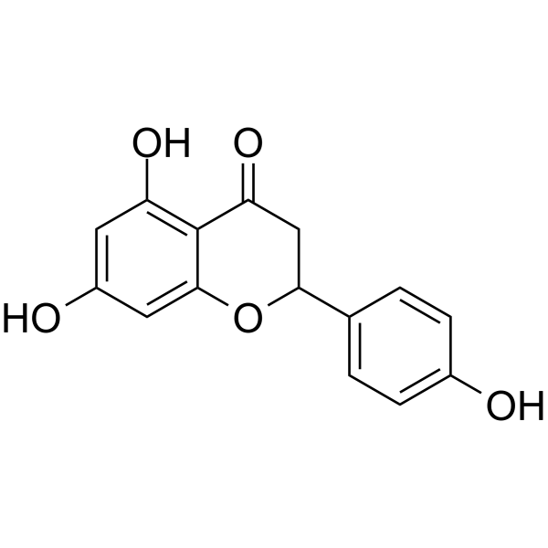
HY-W011641
(±)-NaringeninAnti-inflammatory Agents -
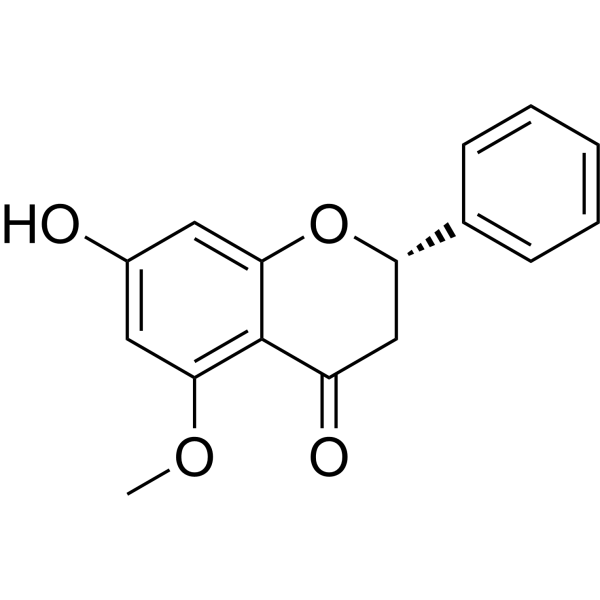
HY-N0625A
AlpinetinPPAR Activator -
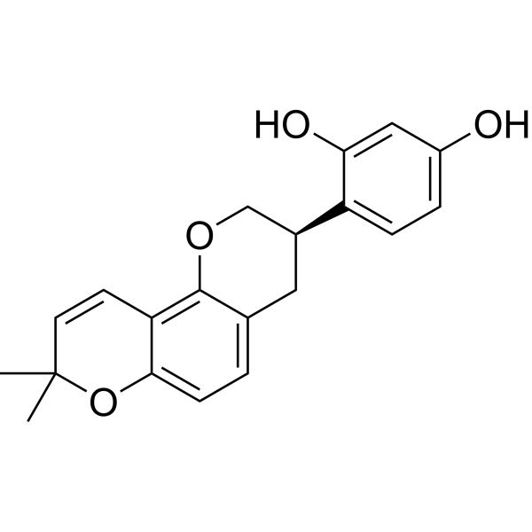
HY-N0393
GlabridinPPARγ Activator -
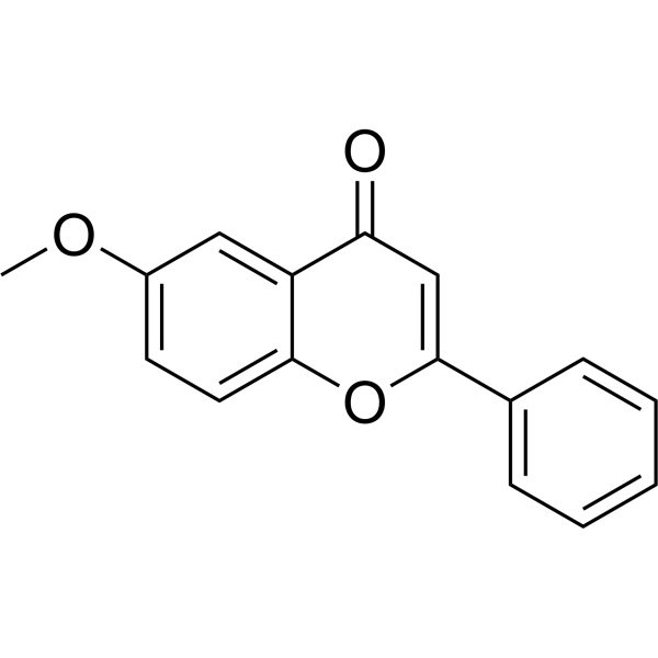
HY-W097625
6-MethoxyflavoneMethoxyflavone -
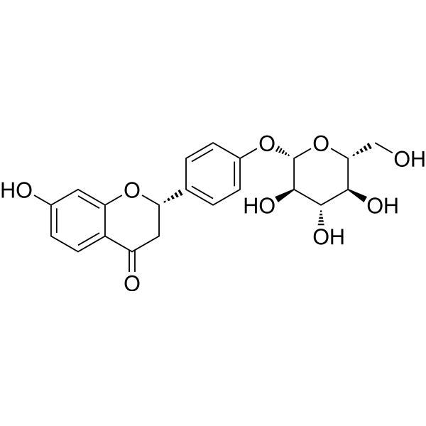
HY-N0376
LiquiritinAKR1C1 Inhibitor -
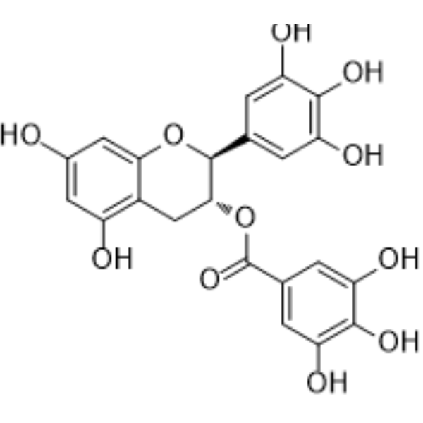
HY-N0522
(-)-Gallocatechin gallateAnticancer Agent -
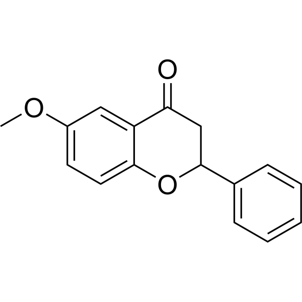
HY-N8852
6-MethoxyflavanoneGABAA Receptor Modulator -
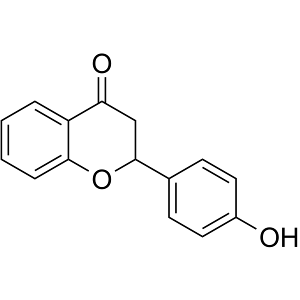
HY-N8495
4'-HydroxyflavanoneSREBP Maturation Inhibitor -
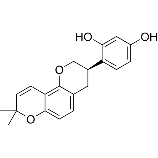
HY-N0393R
Glabridin (Standard)


生物活性
製品説明
IC50 & Target
|
Aromatase 65 μM (IC50) |
IL-6 |
Caspase-7 |
Caspase-3 |
体外実験
7-Hydroxyflavanone (0.5 mg/mL; 14 d) undergoes extensive phase I and phase II microbial metabolism in seven fungi, producing 21 structurally diverse metabolites, with conversion rates varying from 0.08% to 98.2% of 7-Hydroxyflavanone depending on the organism and metabolite[1].
7-hydroxyflavanone (0.01-1000 µM; 6 days) enhances metabolic activity at 1 µM in H9c2 cardiomyoblasts and exhibits cytotoxicity at high concentrations after 6 days[2].
7-hydroxyflavanone (6 days) attenuates Doxorubicin-induced loss of mitochondrial membrane potential and reduction in cellular ATP activity in H9c2 cardiomyoblasts[2].
7-Hydroxyflavanone (6 days) attenuates Doxorubicin (HY-15142A)-induced oxidative stress in H9c2 cardiomyocytes by reducing ROS, lipid peroxidation, and IL-6 while increasing GSH and SOD activity[2].
7-hydroxyflavanone (6 days) improves mitochondrial bioenergetics impaired by Doxorubicin in H9c2 cardiomyoblasts, including maximal respiration rate, ATP-coupled respiration rate, spare respiratory capacity, coupling efficiency, and respiratory control[2].
7-Hydroxyflavanone (6 days) co-treatment in H9c2 cardiomyoblasts upregulates PGC1-α, pAMPK, pMTOR, Beclin-1, PI3K, and pAkt, indicating improved mitochondrial function, autophagic regulation, and cell survival signaling pathways during Doxorubicin exposure[2].
7-Hydroxyflavanone (1 µM; 6 days) reduces Doxorubicin-induced caspase 3/7 activity, apoptosis, and necrosis in H9c2 cardiomyoblasts, thereby increasing cell viability[2].
7-hydroxyflavanone (1 µM; 6 days) does not significantly interfere with the antiproliferative or pro-apoptotic effects of Doxorubicin in MCF-7 breast cancer cells[2].
7-Hydroxyflavanone (100 μM; 24 h) is a relatively weak inhibitor of aromatase activity in H295R human adrenocortical carcinoma cells, with an IC50 of 65 μM, and reduces cell viability by 20%[3].
MedChemExpress (MCE) has not independently confirmed the accuracy of these methods. They are for reference only.
-
Cell Line:rat H9c2 cardiomyoblasts
-
Concentration:0.01, 0.1, 1, 10, 100, 1000 µM
-
Incubation Time:6 days
-
Result:Showed no cytotoxic effects at 0.01, 1, and 10 µM.
Enhanced ATP levels relative to control at 1 µM.
Significantly decreased cardiomyoblast metabolic activity compared with control at 100 µM and 1000 µM.
-
Cell Line:rat H9c2 cardiomyoblasts
-
Concentration:1 µM (co-administered with 0.5 µM doxorubicin)
-
Incubation Time:6 days
-
Result:Significantly reduced doxorubicin-induced caspase 3/7 activity compared with doxorubicin-only treatment.
Reduced early apoptosis, late apoptosis, and necrosis compared with doxorubicin-only treatment.
Increased the number of viable cells compared with doxorubicin-only treatment.
体内実験
7-Hydroxyflavanone (100 mg/kg; p.o.; daily; 7 days post-immunization) has minimal effect on SRBC-induced DTH responses in mice[4].
MedChemExpress (MCE) has not independently confirmed the accuracy of these methods. They are for reference only.
-
Animal Model:Swiss albino mice (randomly bred closed colony; 10-12 weeks old)[4]
-
Dosage:100 mg/kg
-
Administration:p.o.; daily; 14 days
-
Result:Produced 24.13% reduction in primary humoral immune response.
Displayed insignificant suppression in the secondary antibody response.
-
Animal Model:Swiss albino mice (randomly bred closed colony; 10-12 weeks old)[4]
-
Dosage:100 mg/kg
-
Administration:p.o.; daily; 7 days after immunisation
-
Result:Produced a DTH response edema of 2.09 mm, corresponding to a 0.48% reduction in the DTH reaction compared to vehicle control.
- Detection of Apoptosis
- TUNEL staining for apoptotic DNA fragmentation
- Annexin V plus membrane-impermeant dye apoptosis staining
- ROS/oxidative-stress fluorescent staining
- Mitochondrial membrane-potential and mitochondrial mass staining
- Autophagy
- Contact Hypersensitivity Dermatitis
- Apoptosis Solutions
- Research Protocol for Inflammation-related Diseases
- Lysosome and acidic-vesicle live-cell staining
- Macroautophagy Solutions


化学情報
-
CAS 番号 6515-36-2
-
性状 Solid
-
分子量 240.25
-
分子式 C15H12O3
-
Color White to off-white
-
SMILES
O=C1CC(C2=CC=CC=C2)OC3=C1C=CC(O)=C3
-
Structure Classification
-
Initial Source
-
輸送条件
Room temperature in continental US; may vary elsewhere.
-
保管条件
4°C, stored under nitrogen
* In solvent : -80°C, 6 months; -20°C, 1 month (stored under nitrogen)


溶剤 & 溶解度
体外:
DMSO : 125 mg/mL (520.29 mM; Need ultrasonic; Hygroscopic DMSO has a significant impact on the solubility of product, please use newly opened DMSO)
Please refer to the solubility information to select the appropriate solvent. Once prepared, please aliquot and store the solution to prevent product inactivation from repeated freeze-thaw cycles.
Storage method and period of stock solution: -80°C, 6 months; -20°C, 1 month (stored under nitrogen). When stored at -80°C, please use it within 6 months. When stored at -20°C, please use it within 1 month.
Please refer to the solubility information to select the appropriate solvent. Once prepared, please aliquot and store the solution to prevent product inactivation from repeated freeze-thaw cycles.
Storage method and period of stock solution: -80°C, 6 months; -20°C, 1 month (stored under nitrogen). When stored at -80°C, please use it within 6 months. When stored at -20°C, please use it within 1 month.
濃度 (開始) × 体積 (開始) = 濃度 (終了) × 体積 (終了)


純度とドキュメンテーション
-
データシート (289 KB)
-
SDS (393 KB)
- English - EN (393 KB)
- Français - FR (393 KB)
- Deutsch - DE (393 KB)
- Norwegian - NO (393 KB)
- Español - ES (393 KB)
- Swedish - SV (393 KB)
- Italian - IT (393 KB)
- Korean - KR (393 KB)
- Portuguese - PT (393 KB)
-
取扱説明書 (2659 KB)
参考文献


Complete Stock Solution Preparation Table
Please refer to the solubility information to select the appropriate solvent. Once prepared, please aliquot and store the solution to prevent product inactivation from repeated freeze-thaw cycles.
Storage method and period of stock solution: -80°C, 6 months; -20°C, 1 month (stored under nitrogen). When stored at -80°C, please use it within 6 months. When stored at -20°C, please use it within 1 month.
| Optional Solvent | Concentration Solvent Mass | 1 mg | 5 mg | 10 mg | 25 mg |
|---|---|---|---|---|---|
| DMSO | 1 mM | 4.1623 mL | 20.8117 mL | 41.6233 mL | 104.0583 mL |
| 5 mM | 0.8325 mL | 4.1623 mL | 8.3247 mL | 20.8117 mL | |
| 10 mM | 0.4162 mL | 2.0812 mL | 4.1623 mL | 10.4058 mL | |
| 15 mM | 0.2775 mL | 1.3874 mL | 2.7749 mL | 6.9372 mL | |
| 20 mM | 0.2081 mL | 1.0406 mL | 2.0812 mL | 5.2029 mL | |
| 25 mM | 0.1665 mL | 0.8325 mL | 1.6649 mL | 4.1623 mL | |
| 30 mM | 0.1387 mL | 0.6937 mL | 1.3874 mL | 3.4686 mL | |
| 40 mM | 0.1041 mL | 0.5203 mL | 1.0406 mL | 2.6015 mL | |
| 50 mM | 0.0832 mL | 0.4162 mL | 0.8325 mL | 2.0812 mL | |
| 60 mM | 0.0694 mL | 0.3469 mL | 0.6937 mL | 1.7343 mL | |
| 80 mM | 0.0520 mL | 0.2601 mL | 0.5203 mL | 1.3007 mL | |
| 100 mM | 0.0416 mL | 0.2081 mL | 0.4162 mL | 1.0406 mL |


7-Hydroxyflavanone 関連分類
HY-W087008 関連分類
 Powered by Bioz
Keywords
- 7-Hydroxyflavanone
- 6515-36-2
- Cytochrome P450
- Interleukin Related
- Caspase
- Apoptosis
- PGC-1α
- AMPK
- Beclin1
- 20S proteasome
- Dalbergia cochinchinensis
- cardiomyoblasts
- Spatholubus suberectus
- doxorubicin-induced cardiomyopathy
- aromatase
- MCF-7 breast cancer cells
- H295R human adrenocortical carcinoma cells
- H9c2 cardiomyoblasts
- Virola surinamensis
- Inhibitor
- inhibitor
- inhibit