



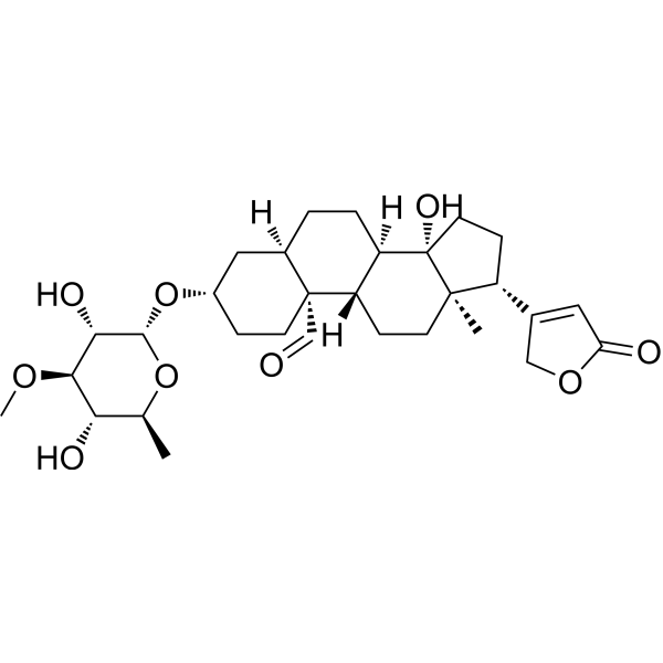
Peruvoside

Based on 1 Customer Validation
Peruvoside is a potent inhibitor of Src, PI3K, JNK, STAT, and EGFR. Peruvoside induces apoptosis and autophagy and possesses a broad spectrum of anticancer activity in breast, lung, liver cancers and leukemia. Peruvoside is a broad-spectrum and potent antiviral activity against positive-sense RNA viruses. Peruvoside sensitizes Gefitinib (HY-50895)-resistant tumour cells (A549, PC9/gef and H1975) to Gefitinib.
商品は「研究用試薬」です。人や動物の医療用・臨床診断用・食品用の製品ではありません。
研究用途以外に使用した場合、当社は一切の責任を負いかねます。
- 純度 : 99.32%
- CAS 番号: 1182-87-2
- 分子式: C30H44O9
- 分子量:548.66
-
保管条件:Powder -20°C, 3 years , 4°C, 2 years ; In solvent -80°C, 6 months , -20°C, 1 month

To place orders, for customer services and technical support, please contact: MedChemExpress USA
Tel: 609-228-6898 E-mail: [email protected] [email protected]


-
 生物活性
生物活性
-
化学情報
-
溶剤 & 溶解度
- プロトコル
- 純度とドキュメンテーション
- 参考文献
-
 Help & FAQs


EGFR アイソフォーム固有の製品をすべて表示
More

生物活性
製品説明
Cellular Effect
|
Cell Line

|
Type | Value | Description | References |
|---|---|---|---|---|
| L02 | IC50 |
>10 μM
Compound: 29
|
Cytotoxicity against human LO2 cells after 48 hrs by MTT assay
Cytotoxicity against human LO2 cells after 48 hrs by MTT assay
|
[PMID: 26714048] |
| MGC-803 | IC50 |
0.02 μM
Compound: 29
|
Cytotoxicity against human MGC803 cells after 48 hrs by MTT assay
Cytotoxicity against human MGC803 cells after 48 hrs by MTT assay
|
[PMID: 26714048] |
| SW1990 | IC50 |
0.02 μM
Compound: 29
|
Cytotoxicity against human SW1990 cells after 48 hrs by MTT assay
Cytotoxicity against human SW1990 cells after 48 hrs by MTT assay
|
[PMID: 26714048] |
体外実験
Peruvoside (50-1000 nM, 24 h) inhibits the viability and proliferation in PC9, PC9/gef, H3255, and H1975 cell lines[1].
Peruvoside (0.005-0.5 μM, 72 h) sensitizes A549, PC9/gef and H1975 to Gefitinib when in combination with Gefitinib (0.01~0.5 μM)[1].
Peruvoside (0-100 μM, 24 h) induces cell cycle arrest and apoptosis in MCF-7, HpG2, and A549 cells[2.
MedChemExpress (MCE) has not independently confirmed the accuracy of these methods. They are for reference only.
-
Cell Line:PC9, PC9/gef, H3255, and H1975 cell lines
-
Concentration:0, 0.01, 0.05, 0.1, 0.5, 1 μM
-
Incubation Time:24, 48, 72, 96 h.
-
Result:Inhibited the viabilities of TKI-sensitive and TKI-resistant cell lines at all tested time points.
Inhibited the EGFR-mutant lung cancer cell viability and proliferation with 24 h IC50s of 48 nM, 74 nM, 67 nM, 143 nM, 277 nM and 428 nM for A549, PC9, PC9/gef, H3255, H1975 and BEAS-2B cells, respectively.
Significantly inhibited the proliferation of A549 (48, 72, and 96 h) and H3255 (24, 48, 72, and 96 h) lungcancer cells.
-
Cell Line:A549, PC9/gef and H1975 cell lines
-
Concentration:0.005, 0.0075, 0.01, 0.025, 0.05, 0.5 μM (in combination with 0.01, 0.05, 0.1, 0.25, 0.5 μM Gefitinib)
-
Incubation Time:72 h.
-
Result:Had synergistic effects on A549 cells at the combination of 0.005, 0.075, or 0.01 μM and a low dose of gefitinib (0.01 or 0.05 μM).
Increased the sensitivity of PC9/gef and H1975 cells to Gefitinib at 0.025 or 0.05 μM.
-
Cell Line:MCF-7, A549 and HepG2 cell lines
-
Concentration:0-100 μM
-
Incubation Time:24 h
-
Result:Induced cell cycle arrest and apoptosis with lethal concentrations of IC50 for (MCF-7 - 100 nM), (A549 – 100 nM) and (HepG2 - 100 nM), respec-tively.
Arrested cell cycle at G0/G1 in MCF-7, A549 and HepG2 cells.
Significantly decreased the transcription of Chk1, Chk2, CDK6 and Cyclin D1 cell cycle genes in MCF-7, A549, and HepG2 cells.
体内実験
Peruvoside (0.59 mg/kg for i.p; once daily for 7 days) reduces mortality in EV-A71-infected mice model[4].
MedChemExpress (MCE) has not independently confirmed the accuracy of these methods. They are for reference only.
-
Animal Model:Lung cancer mouse model[1]
-
Dosage:0.1 mg/kg
-
Administration:Intraperitoneal injection (i.p.); Once daily for 28 days
-
Result:Reduced the tumour size to 129.9 mm3, which was significantly smaller than the control group (348 mm3).
Significantly decreased levels of phosphorylated Src Y419 in tumour tissues compared with control tissues.
-
Animal Model:EV-A71-infected mice model[4]
-
Dosage:0.59 mg/kg
-
Administration:Intraperitoneal injection (i.p.); Once daily for 7 days
-
Result:Substantially reduced clinical scores based on physical symptoms of body weight, activity,breathing, movement, and dehydration.
Showed about 6 log reduction in viral titre with 99.9% efficacy in inhibiting virus.


化学情報
-
CAS 番号 1182-87-2
-
性状 Solid
-
分子量 548.66
-
分子式 C30H44O9
-
Color White to off-white
-
SMILES
O=C[C@@]([C@](C1)([H])CC2)(CC[C@@H]1O[C@H](O[C@H]3C)[C@H]([C@@H]([C@H]3O)OC)O)[C@](CC[C@@]45C)([H])[C@]2([H])[C@]4(CC[C@@H]5C(CO6)=CC6=O)O
-
別名
Encordin
-
Structure Classification
-
Initial Source
-
輸送条件
Room temperature in continental US; may vary elsewhere.
-
保管条件
Powder -20°C 3 years 4°C 2 years In solvent -80°C 6 months -20°C 1 month


溶剤 & 溶解度
体外:
DMSO : ≥ 250 mg/mL (455.66 mM; Hygroscopic DMSO has a significant impact on the solubility of product, please use newly opened DMSO)
* "≥" means soluble, but saturation unknown.
Please refer to the solubility information to select the appropriate solvent. Once prepared, please aliquot and store the solution to prevent product inactivation from repeated freeze-thaw cycles.
Storage method and period of stock solution: -80°C, 6 months; -20°C, 1 month. When stored at -80°C, please use it within 6 months. When stored at -20°C, please use it within 1 month.
Please refer to the solubility information to select the appropriate solvent. Once prepared, please aliquot and store the solution to prevent product inactivation from repeated freeze-thaw cycles.
Storage method and period of stock solution: -80°C, 6 months; -20°C, 1 month. When stored at -80°C, please use it within 6 months. When stored at -20°C, please use it within 1 month.
濃度 (開始) × 体積 (開始) = 濃度 (終了) × 体積 (終了)


純度とドキュメンテーション
-
データシート (281 KB)
-
SDS (393 KB)
- English - EN (393 KB)
- Français - FR (393 KB)
- Deutsch - DE (393 KB)
- Norwegian - NO (393 KB)
- Español - ES (393 KB)
- Swedish - SV (393 KB)
- Italian - IT (393 KB)
- Korean - KR (393 KB)
- Portuguese - PT (393 KB)
-
取扱説明書 (2659 KB)
参考文献
[1]. Lai Y, et al. Peruvoside is a novel Src inhibitor that suppresses NSCLC cell growth and motility by downregulating multiple Src-EGFR-related pathways. Am J Cancer Res. 2022 Jun 15;12(6):2576-2593. [Content Brief]
[2]. Reddy D, et al. Peruvoside targets apoptosis and autophagy through MAPK Wnt/β-catenin and PI3K/AKT/mTOR signaling pathways in human cancers. Life Sci. 2020 Jan 15;241:117147. doi: 10.1016/j.lfs.2019.117147. Epub 2019 Dec 9. PMID: 31830480. [Content Brief]


Complete Stock Solution Preparation Table
Please refer to the solubility information to select the appropriate solvent. Once prepared, please aliquot and store the solution to prevent product inactivation from repeated freeze-thaw cycles.
Storage method and period of stock solution: -80°C, 6 months; -20°C, 1 month. When stored at -80°C, please use it within 6 months. When stored at -20°C, please use it within 1 month.
| Optional Solvent | Concentration Solvent Mass | 1 mg | 5 mg | 10 mg | 25 mg |
|---|---|---|---|---|---|
| DMSO | 1 mM | 1.8226 mL | 9.1131 mL | 18.2262 mL | 45.5656 mL |
| 5 mM | 0.3645 mL | 1.8226 mL | 3.6452 mL | 9.1131 mL | |
| 10 mM | 0.1823 mL | 0.9113 mL | 1.8226 mL | 4.5566 mL | |
| 15 mM | 0.1215 mL | 0.6075 mL | 1.2151 mL | 3.0377 mL | |
| 20 mM | 0.0911 mL | 0.4557 mL | 0.9113 mL | 2.2783 mL | |
| 25 mM | 0.0729 mL | 0.3645 mL | 0.7290 mL | 1.8226 mL | |
| 30 mM | 0.0608 mL | 0.3038 mL | 0.6075 mL | 1.5189 mL | |
| 40 mM | 0.0456 mL | 0.2278 mL | 0.4557 mL | 1.1391 mL | |
| 50 mM | 0.0365 mL | 0.1823 mL | 0.3645 mL | 0.9113 mL | |
| 60 mM | 0.0304 mL | 0.1519 mL | 0.3038 mL | 0.7594 mL | |
| 80 mM | 0.0228 mL | 0.1139 mL | 0.2278 mL | 0.5696 mL | |
| 100 mM | 0.0182 mL | 0.0911 mL | 0.1823 mL | 0.4557 mL |


Peruvoside 関連分類
HY-108016 関連分類
 Powered by Bioz