



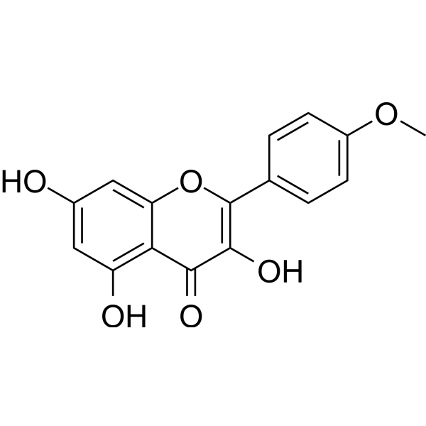
Kaempferide
Based on 7 publication(s) in Google Scholar
Kaempferide is an orally active flavonol isolated from Hippophae rhamnoides L. Kaempferide has anticancer, anti-inflammatory, antioxidant, antidiabetic, antiobesity, antihypertensive, and neuroprotective activities. Kaempferide induces apoptosis. Kaempferide promotes osteogenesis through antioxidants and can be used in osteoporosis research.
For research use only. We do not sell to patients.
- Purity : 99.80%
- CAS No.: 491-54-3
- Formula: C16H12O6
- Molecular Weight:300.26
-
Storage:Powder -20°C, 3 years , 4°C, 2 years ; In solvent -80°C, 2 years , -20°C, 1 year

To place orders, for customer services and technical support, please contact: MedChemExpress USA
Tel: 609-228-6898 E-mail: [email protected] [email protected]


-
 Biological Activity
-
Chemical Information
-
Solvent & Solubility
- Protocols
- Purity & Documentation
- References
-
 Help & FAQs


-
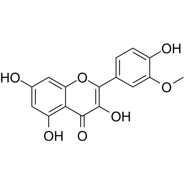
HY-N0776
IsorhamnetinEndogenous Metabolite -
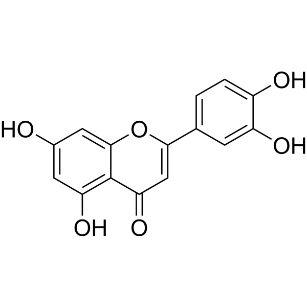
HY-N0162
LuteolinNrf2 Inhibitor -
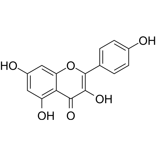
HY-14590
KaempferolEstrogen Receptor α Inhibitor -
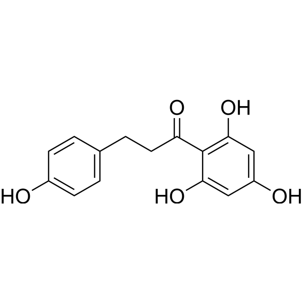
HY-N0142
PhloretinAnti-inflammatory Agent -
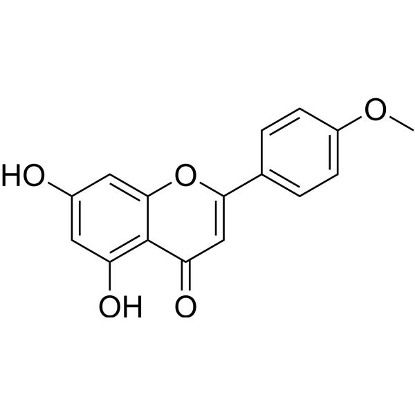
HY-N0451
AcacetinAutophagy Inducer -
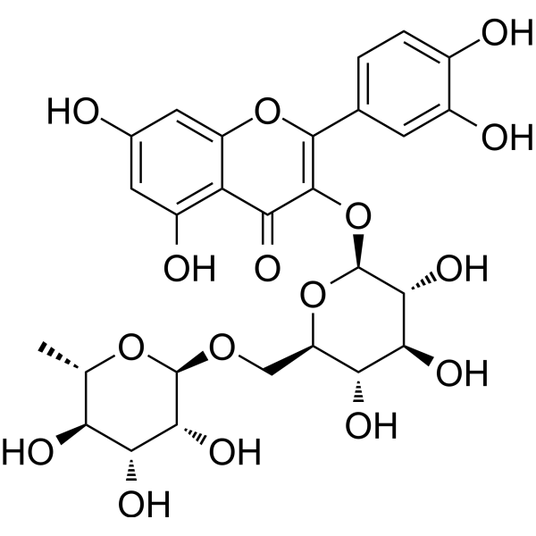
HY-N0148
RutinCBR1 Inhibitor -
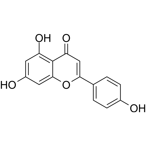
HY-N1201
ApigeninCytochrome P450 Inhibitor -
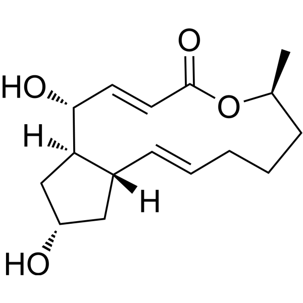
HY-16592
Brefeldin AProtein Transport Inhibitor -
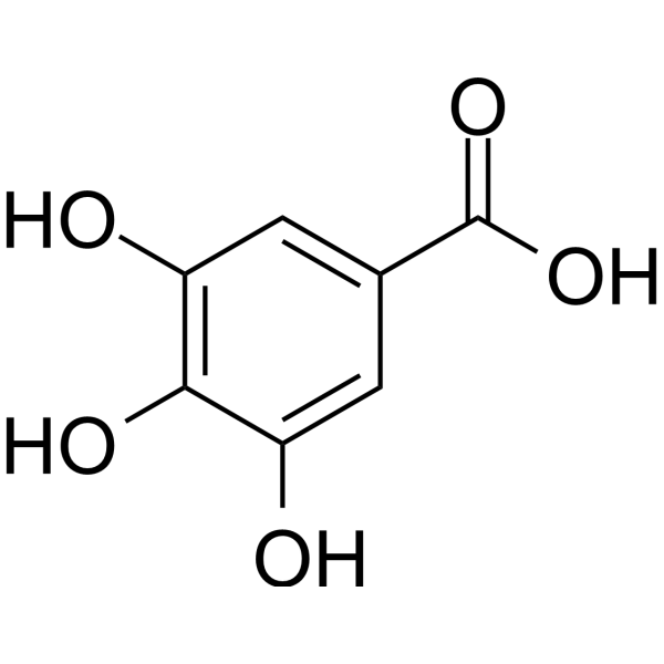
HY-N0523
Gallic acidBioactive Polyhydroxyphenolic Compound -
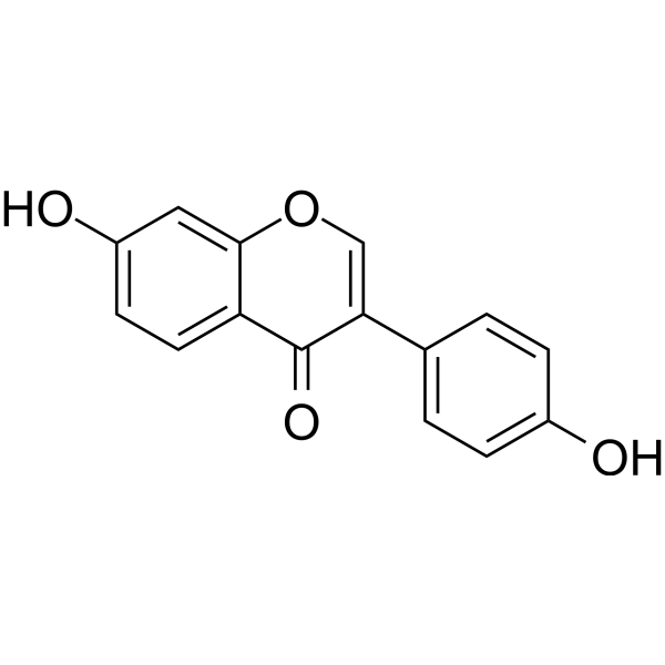
HY-N0019
DaidzeinEndogenous Metabolite -
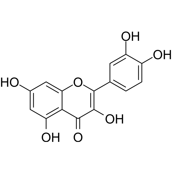
HY-18085
QuercetinFlavonoid Antioxidant -
HY-14393
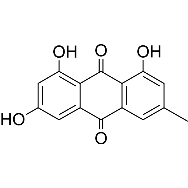
EmodinNatural Anthraquinone
Publications Citing Use of MedChemExpress (MCE) Kaempferide
More- Acta Pharm Sin B. 2021 Jan;11(1):143-155. [Abstract]
- Food Chem. 2025 Dec 30:497:146992. [Abstract]
- Food Chem. 2025 Oct 15:489:144992. [Abstract]
- Ind Crops Prod. 2025 Dec 11;239:122458.
- Int Immunopharmacol. 2026 Jun 15:179:116601. [Abstract]
- Vet Microbiol. 2026 May:316:110992. [Abstract]
- Evid Based Complement Alternat Med. 2021 Aug 26:2021:7169211. [Abstract]


Biological Activity
Description
Cellular Effect
|
Cell Line

|
Type | Value | Description | References |
|---|---|---|---|---|
| A549 | IC50 |
2.2 x 10⁻⁵ M
Compound: 13
|
Cytotoxicity against human A549 cells after 72 hrs by XTT assay
Cytotoxicity against human A549 cells after 72 hrs by XTT assay
|
[PMID: 20619940] |
| B16 | IC50 |
16 μM
Compound: 5
|
Inhibition of melanogenesis in theophylline-stimulated mouse B16-4A5 cells after 72 hrs
Inhibition of melanogenesis in theophylline-stimulated mouse B16-4A5 cells after 72 hrs
|
[PMID: 19615910] |
| B16-4A5 | IC50 |
>100 μM
Compound: 5
|
Inhibition of theophylline-stimulated mouse B16-4A5 cell proliferation assessed as cell viability after 68 hrs by WST8 assay
Inhibition of theophylline-stimulated mouse B16-4A5 cell proliferation assessed as cell viability after 68 hrs by WST8 assay
|
[PMID: 19615910] |
| HeLa | IC50 |
2 μg/mL
Compound: 7
|
Antiallergic activity in Ca(2+)-stimulated differentiated human HeLa cells assessed as inhibition of cys-leukotriene release after 6 days by ELISA
Antiallergic activity in Ca(2+)-stimulated differentiated human HeLa cells assessed as inhibition of cys-leukotriene release after 6 days by ELISA
|
[PMID: 19942440] |
| HeLa | IC50 |
2.3 x 10⁻⁵ M
Compound: 13
|
Cytotoxicity against human HeLa cells after 72 hrs by XTT assay
Cytotoxicity against human HeLa cells after 72 hrs by XTT assay
|
[PMID: 20619940] |
| HT-1080 | ED50 |
<8 μg/mL
Compound: Kaempferide
|
Cytotoxicity against human HT1080 cells by MTT assay
Cytotoxicity against human HT1080 cells by MTT assay
|
[PMID: 11000036] |
| HT-1080 | ED50 |
2.91 μg/mL
Compound: 20
|
Cytotoxicity against human HT1080 cells after 24 hrs by MTT assay
Cytotoxicity against human HT1080 cells after 24 hrs by MTT assay
|
[PMID: 9677271] |
| HT-29 | IC50 |
3.2 x 10⁻⁵ M
Compound: 13
|
Cytotoxicity against human HT-29 cells after 72 hrs by XTT assay
Cytotoxicity against human HT-29 cells after 72 hrs by XTT assay
|
[PMID: 20619940] |
| Huh-7 | CC50 |
>50 μM
Compound: 30
|
Cytotoxicity against human Huh7.5.1 cells by MTT assay
Cytotoxicity against human Huh7.5.1 cells by MTT assay
|
[PMID: 22445328] |
| KB | ED50 |
20 μg/mL
Compound: NSC-407294
|
Cytotoxicity against human KB cells
Cytotoxicity against human KB cells
|
[PMID: 469554] |
| L929 | EC50 |
129 μM
Compound: kaempferide
|
Inhibition of recombinant human TNF-alpha-induced cytotoxicity of mouse L929 cells assessed as survivality preincubated for 15 mins before TNFalpha addition measured after 24 hrs by crystal violet staining
Inhibition of recombinant human TNF-alpha-induced cytotoxicity of mouse L929 cells assessed as survivality preincubated for 15 mins before TNFalpha addition measured after 24 hrs by crystal violet staining
|
[PMID: 9287415] |
| L929 | EC50 |
140 μM
Compound: kaempferide
|
Inhibition of recombinant human TNF-alpha-induced cytotoxicity of mouse L929 cells assessed as survivality preincubated for 15 mins before TNFalpha addition measured after 24 hrs by [methyl-3H]thymidine incorporation assay
Inhibition of recombinant human TNF-alpha-induced cytotoxicity of mouse L929 cells assessed as survivality preincubated for 15 mins before TNFalpha addition measured after 24 hrs by [methyl-3H]thymidine incorporation assay
|
[PMID: 9287415] |
| Monocyte | IC50 |
5.7 μM
Compound: kaempferide
|
Inhibition of procoagulant activity in monocyte from human blood assessed as counteraction of IL1-induced tissue factor expression after 18 hrs
Inhibition of procoagulant activity in monocyte from human blood assessed as counteraction of IL1-induced tissue factor expression after 18 hrs
|
[PMID: 8882428] |
| Monocyte | IC50 |
5.7 x 10⁻⁶ M
Compound: kaempferide
|
Inhibition of procoagulant activity in monocyte from human blood assessed as counteraction of IL1-induced tissue factor expression after 18 hrs
Inhibition of procoagulant activity in monocyte from human blood assessed as counteraction of IL1-induced tissue factor expression after 18 hrs
|
[PMID: 8882428] |
In Vitro
Kaempferid is toxic in HCC cell lines (HepG2:IC50 = 27.94 µM; Huh7: IC50 = 25.65 µM; N1S1: IC50 = 15.18 µM)[1].
Kaempferid (5, 20 μM; 48 h) reduces lipid accumulation and oxidative stress in HepG2 cells induced by oral acid (OA) (0.5 mM) (HY-N1446)[2].
Kaempferide promotes osteogenesis through the FoxO1/β-catenin signaling pathway [3].
Kaempferide (10-15 μM; 24 h) is toxic in HeLa cells with an IC50 of 16 μM and can induce apoptosis[6].
MedChemExpress (MCE) has not independently confirmed the accuracy of these methods. They are for reference only. Further protocols information, click here.
-
Cell Line:HepG2
-
Concentration:5 μM, 10 μM, 20 μM . Before treatment with OA (HY-N1446) (0.5 mM; 48 h)
-
Incubation Time:48 h
-
Result:Lowered the expression of proteins related to fat production, including sterol regulatory element-binding protein 1 (SREBP1), fatty acid synthase (FAS), and stearoyl-CoA desaturase 1 (SCD-1).
Reduced the expression of two adipogenic transcription factors, peroxisome proliferator-activated receptor gamma (PPARγ) and CCAAT enhancer-binding protein β (C/EBPβ).
Enhanced the expression of heme oxygenase-1 (HO-1) and nuclear factor erythroid 2-related factor 2 (Nrf2).
In Vivo
Kaempferide (0.1-0.3 mg/kg; injection, single dose) alleviates myocardial ischemia/reperfusion injury by activating PI3K/Akt/GSK-3β pathway in Sprague Dawley (I/R)-induced rats[5].
MedChemExpress (MCE) has not independently confirmed the accuracy of these methods. They are for reference only.
-
Animal Model:High-fat diet male C57BL/6J mice model[4]
-
Dosage:10 mg/kg
-
Administration:Supplemented in daily diet, once daily for16 weeks
-
Result:Reduced the weight, organ weight, and index of mice.
Lowered the levels of glycolipids in mouse serum.
Decreased the expression levels of inflammatory-related genes, including NF-κB, IL-6, ICAM-1, VCAM-1, and TNF-α.
-
Animal Model:Ischemia/Reperfusion (I/R) SD rat model[5].
-
Dosage:0.1 mg/kg, 0.3 mg/kg, 3 mg/kg
-
Administration:Injection, Single dose. Before the I/R injury induced by Coronary Artery Ligation (CAL) in SD rats.
-
Result:Significantly improved heart function, reduced myocardial injury by reducing myocardial enzyme levels, and dose-dependently reduced the area of myocardial infarction in rats.
Significantly decreased serum levels of TNF-α, IL-6, C-reactive protein (CRP), MDA, and ROS, while increasing serum levels of SOD.
Downregulated the expression levels of nuclear factor erythroid 2-related factor 2 (Nrf2) and cleaved caspase-3, and upregulated the phosphorylation expression levels of phospho-Akt (p-Akt) and phospho-glycogen synthase kinase-3β (p-GSK-3β).


Chemical Information
-
CAS No. 491-54-3
-
Appearance Solid
-
Molecular Weight 300.26
-
Formula C16H12O6
-
Color Light yellow to yellow
-
SMILES
O=C1C(O)=C(C2=CC=C(OC)C=C2)OC3=CC(O)=CC(O)=C13
-
Synonyms
Kaempferol 4'-O-methyl ether
-
Structure Classification
-
Initial Source
-
Shipping
Room temperature in continental US; may vary elsewhere.
-
Storage
Powder -20°C 3 years 4°C 2 years In solvent -80°C 2 years -20°C 1 year


Publications (7)
-
Journal Impact Factor
-
Most Recent
-
Acta Pharm Sin B
Chrysin serves as a novel inhibitor of DGK α/FAK interaction to suppress the malignancy of esophageal squamous cell carcinoma (ESCC). [Abstract]2021 Jan;11(1):143-155. PMID: 33532186 -
Food Chem
Effects of sun drying combined with baking processes on the flavor quality of Chongqing Tuocha raw tea. [Abstract]2025 Dec 30:497:146992. PMID: 41285060 -
Food Chem
Flavonoid-mediated metabolic underpinning quality variation in red bud-sport pear mutants. [Abstract]2025 Oct 15:489:144992. PMID: 40466530 -
-
Int Immunopharmacol
Kaempferide alleviates glucocorticoid-induced osteoporosis by targeting ferroptosis through the GSK3β/Nrf-2/GPX4 pathway. [Abstract]2026 Jun 15:179:116601. PMID: 41962468 -
Vet Microbiol
The Chinese medicine monomer Schisandrin C inhibits PRRSV infection by regulating the OGT-PI3K/AKT/mTOR signaling pathway. [Abstract]2026 May:316:110992. PMID: 41865607 -
Evid Based Complement Alternat Med
Identifying Active Compounds and Mechanism of Camellia nitidissima Chi on Anti-Colon Cancer by Network Pharmacology and Experimental Validation. [Abstract]2021 Aug 26:2021:7169211. PMID: 34484402


Solvent & Solubility
In Vitro:
DMSO : 20 mg/mL (66.61 mM; Need ultrasonic; Hygroscopic DMSO has a significant impact on the solubility of product, please use newly opened DMSO)
Please refer to the solubility information to select the appropriate solvent. Once prepared, please aliquot and store the solution to prevent product inactivation from repeated freeze-thaw cycles.
Storage method and period of stock solution: -80°C, 2 years; -20°C, 1 year. When stored at -80°C, please use it within 2 years. When stored at -20°C, please use it within 1 year.
Please refer to the solubility information to select the appropriate solvent. Once prepared, please aliquot and store the solution to prevent product inactivation from repeated freeze-thaw cycles.
Storage method and period of stock solution: -80°C, 2 years; -20°C, 1 year. When stored at -80°C, please use it within 2 years. When stored at -20°C, please use it within 1 year.
Concentration (start) × Volume (start) = Concentration (final) × Volume (final)
In Vivo:
Select the appropriate dissolution method based on your experimental animal and administration route.
- For the following dissolution methods, please ensure to first prepare a clear stock solution using an In Vitro approach and then sequentially add co-solvents:
- To ensure reliable experimental results, the clarified stock solution can be appropriately stored based on storage conditions. As for the working solution for In Vivo experiments, it is recommended to prepare freshly and use it on the same day.
- The percentages shown for the solvents indicate their volumetric ratio in the final prepared solution. If precipitation or phase separation occurs during preparation, heat and/or sonication can be used to aid dissolution.
Add each solvent one by one: 10% DMSO 40% PEG300 5% Tween-80 45% Saline
Solubility: 2 mg/mL (6.66 mM); Suspended solution; Need ultrasonic
This protocol yields a suspended solution of 2 mg/mL. Suspended solution can be used for oral and intraperitoneal injection.
Taking 1 mL working solution as an example, add 100 μL DMSO stock solution (20.0 mg/mL) to 400 μL PEG300, and mix evenly; then add 50 μL Tween-80 and mix evenly; then add 450 μL Saline to adjust the volume to 1 mL.
Preparation of Saline: Dissolve 0.9 g sodium chloride in ddH₂O and dilute to 100 mL to obtain a clear Saline solution.
In Vivo Dissolution Calculator
Please enter the basic information of animal experiments:
-
-
-
-
Recommended: Prepare an additional quantity of animals to account for potential losses during experiments.
Please enter your animal formula composition:
-
%DMSO +
Recommended: Keep the proportion of DMSO in working solution below 2% if your animal is weak.
-
%+
-
+%Tween-80 + +
-
%Saline +
The co-solvents required include: DMSO, . All of co-solvents are available by MedChemExpress (MCE). , Tween 80. All of co-solvents are available by MedChemExpress (MCE).
Working solution concentration: 0.22 mg/mL
Method for preparing stock solution: mg drug dissolved in μL DMSO. Stock solution concentration: mg/mL.
1. Take μL DMSO stock solution;
2. Add μL .
μL , mix evenly;
3. Then add μL Tween 80, mix evenly;
4. Then add μL
Please ensure that the stock solution in the first step is dissolved to a clear state, and add co-solvents in sequence. You can use ultrasonic heating (ultrasonic cleaner, recommended frequency 20-40 kHz), vortexing, etc. to assist dissolution.

Protocols
-
Apoptosis
Apoptosis, also called programmed cell death, is generally characterized by distinct morphological characteristics.
-
TUNEL staining for apoptotic DNA fragmentation
TUNEL staining detects DNA strand breaks by using terminal deoxynucleotidyl transferase to add labeled nucleotides to exposed 3′-OH DNA termini, generating either microscopic staining in fixed cells or tissue sections, or fluorescence/cytometric signal in cell suspensions. TUNEL positivity reflects DNA fragmentation but should not be interpreted alone as definitive apoptosis, because TUNEL can also label necrotic, autolytic, mechanically damaged, or DNA-repair-associated DNA breaks.
-
Annexin V plus membrane-impermeant dye apoptosis staining
Annexin V-based apoptosis assays rely on the detection of phosphatidylserine (PS) externalization from the inner leaflet of the plasma membrane to the outer leaflet, an early biochemical hallmark of apoptosis. Fluorescently labeled Annexin V binds PS in a calcium-dependent manner, enabling identification of early apoptotic cells by flow cytometry or fluorescence microscopy. When combined with a membrane-impermeant DNA-binding dye (e. g. , propidium iodide), this approach allows discrimination between viable (Annexin V−/dye−), early apoptotic (Annexin V+/dye−), and late apoptotic or necrotic (Annexin V+/dye+) cell populations by assessing membrane integrity and PS exposure.
-
Mesenchymal stromal/stem cell osteogenic differentiation
Mesenchymal stromal/stem cells can be induced toward an osteoblast-like lineage in vitro by culture in osteogenic medium containing dexamethasone, ascorbic acid or ascorbate-2-phosphate, and β-glycerophosphate; the differentiation process is commonly evaluated by alkaline phosphatase activity, osteogenic marker expression, collagenous matrix formation, and calcium-rich matrix mineralization. The main readouts are alkaline phosphatase activity as an early osteogenic marker and Alizarin Red S staining as a calcium-deposit readout for mineralized extracellular matrix; Alizarin Red S can be inspected microscopically or extracted and measured colorimetrically at 405 nm.
-
Apoptosis Solutions
Apoptosis is a regulated, generally non-lytic cell-death pathway that removes unwanted, damaged, infected, or abnormal cells through coordinated morphological changes, caspase activation, DNA fragmentation, and membrane remodeling. The intrinsic apoptosis pathway is controlled mainly by mitochondrial outer membrane permeabilization, BCL-2 family proteins, cytochrome c release, apoptosome formation, caspase-9 activation, and downstream executioner caspase-3/7 activation. The extrinsic apoptosis pathway is initiated by death receptors such as Fas, TNFR, and TRAIL receptors, which recruit adaptor proteins and activate caspase-8 before engaging executioner caspases or mitochondrial amplification through BID cleavage. Apoptosis is linked to many phenotypes, including cancer cell killing, tissue homeostasis, immune regulation, neurodegeneration, infection response, and treatment-induced cytotoxicity; unresolved questions include how apoptosis interacts with necroptosis, pyroptosis, ferroptos
-
Research Protocol for Inflammation-related Diseases
The NLRP3 inflammasome is a cytosolic innate immune signaling platform that integrates priming signals and danger-signal activation to promote caspase-1 activation, maturation of IL-1β and IL-18, and gasdermin D-mediated pyroptotic cell death. The core experimental logic is to determine whether inflammatory disease phenotypes are driven by increased NLRP3 expression, ASC-containing inflammasome assembly, caspase-1 cleavage, GSDMD cleavage, and extracellular release of IL-1β/IL-18 rather than by nonspecific cell injury alone. The pathway is strongly linked to inflammation-related disease phenotypes because monosodium urate crystals activate NALP3/NLRP3 inflammasome signaling in gout-like crystal inflammation, cholesterol crystals activate NLRP3 inflammasomes in atherogenesis models, and DSS-induced intestinal inflammation has been reported to involve NLRP3 inflammasome activity. However, experimental colitis studies also show context-dependent protective effects of NLRP3 inflammasome co


Purity & Documentation
-
Data Sheet (282 KB)
-
SDS (396 KB)
- English - EN (396 KB)
- Français - FR (396 KB)
- Deutsch - DE (396 KB)
- Norwegian - NO (396 KB)
- Español - ES (396 KB)
- Swedish - SV (396 KB)
- Italian - IT (396 KB)
- Korean - KR (396 KB)
- Portuguese - PT (396 KB)
-
Handling Instructions (2659 KB)
References
[2]. Tie F, et al. Kaempferol and kaempferide attenuate oleic acid-induced lipid accumulation and oxidative stress in HepG2 cells[J]. International Journal of Molecular Sciences, 2021, 22(16): 8847. [Content Brief]
[3]. Ma X, et al. Kaempferide enhances antioxidant capacity to promote osteogenesis through FoxO1/β-catenin signaling pathway[J]. European Journal of Pharmacology, 2021, 911: 174555. [Content Brief]
[4]. Tang H, et al. Kaempferide improves oxidative stress and inflammation by inhibiting the TLR4/IκBα/NF-κB pathway in obese mice[J]. Iranian Journal of Basic Medical Sciences, 2021, 24(4): 493. [Content Brief]
[5]. Wang D, et al. Kaempferide Protects against Myocardial Ischemia/Reperfusion Injury through Activation of the PI3K/Akt/GSK-3β Pathway. Mediators Inflamm. 2017;2017:5278218. [Content Brief]


Complete Stock Solution Preparation Table
Please refer to the solubility information to select the appropriate solvent. Once prepared, please aliquot and store the solution to prevent product inactivation from repeated freeze-thaw cycles.
Storage method and period of stock solution: -80°C, 2 years; -20°C, 1 year. When stored at -80°C, please use it within 2 years. When stored at -20°C, please use it within 1 year.
| Optional Solvent | Concentration Solvent Mass | 1 mg | 5 mg | 10 mg | 25 mg |
|---|---|---|---|---|---|
| DMSO | 1 mM | 3.3304 mL | 16.6522 mL | 33.3045 mL | 83.2612 mL |
| 5 mM | 0.6661 mL | 3.3304 mL | 6.6609 mL | 16.6522 mL | |
| 10 mM | 0.3330 mL | 1.6652 mL | 3.3304 mL | 8.3261 mL | |
| 15 mM | 0.2220 mL | 1.1101 mL | 2.2203 mL | 5.5507 mL | |
| 20 mM | 0.1665 mL | 0.8326 mL | 1.6652 mL | 4.1631 mL | |
| 25 mM | 0.1332 mL | 0.6661 mL | 1.3322 mL | 3.3304 mL | |
| 30 mM | 0.1110 mL | 0.5551 mL | 1.1101 mL | 2.7754 mL | |
| 40 mM | 0.0833 mL | 0.4163 mL | 0.8326 mL | 2.0815 mL | |
| 50 mM | 0.0666 mL | 0.3330 mL | 0.6661 mL | 1.6652 mL | |
| 60 mM | 0.0555 mL | 0.2775 mL | 0.5551 mL | 1.3877 mL |


Kaempferide Related Classifications
HY-15449 Related Classifications
 Powered by Bioz
Keywords
- Kaempferide
- 491-54-3
- Kaempferol 4'-O-methyl ether
- Estrogen Receptor/ERR
- Autophagy
- Bacterial
- Influenza Virus
- Apoptosis
- PI3K
- Akt
- GSK-3
- FOXO
- β-catenin
- HCC
- osteoporosis
- osteogenesis
- antioxidant
- non-alcoholic fatty liver disease (NAFLD)
- HepG2
- HeLa
- Huh7
- N1S1
- apoptosis
- diabetes
- obesity
- hypertension
- neuroprotection
- SD (Sprague Dawley)
- C57BL /6J
- myocardial ischemia/reperfusion injury
- Inhibitor
- inhibitor
- inhibit