



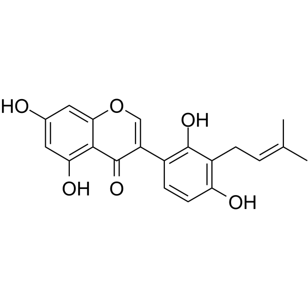
Licoisoflavone A
Based on 2 publication(s) in Google Scholar
Licoisoflavone A is an orally active isoflavone. Licoisoflavone A inhibits proliferation, induces apoptosis, and causes G1/S phase arrest in colorectal cancer (CRC) cells. Licoisoflavone A inhibits the CDK2-Cyclin E1 axis. Licoisoflavone A inhibits lipid peroxidation with an IC50 of 7.2 μM. Licoisoflavone A shows a dose-dependent inhibition effect on SARS-CoV-2 infection. Licoisoflavone A exhibits significant anti-tumor efficacy in mice bearing CT26 cell subcutaneous xenografts. Licoisoflavone A can be used for the study of colorectal cancer and SARS-CoV-2 infection.
For research use only. We do not sell to patients.
- Purity : 99.53%
- CAS No.: 66056-19-7
- Formula: C20H18O6
- Molecular Weight:354.35
-
Storage:Powder -20°C, 3 years , 4°C, 2 years ; In solvent -80°C, 6 months , -20°C, 1 month

To place orders, for customer services and technical support, please contact: MedChemExpress USA
Tel: 609-228-6898 E-mail: [email protected] [email protected]


-
 Biological Activity
-
Chemical Information
-
Solvent & Solubility
- Protocols
- Purity & Documentation
- References
-
 Help & FAQs


Publications Citing Use of MedChemExpress (MCE) Licoisoflavone A
More

Biological Activity
Description
In Vitro
Licoisoflavone A (0-100 μM) inhibits RNA-dependent RNA polymerase (RdRp) and cAMP-specific 3’,5’-cyclic phosphodiesterase 4 (PDE4) with IC50 values of 47.31 μM and 1.77 μM, respectively[1].
Licoisoflavone A (5 μM, 2 days) significantly reduces the cross-sectional area of Ang II-induced hypertrophic H9c2 cells by approximately 46%[2].
Licoisoflavone A (15 μM, 48 h) upregulates Sirt3 expression and reduces MnSOD acetylation at K68 in PE-induced neonatal rat cardiomyocytes[2].
Licoisoflavone A (15 μM, 48 h) decreases relative ANF and BNP levels in Phenylephrine (PE) (HY-B0769)-induced hypertrophic cardiac cells, but this effect is blocked when co-treated with Sirt3 inhibitor 3-TYP (HY-108331)[2].
Licoisoflavone A ( 6.25-25 μM, 48 h) significantly inhibits proliferation, induces apoptosis, and causes G1/S phase arrest in HCT116 and SW480 colorectal cancer (CRC) cells[4].
Licoisoflavone A (6.25-25 μM, 48 h) downregulates CDK2, Cyclin E1, and phosphorylated Rb (p-Rb) while upregulating p27 in HCT116 and SW480 cells, inhibiting the CDK2-Cyclin E1 axis[4].
Licoisoflavone A (1-1000000 nM, 3 days) dose-dependently inhibits viability of 5 patient-derived CRC organoids (CRCOs) with IC50 values ranging from 7.02 to 82.98 μM, and shows lower toxicity to normal colorectal organoids (CNOs)[4].
MedChemExpress (MCE) has not independently confirmed the accuracy of these methods. They are for reference only. Further protocols information, click here.
-
Cell Line:PE-induced hypertrophic cardiac cells
-
Concentration:15 μM
-
Incubation Time:48 h
-
Result:Decreased relative ANF and BNP levels in PE-induced hypertrophic cardiac cells, but this effect is blocked when co-treated with Sirt3 inhibitor 3-TYP.
-
Cell Line:HCT116 and SW480 colorectal cancer (CRC) cells
-
Concentration:6.25, 12.5, 25 μM
-
Incubation Time:48 h
-
Result:CausesdG1/S phase arrest in HCT116 and SW480 colorectal cancer (CRC) cells.
-
Cell Line:HCT116 and SW480 colorectal cancer (CRC) cells
-
Concentration:6.25, 12.5, 25 μM
-
Incubation Time:48 h
-
Result:Induced apoptosis in HCT116 and SW480 colorectal cancer (CRC) cells.
-
Cell Line:HCT116 and SW480 colorectal cancer (CRC) cells
-
Concentration:6.25, 12.5, 25 μM
-
Incubation Time:48 h
-
Result:Downregulated CDK2, Cyclin E1, and phosphorylated Rb (p-Rb).
Upregulated p27.
In Vivo
MedChemExpress (MCE) has not independently confirmed the accuracy of these methods. They are for reference only.
-
Animal Model:CT26.WT cells (106 in 100 μL) were subcutaneously inoculated into the right axillary region of 6-week-old male BALB/c SPF mice[4].
-
Dosage:25, 50 mg/kg
-
Administration:p.o. daily for 2 weeks
-
Result:Achieved dose-dependent tumor growth inhibition in BALB/c mice bearing CT26 cell xenografts, with reduced tumor weight and volume at 25 mg/kg and 50 mg/kg doses.
Showed no significant toxicity, with no abnormal changes in liver and spleen histopathology and serum ALT/AST levels.
Reduced the expression of CDK2 and Cyclin E1 in tumor tissues, while increased p27 expression.
Inhibited phosphorylation of Rb in tumor tissues.


Chemical Information
-
CAS No. 66056-19-7
-
Appearance Solid
-
Molecular Weight 354.35
-
Formula C20H18O6
-
Color Light yellow to yellow
-
SMILES
O=C1C(C2=C(C(C/C=C(C)/C)=C(O)C=C2)O)=COC3=CC(O)=CC(O)=C13
-
Structure Classification
-
Initial Source
-
Shipping
Room temperature in continental US; may vary elsewhere.
-
Storage
Powder -20°C 3 years 4°C 2 years In solvent -80°C 6 months -20°C 1 month


Publications (2)
-
Journal Impact Factor
-
Most Recent
-
Food Chem
Effects of sun drying combined with baking processes on the flavor quality of Chongqing Tuocha raw tea. [Abstract]2025 Dec 30:497:146992. PMID: 41285060 -
Food Chem
Flavonoid-mediated metabolic underpinning quality variation in red bud-sport pear mutants. [Abstract]2025 Oct 15:489:144992. PMID: 40466530


Solvent & Solubility
In Vitro:
DMSO : 100 mg/mL (282.21 mM; Need ultrasonic; Hygroscopic DMSO has a significant impact on the solubility of product, please use newly opened DMSO)
Please refer to the solubility information to select the appropriate solvent. Once prepared, please aliquot and store the solution to prevent product inactivation from repeated freeze-thaw cycles.
Storage method and period of stock solution: -80°C, 6 months; -20°C, 1 month. When stored at -80°C, please use it within 6 months. When stored at -20°C, please use it within 1 month.
Please refer to the solubility information to select the appropriate solvent. Once prepared, please aliquot and store the solution to prevent product inactivation from repeated freeze-thaw cycles.
Storage method and period of stock solution: -80°C, 6 months; -20°C, 1 month. When stored at -80°C, please use it within 6 months. When stored at -20°C, please use it within 1 month.
Concentration (start) × Volume (start) = Concentration (final) × Volume (final)
In Vivo:
Select the appropriate dissolution method based on your experimental animal and administration route.
- For the following dissolution methods, please ensure to first prepare a clear stock solution using an In Vitro approach and then sequentially add co-solvents:
- To ensure reliable experimental results, the clarified stock solution can be appropriately stored based on storage conditions. As for the working solution for In Vivo experiments, it is recommended to prepare freshly and use it on the same day.
- The percentages shown for the solvents indicate their volumetric ratio in the final prepared solution. If precipitation or phase separation occurs during preparation, heat and/or sonication can be used to aid dissolution.
Add each solvent one by one: 10% DMSO 40% PEG300 5% Tween-80 45% Saline
Solubility: ≥ 2.5 mg/mL (7.06 mM); Clear solution
This protocol yields a clear solution of ≥ 2.5 mg/mL (saturation unknown).
Taking 1 mL working solution as an example, add 100 μL DMSO stock solution (25.0 mg/mL) to 400 μL PEG300, and mix evenly; then add 50 μL Tween-80 and mix evenly; then add 450 μL Saline to adjust the volume to 1 mL.
Preparation of Saline: Dissolve 0.9 g sodium chloride in ddH₂O and dilute to 100 mL to obtain a clear Saline solution.
Add each solvent one by one: 10% DMSO 90% (20% SBE-β-CD in Saline)
Solubility: ≥ 2.5 mg/mL (7.06 mM); Clear solution
This protocol yields a clear solution of ≥ 2.5 mg/mL (saturation unknown).
Taking 1 mL working solution as an example, add 100 μL DMSO stock solution (25.0 mg/mL) to 900 μL 20% SBE-β-CD in Saline, and mix evenly.
Preparation of 20% SBE-β-CD in Saline (4°C, storage for one week): 2 g SBE-β-CD powder is dissolved in 10 mL Saline, completely dissolve until clear.
In Vivo Dissolution Calculator
Please enter the basic information of animal experiments:
-
-
-
-
Recommended: Prepare an additional quantity of animals to account for potential losses during experiments.
Please enter your animal formula composition:
-
%DMSO +
Recommended: Keep the proportion of DMSO in working solution below 2% if your animal is weak.
-
%+
-
+%Tween-80 + +
-
%Saline +
The co-solvents required include: DMSO, . All of co-solvents are available by MedChemExpress (MCE). , Tween 80. All of co-solvents are available by MedChemExpress (MCE).
Working solution concentration: 0.22 mg/mL
Method for preparing stock solution: mg drug dissolved in μL DMSO. Stock solution concentration: mg/mL.
1. Take μL DMSO stock solution;
2. Add μL .
μL , mix evenly;
3. Then add μL Tween 80, mix evenly;
4. Then add μL
Please ensure that the stock solution in the first step is dissolved to a clear state, and add co-solvents in sequence. You can use ultrasonic heating (ultrasonic cleaner, recommended frequency 20-40 kHz), vortexing, etc. to assist dissolution.

Protocols
-
Research Protocol for Infectious Diseases
Infectious-disease experiments test how pathogens interact with host barriers, innate immune receptors, inflammatory signaling, pathogen replication, and tissue injury; pattern-recognition receptors such as TLRs, RIG-I-like receptors, NOD-like receptors, and inflammasomes detect microbial molecules and activate NF-κB, interferon, and cytokine responses. The central hypothesis is that infection severity reflects the balance between pathogen burden and host response: protective inflammation restricts pathogen growth, whereas excessive or mislocalized inflammation contributes to tissue damage and disease phenotype. Unresolved questions include which host pathways are protective versus pathogenic, why some infection models fail to translate to human disease, and which combined readouts best predict clinically relevant infection outcomes.
-
Annexin V plus membrane-impermeant dye apoptosis staining
Annexin V-based apoptosis assays rely on the detection of phosphatidylserine (PS) externalization from the inner leaflet of the plasma membrane to the outer leaflet, an early biochemical hallmark of apoptosis. Fluorescently labeled Annexin V binds PS in a calcium-dependent manner, enabling identification of early apoptotic cells by flow cytometry or fluorescence microscopy. When combined with a membrane-impermeant DNA-binding dye (e. g. , propidium iodide), this approach allows discrimination between viable (Annexin V−/dye−), early apoptotic (Annexin V+/dye−), and late apoptotic or necrotic (Annexin V+/dye+) cell populations by assessing membrane integrity and PS exposure.
-
BrdU Incorporation Assay
Bromodeoxyuridine (BrdU) incorporation assay is based on the principle that BrdU, a thymidine analog, is incorporated into newly synthesized DNA during the S phase of the cell cycle, thereby serving as a marker of DNA replication and cellular proliferation. Incorporated BrdU can be detected using anti-BrdU antibodies following DNA denaturation, enabling visualization or quantification of proliferating cells through immunochemical detection methods such as immunofluorescence or immunohistochemistry.
-
Protocol for Cell Cycle
Cell-cycle analysis by flow cytometry measures DNA content in single cells to estimate the fraction of cells in G0/G1, S, and G2/M phases. Propidium iodide intercalates into DNA, and after RNA removal with RNase, fluorescence intensity reflects cellular DNA content: 2N cells are assigned to G0/G1, cells between 2N and 4N to S phase, and 4N cells to G2/M. DNA-content analysis alone cannot reliably separate G0 from G1 or G2 from M. Ki-67 can distinguish quiescent G0 cells from cycling cells, EdU or BrdU incorporation marks active DNA synthesis in S phase, and phospho-histone H3 staining identifies mitotic cells within the 4N population.
-
Cell Viability Determination by MTT Colorimetric Assay
The following protocol uses the MTT colorimetric assay as a classic literature-established method for assessing cell viability/metabolic activity in cultured mammalian cells. MTT[3-(4,5-dimethylthiazol-2-yl)-2,5-diphenyltetrazolium bromide] is reduced by metabolically active cells to a colored formazan product; the amount of formazan is quantified spectrophotometrically and provides an indirect measure of metabolically active viable cells. Importantly, MTT reduction reflects cellular oxidoreductase/metabolic activity rather than an absolute direct count of living cells, so changes in cellular metabolism can alter the signal independently of cell number.
-
How to Select the Route of Administration for Mammals
Route-of-administration selection in mammals is a pharmacokinetic, pharmacodynamic, formulation, animal-welfare, and translational decision, not a default technical choice. The selected route should match the study goal: intravenous dosing is most useful when complete systemic exposure and rapid onset are required, oral dosing is most translational for orally intended medicines but is affected by absorption and first-pass metabolism, subcutaneous or intramuscular dosing can provide slower systemic exposure, and intraperitoneal dosing can be useful in rodent proof-of-concept studies but may have limited clinical translation. Published route-comparison studies show that the same compound can produce different exposure, onset, bioavailability, tissue distribution, and tolerability depending on route; therefore, route choice should be supported by pilot pharmacokinetic or pharmacodynamic evidence when the literature is insufficient. Unresolved questions include how to standardize route sel
-
EdU Incorporation Assay (Click Chemistry-Based DNA Synthesis Measurement)
The EdU incorporation assay measures DNA synthesis by adding the thymidine analog 5-ethynyl-2′-deoxyuridine to cells or tissues, where it is incorporated into newly synthesized DNA during S phase. Incorporated EdU is detected by copper-catalyzed azide-alkyne cycloaddition, in which a fluorescent azide covalently reacts with the ethynyl group on EdU, allowing S-phase cells to be detected by fluorescence microscopy, flow cytometry, or high-content imaging. EdU detection does not require DNA denaturation or anti-BrdU antibody access, which preserves sample structure and improves compatibility with immunostaining and multiparameter cytometry compared with BrdU-based detection. EdU can be cytotoxic in a cell-type- and exposure-dependent manner, so pulse duration, concentration, and continuous-labeling designs should be validated for each cell type.
-
Apoptosis
Apoptosis, also called programmed cell death, is generally characterized by distinct morphological characteristics.
-
TUNEL staining for apoptotic DNA fragmentation
TUNEL staining detects DNA strand breaks by using terminal deoxynucleotidyl transferase to add labeled nucleotides to exposed 3′-OH DNA termini, generating either microscopic staining in fixed cells or tissue sections, or fluorescence/cytometric signal in cell suspensions. TUNEL positivity reflects DNA fragmentation but should not be interpreted alone as definitive apoptosis, because TUNEL can also label necrotic, autolytic, mechanically damaged, or DNA-repair-associated DNA breaks.
-
Flow cytometric DNA-content cell-cycle staining
Flow cytometric DNA-content cell-cycle staining measures the fluorescence intensity of DNA-bound fluorochromes in single cells or nuclei to estimate DNA content distributions, allowing assignment of populations to G0/G1, S, and G2/M phases by DNA histogram deconvolution. Propidium iodide (PI) intercalates into DNA, and PI fluorescence is proportional to cellular DNA content when staining is performed under conditions that make DNA accessible and minimize non-DNA signal. Cells with G2/M DNA content are expected to show approximately twice the fluorescence intensity of G0/G1 cells, while S-phase cells occupy intermediate fluorescence values. PI-based DNA-content analysis can also detect cells with fractional DNA content, often reported as sub-G1, when DNA fragmentation and extraction during staining reduce retained DNA signal in apoptotic cells. DAPI is an alternative DNA fluorochrome for univariate DNA-content analysis, while bivariate approaches combining DNA content with proliferation
-
Cell Cytotoxicity Assay
Cytotoxicity assays are usually based on the assessment of cell membrane damage, which can also be indirectly detected by measuring cell viability. Detection methods include MTT assay, CKK-8 assay, LDH assay and ATP assay, etc.
-
Apoptosis Solutions
Apoptosis is a regulated, generally non-lytic cell-death pathway that removes unwanted, damaged, infected, or abnormal cells through coordinated morphological changes, caspase activation, DNA fragmentation, and membrane remodeling. The intrinsic apoptosis pathway is controlled mainly by mitochondrial outer membrane permeabilization, BCL-2 family proteins, cytochrome c release, apoptosome formation, caspase-9 activation, and downstream executioner caspase-3/7 activation. The extrinsic apoptosis pathway is initiated by death receptors such as Fas, TNFR, and TRAIL receptors, which recruit adaptor proteins and activate caspase-8 before engaging executioner caspases or mitochondrial amplification through BID cleavage. Apoptosis is linked to many phenotypes, including cancer cell killing, tissue homeostasis, immune regulation, neurodegeneration, infection response, and treatment-induced cytotoxicity; unresolved questions include how apoptosis interacts with necroptosis, pyroptosis, ferroptos
-
Ferroptosis Solutions
Ferroptosis is an iron-dependent, non-apoptotic form of regulated cell death characterized by lethal lipid peroxidation and sensitivity to suppression by iron chelators or lipophilic radical-trapping antioxidants. The core pathway links cystine uptake through system Xc−, glutathione availability, GPX4-dependent detoxification of phospholipid hydroperoxides, iron-dependent oxidative reactions, and polyunsaturated-phospholipid metabolism into a cell-death program that is biochemically and morphologically distinct from apoptosis, necrosis, and autophagy. The ferroptosis pathway is experimentally linked to phenotype through chemical and genetic perturbation. Erastin induces ferroptosis by inhibiting cystine uptake through system Xc− and weakening antioxidant defenses, while GPX4 inhibition or depletion causes lipid peroxide accumulation and ferroptotic cancer-cell death. ACSL4 and oxidizable arachidonoyl- or adrenoyl-containing phosphatidylethanolamines shape ferroptosis sensitivity by con


Purity & Documentation
-
Data Sheet (280 KB)
-
SDS (393 KB)
- English - EN (393 KB)
- Français - FR (393 KB)
- Deutsch - DE (393 KB)
- Norwegian - NO (393 KB)
- Español - ES (393 KB)
- Swedish - SV (393 KB)
- Italian - IT (393 KB)
- Korean - KR (393 KB)
- Portuguese - PT (393 KB)
-
Handling Instructions (2659 KB)
References
[1]. Xu H, et al. Bioactive compounds from Huashi Baidu decoction possess both antiviral and anti-inflammatory effects against COVID-19. Proc Natl Acad Sci U S A. 2023 May 2;120(18):e2301775120. [Content Brief]
[2]. Guo R, et al. High content screening identifies licoisoflavone A as a bioactive compound of Tongmaiyangxin Pills to restrain cardiomyocyte hypertrophy via activating Sirt3. Phytomedicine. 2020 Mar;68:153171. [Content Brief]
[4]. Cui J, et al. Licoisoflavone A inhibits colorectal cancer cell proliferation through targeting CDK2-Cyclin E1 axis-mediated cell cycle transition. Biochem Pharmacol. 2025 Oct;240:117124. [Content Brief]


Complete Stock Solution Preparation Table
Please refer to the solubility information to select the appropriate solvent. Once prepared, please aliquot and store the solution to prevent product inactivation from repeated freeze-thaw cycles.
Storage method and period of stock solution: -80°C, 6 months; -20°C, 1 month. When stored at -80°C, please use it within 6 months. When stored at -20°C, please use it within 1 month.
| Optional Solvent | Concentration Solvent Mass | 1 mg | 5 mg | 10 mg | 25 mg |
|---|---|---|---|---|---|
| DMSO | 1 mM | 2.8221 mL | 14.1103 mL | 28.2207 mL | 70.5517 mL |
| 5 mM | 0.5644 mL | 2.8221 mL | 5.6441 mL | 14.1103 mL | |
| 10 mM | 0.2822 mL | 1.4110 mL | 2.8221 mL | 7.0552 mL | |
| 15 mM | 0.1881 mL | 0.9407 mL | 1.8814 mL | 4.7034 mL | |
| 20 mM | 0.1411 mL | 0.7055 mL | 1.4110 mL | 3.5276 mL | |
| 25 mM | 0.1129 mL | 0.5644 mL | 1.1288 mL | 2.8221 mL | |
| 30 mM | 0.0941 mL | 0.4703 mL | 0.9407 mL | 2.3517 mL | |
| 40 mM | 0.0706 mL | 0.3528 mL | 0.7055 mL | 1.7638 mL | |
| 50 mM | 0.0564 mL | 0.2822 mL | 0.5644 mL | 1.4110 mL | |
| 60 mM | 0.0470 mL | 0.2352 mL | 0.4703 mL | 1.1759 mL | |
| 80 mM | 0.0353 mL | 0.1764 mL | 0.3528 mL | 0.8819 mL | |
| 100 mM | 0.0282 mL | 0.1411 mL | 0.2822 mL | 0.7055 mL |


Licoisoflavone A Related Classifications
HY-N3389 Related Classifications
 Powered by Bioz